Fetal echocardiography
(fetal echocardiography) is a special ultrasound technique that allows you to study the morphological and functional features of the fetal heart during pregnancy. Allows non-invasive detection of congenital heart pathology even before birth. Usually performed at 18-24 weeks of gestation. Echocardiography of the fetus makes it possible to assess the condition of the cavities of the heart, the interatrial septa of the heart valves, and the great vessels. Fetal echocardiography is used for prenatal (prenatal) diagnosis of congenital heart defects and heart rhythm disorders in the fetus, which makes it possible to optimally plan further tactics for their treatment.
Fetal echocardiography is a detailed study of the anatomy and functioning of the fetal heart using high-frequency ultrasound waves. Assessment of fetal cardiac activity can be carried out through auscultation, phonocardiography, cardiotocography. Fetal echocardiography is not a mandatory ultrasound scan during pregnancy and is prescribed only in cases where other studies give reason to suspect that the child has congenital heart disease or severe arrhythmia. If cardiac defects incompatible with life are detected in the fetus, it is recommended to terminate the pregnancy. In cases where congenital heart defects are subject to surgical correction, the operation can be performed shortly after birth. Fetal echocardiography in Moscow, as a prenatal diagnostic technique, is indispensable for developing a strategy for managing pregnancy, childbirth and the early neonatal period.
When and how is the examination carried out?
The examination is carried out at 20–25 obstetric weeks of pregnancy.
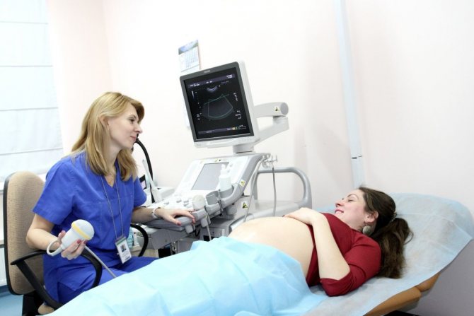
Echocardiography is a non-invasive procedure, which means it will not have any effect on the child. The pregnant mother lies quietly for 20 minutes, the doctor examines the baby’s heart with a special ultrasound sensor through the mother’s anterior abdominal wall. Next, using a computer program, the specialist processes the images and produces the result. All in one patient visit.
A condition for qualitative analysis is the presence of cardiovascular programs (programs for examining cardiovascular activity) on the device, equipped with a special sensor with a scanning frequency of at least 5 MHz, color mapping of blood flow and Doppler.
Varieties of echo kg fetus
Many expectant mothers are interested in the question of how to make an echo of a kg fetus. The method includes several different types of techniques, the choice of which is made based on the purpose of the study.
These can be the following types of echocardiography method:
- A two-dimensional echocardiogram (or 2-D) allows you to study the anatomy of the main structures of the heart (valves, chambers, vascular system).
- M-mode (one-dimensional) allows you to observe the state of the leaflets, valves and walls of the heart during their operation.
- Pulsed wave Doppler echocardiography makes it possible to study and evaluate hemodynamics and heart rhythm.
Who carries out this research?
Fetal echocardiography is performed by prenatal diagnostic experts who have undergone special training and are certified to perform this procedure. Of course, these are obstetricians-gynecologists or cardiologists, specialists with many years of practice and established clinical thinking in this field.
Some fetal defects manifest themselves during pregnancy and a competent doctor can suspect important markers in time. Therefore, it is important to listen to his instructions about the need for additional research within the time frame specified in the recommendations of the conclusions.
Medical certificate
EchoCG (or otherwise – fetal echocardiography) is a non-invasive method based on harmless ultrasound waves, which guarantees the safety of the procedure. It is carried out by placing a sensor on the pregnant woman’s stomach or using the intravaginal method (in the early stages) and allows you to determine in the unborn baby:
- direction and speed of blood flow in the vessels;
- vascular patency;
- the ratio of the volume of blood entering and exiting the fetal heart;
- heart rate.
Echocardiography of the fetus is not included in the mandatory examination during pregnancy, but is prescribed by a gynecologist or cardiologist to confirm/exclude suspicions after a planned ultrasound at 10-12 weeks.
The optimal visual effect of the procedure is achieved between 18 and 24 weeks. Research before this period will not be comprehensive due to the small size of the fetal heart. In the later stages, echocardiography during pregnancy is difficult due to the large volume of the expectant mother’s abdomen.
Important! It is much more difficult for obese women to perform a high-quality echocardiography of the fetus due to the thickness of the fat layer.
Fetal echocardiography procedure
To understand how fetal echocardiography is performed, it is worth clarifying the sequence of stages of the procedure:
- the position of the fetus in the uterus is determined;
- the axis of the heart is located;
- the ratio of the anteroposterior cardiac size to the chest circumference is determined;
- sections of the heart are examined (from 4 to 10, depending on the equipment);
- The echogenicity and condition of the septa, valves and vessels are determined.
Next, the data obtained during the study is analyzed, and a decision is made on the need for further observation and/or treatment.
Indications
This examination is indicated for many expectant mothers, namely women:
- with a high risk of chromosomal abnormalities in the fetus, which are detected as early as 12-14 weeks of pregnancy during a comprehensive test
- who have already given birth to children with heart defects or CA or who have them themselves;
- with a diagnosis of recurrent miscarriage;
- with a complicated course of this pregnancy with progressive placental insufficiency and fetal growth retardation;
- with multiple pregnancies or those resulting from assisted reproductive technologies
- pregnant women who suffered from ARVI during the period of 5–8 weeks. It is important to understand how the cardiovascular system of the human embryo is formed: the fetal heart begins to form at the end of the 4th week of pregnancy, so mothers who were in contact with the infection during this period need to undergo this examination.
Normal indicators and deviations
There is a certain standard for EchoCG indicators, as for any medical study. Deviations from certain values require additional examinations and, possibly, treatment. Heart rate is the first indicator for assessing the baby’s health. In the early stages (6-8 weeks), the heart rate should be 110-130 beats per minute. The norm for a period of 9-12 weeks is 170-190 beats, for later periods (13-40 weeks) the optimal indicator is 140-160 beats per minute. It is also worth paying attention to the nature of the heartbeat:
- if the value is less than necessary, this indicates bradycardia (slowing the rhythm), and if it is more, it indicates tachycardia (accelerated rhythm);
- the blows must be repeated at regular intervals. Rhythm disturbances indicate oxygen deficiency in the baby or congenital heart defects;
- The heartbeat should be clearly audible and as clear as possible. If the sound is dull, this indicates fetal hypoxia.
Fetal heart rate
Obviously, the discrepancy between the indicators and the norm is a sign of deviations and possible pathologies. EchoCG can also detect heart defects. They are a violation of the structure of the heart, which negatively affects blood flow. In addition to defects, a disease such as cardiomyopathy can be detected (the size of the heart and its walls increases). Tumors are quite rare and are often benign. These formations can also be seen on EchoCG.
Developmental abnormalities detected by echocardiography may require termination of pregnancy if the abnormalities are not compatible with life; but such a sad outcome is very rare. Basically, echocardiography makes it possible to: identify the problem and solve it while the baby is in the womb or immediately intervene after the baby is born. Such efficiency can save his life and health. Ultrasound examination of the heart is not cheap, but it is very effective. It makes sense to spend money on this, because EchoCG gives very accurate indicators. It is especially important to undergo this procedure if, during a routine ultrasound, the doctor has identified some abnormalities that require a more detailed study.
Gallery of pictures
Types of echocardiography
When echocardiography of the fetal heart is used, 3 research techniques are used, which differ in their purpose:
- Doppler echocardiography. The most advanced technique that makes it possible to study hemodynamics, contractility and rhythm of the heart muscle along with the direction of blood flow through the vessels.
- The two-dimensional scanning mode examines the basic anatomical structure of the organ. At the same time, the ultrasound machine clearly shows the slightest changes in the structure of the cardiovascular system.
- A one-dimensional study reveals the normal functioning, parameters, and anatomical structure of the valves, walls and leaflets of the heart valves. With such an echocardiography of the fetus, cardiac activity is visualized in dynamics on the device’s screen.
The listed types of ultrasound examination modes assess the amount of circulating blood, emission per minute and the degree of narrowing of the arteries. These indicators affect the development and future life of the unborn child.
How to prepare for research
No special preparation is required for diagnosis. There are general recommendations that are followed during any ultrasound examination:
- It is recommended to rest for 5–10 minutes before fetal echocardiography. This stabilizes the functioning of the cardiovascular and respiratory systems.
- The day before the study, it is prohibited to take sedative medications that affect the functions of the heart and blood vessels.
- You should eat lightly 2 hours before the ultrasound. It is better to avoid foods that cause gas. This makes diagnosis difficult. The result is interpreted incorrectly in some cases.
- They come for analysis in clothes that will allow you to quickly and easily expose your stomach. This is especially true for women in the last stages of pregnancy.
How to do an ultrasound of a baby's heart
Echocardiography takes no more than 30 minutes. The woman is laid on the couch. Afterwards, the sonologist applies a special ultrasound gel to the pregnant woman’s bare belly. The substance allows the ultrasonic waves of the sensor to penetrate deeper to visualize an accurate result.
Transabdominal ultrasound of the fetal cardiovascular system, through the abdominal cavity, is prescribed after 20 weeks. In the early stages, it is more informative to perform transvaginal echocardiography.
For accurate visualization, the doctor asks the woman to take different positions. It is recommended to relax as much as possible.
A gynecologist will tell you more about echocardiography in his video review:
What is EchoCG
Echo-KG is a modern diagnostic method for determining defects in the structure of the cardiovascular system. In turn, the presence of such pathologies negatively affects the functioning of the organ. The study is carried out using ultrasonic waves.
Echocardiotocography allows you to study the condition of the heart not only in a pregnant woman, but also in a developing child in the womb.
Many women who have been referred for this procedure doubt whether it is possible to do a fetal echo during pregnancy.
Echocardiography is safe for both mother and developing baby. It can be carried out during the perinatal period, and it almost never causes complications.
Options:
- frequency of contractions of the heart muscles;
- absence of vascular plaques and vascular patency;
- blood flow speed;
- the ratio of the amount of blood that enters and leaves the aortic cavity.
Prescribed after detection of suspected defects on a screening ultrasound examination.
Significance of the Study
An echo kg is prescribed for suspected pathologies in the heart and blood vessels or for diagnosing existing diseases.
Timely detection of heart disease using this research method allows the woman to be provided with the correct timely treatment.
Signs for which echocardiotocography is prescribed:
- respiratory failure with minimal physical exertion;
- pain in the heart area;
- increased heart rate;
- a history of heart and vascular pathologies in a pregnant woman;
- constant feeling of fatigue and general malaise;
- a history of heart surgery;
- heart rhythm disturbances.
If you notice any alarming symptoms of a cardiovascular system disorder, you should immediately inform your doctor.
Such pathologies have an extremely negative impact on the course of pregnancy and the development of the child.
Significance of the study for the fetus
Echo-kg is performed not only on women during pregnancy, but also on the fetus during its intrauterine development. With the help of an examination, you can determine the correct functioning of the child’s heart.
Echocardiotocography is prescribed only if, during a routine ultrasound, there are suspicions of pathology of the baby’s heart and blood vessels.
Echo-kg allows you to determine the presence of organ malformations with a high degree of accuracy. Based on this study, doctors can select treatment that can be carried out already in the perinatal period.
In some cases, the echo-kg technique is used when performing intrauterine heart surgery on a child and thereby allows saving his life.
Allows you not only to determine the presence of a particular pathology, but also to plan further management of pregnancy.
Decoding the results
The main criterion for the normal development of the cardiac activity of the unborn child is heart rate, heart rate. It changes with each week of pregnancy. Let's consider the norms:
- at week 5, the fetal heart beats approximately 85 beats per minute;
- starting from the 6th week, the frequency increases to 120;
- from the 7th to the 11th week, the heart rate is in the range of 130–180 beats per minute;
- at the end of the first trimester and until the end of pregnancy, the baby's heart beats 145–170 beats/min.
The interval between heartbeats should be the same. Otherwise, the doctor will suspect a congenital malformation or hypoxia or oxygen starvation in the fetus. During the examination, no extraneous sounds are normally heard. The beats are clear and rhythmic.
At approximately the 6th month of pregnancy, the fetus has clearly visible 2 cardiac ventricles, 2 atria, the interventricular and interatrial septa and the valve of the foramen ovale.
Often, echocardiography diagnoses a hyperechoic focus in the fetal heart when small noises and a characteristic process appear on the image. This means that an extra chord has formed in the left ventricle.
In this case, the child is prescribed a postpartum fetal echocardioscopy to monitor the condition. GEF is not scary: most people live with this structural feature of the heart.
Some congenital diseases identified by fetal echocardiography are treated after the birth of the child. Sometimes doctors, if a possible pathology is detected on echoCG, recommend waiting 2–3 months after birth in order to appropriately assess the situation. Some diseases diagnosed in the womb go away with age.
In the video, heart pathology in the fetus: