Cerebral ischemia in a newborn is a reaction to oxygen deficiency, to which the brain is most susceptible. The disease can develop both in utero and be acquired during childbirth. The danger lies in the possible occurrence of partial or complete death of the brain, and in the best case – hypotension of the baby’s muscles. Often, damage to the central nervous system (CNS) occurs in premature babies, under the influence of intrauterine infections and factors preceding pregnancy (abortion, hormonal disorders, bad habits, etc.).
Hypoxia is a terrible diagnosis that every late pregnant woman or mother who has just given birth to a baby can hear.
Causes of coronary disease
Brain damage in infants is caused by:
- severe illness of the mother during pregnancy;
- bad habits when carrying a child;
- immoral lifestyle of a pregnant woman;
- critical age of the expectant mother (under 18 or over 35 years);
- pathologies during pregnancy;
- birth of premature babies;
- difficult childbirth (long passage of the head through the pelvic bones of the woman in labor, cesarean section, large fetus, rapid labor);
- premature aging of the placenta.
Ischemic stroke is often diagnosed in infants. We’ll tell you in more detail what it is and how to treat it. Stroke in children occurs for two reasons: due to a blood clot blocking the lumen in the vessels, which leads to the cessation of blood access to the brain, and due to hemorrhage in the brain due to a ruptured vessel. Hence there are 2 types of strokes.
Cerebral ischemia in full-term and premature infants
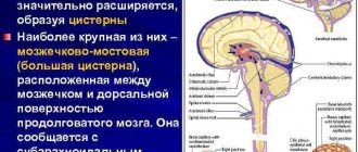
The nature of brain damage due to asphyxia differs between children born at term and premature babies. The earlier a child is born, the greater the risk of periventricular leukomalacia (PVL).
This term means necrosis of the white matter of the brain located near special cavities (ventricles). Cysts form in place of dead cells.
https://www.youtube.com/watch?v=5X88KilRGlc
It is PVL that is responsible for most cases of cerebral palsy and dementia in children born before 31 weeks of pregnancy.
In full-term infants, the cerebral cortex - the gray matter - is more often damaged. The health consequences will depend on the volume and location of damaged neurons. If the asphyxia was severe and acute, the brain stem, which is responsible for breathing and heartbeat, may be damaged. This poses a direct threat to the baby's life.
Types of hypoemia
Depending on the cause, the disease is divided:
- hemorrhagic (with hemorrhage);
- ischemic (when blood supply is cut off).
Depending on the degree of damage and timely prescribed preventive measures for mild and moderate forms of cerebral ischemia, the prognosis for the course of the disease can be quite optimistic
Attention! When brain power is cut off, it dies within 4 minutes, so resuscitation measures in this case are extremely important. In addition, when cells die, irreversible processes occur associated with the loss of motor, mental and other functions.
Remember that ischemic changes in the baby’s brain can be stopped by timely detection and immediate treatment. It is necessary to prevent hypoxia in order to help the baby recover as quickly as possible.
Possible consequences and prevention of the disease
In some cases, cerebral ischemia can become chronic, and this will entail a number of problems for the child:
- Hot temper and irritability over trifles.
- Problems in development and learning.
- Frequent headaches.
- Sleep disturbance.
- Epilepsy.
- Silence.
It is very important to reduce the risk of such a depressing disease. The expectant mother should think about this even at the pregnancy planning stage, not to mention when the fetus begins to form.
To prevent ischemia in newborns during pregnancy, it is necessary:
- Walk as much as possible.
- Eliminate all bad habits.
- Avoid any stressful situations and strenuous physical activity.
- Stick to your daily routine.
- Always get enough sleep.
- Eat a balanced diet.
- Follow your doctor's recommendations.
- Complete all stages of the examination on time.
If trouble nevertheless overtakes you, and your baby is faced with this disease, then remember that ischemia in newborns is curable with the right approach to treatment. Even severe cerebral ischemia in a newborn can be stopped by 90%. It all depends on your mood and the professionalism of the doctors. Therefore, choose your doctor carefully and never give up. Scientists have proven that a mother’s positive emotional state has a beneficial effect on the baby and speeds up the healing process.
Symptoms of ischemia in newborns
Signs of cerebral ischemia in infants include:
- increase in head volume;
- enlarged fontanel;
- lethargy;
- muscle hypotonicity;
- weakening of conditioned reflexes;
Cerebral ischemia in adults and newborns is a serious condition that should not be taken lightly
- convulsive contractions of the limbs;
- tremor of the arms, legs, chin;
- coma;
- restless sleep;
- causeless crying.
In the first moments of a baby’s life, a neonatologist assesses his condition using the Apgar scale and identifies possible physiological complications. If the doctor identifies the prerequisites for coronary disease, the child is sent for examination using a neurosonogram.
Reasons for development
Ischemia in newborns occurs as a result of hypoxia, which can occur at different stages of fetal and infant development:
- Intrauterine hypoxia, or asphyxia (suffocation), develops as a result of:
- disorders of placental circulation;
- too fast or prolonged labor;
- umbilical cord damage during childbirth.
- Postpartum hypoxia develops as a result of:
- infection of the newborn;
- prematurity of the child;
- heart defect in a newborn.
- Hypoxia due to low blood pressure in an infant can be caused by:
- bacterial infection;
- bleeding associated with liver injuries, bleeding disorders, etc.
- the woman belongs to the age category under 18 or after 35 years;
- during pregnancy, the woman suffered acute illnesses or had chronic disorders, such as pathologies of the heart and blood vessels, anemia, hypertension;
- the woman suffers from pathologies associated with poor blood clotting or thrombosis;
- a woman suffers from alcohol or nicotine addiction;
- the pregnant woman experienced bleeding at any stage;
- the pregnant woman had preeclampsia, characterized by swelling, high blood pressure and protein in the urine;
- turbidity of the amniotic fluid has occurred;
- Polyhydramnios was diagnosed during pregnancy;
- there was a malnutrition of the placenta or it detached too early;
- blood circulation between the uterus and placenta is disrupted;
- multiple pregnancy;
- at the time of birth, the woman in labor had a temperature above 38°C;
- birth occurred at less than 37 or more than 42 weeks of pregnancy;
- an emergency caesarean section was performed;
- complications arose during childbirth that required the use of drug stimulation;
- the fetus became entangled with the umbilical cord during intrauterine development or during childbirth.
The development of ischemic encephalopathy in newborns is often associated with factors depending on the mother: her age, lifestyle during pregnancy, the nature of gestation, etc.
The risk of developing encephalopathy in a newborn increases if:
Degrees of the disease
There are 3 degrees of cerebral ischemia in a newborn:
- Stage 1 (mild) – the initial stage of the disease, at which outpatient treatment is prescribed. The therapy gives good results. Signs of 1st degree of ischemia are restless sleep, lethargy, loss of appetite, tremors and frequent shuddering of the baby. Pay special attention to the duration of crying, which often signals a headache.
- 2nd degree (medium). Symptoms of stage 2 are similar to the previous ones, but are longer and more painful in nature. Due to ischemic changes in the cells of the organ and increased intracranial pressure, the baby’s skin becomes “marbled” and dysfunction of the gastrointestinal tract appears. Can be completely cured while in hospital.
Lack of proper treatment for ischemia can lead to disability of the child and hinder his social adaptation
- Grade 3 (severe) is the most serious and causes irreversible processes in the body. Most often, grade 3 cerebral ischemia is diagnosed in premature infants who need to be admitted to the intensive care unit for emergency care. A severe form of the disease occurs under the influence of prolonged oxygen starvation and intrauterine asphyxia. It is characterized by such manifestations as:
- coma,
- respiratory arrest,
- delayed psychomotor and psychoemotional development,
- strabismus,
- convulsions.
Treatment is provided through surgery, removal of the blood clot and restoration of the circulatory systems. In case of delay, death is possible.
Symptoms and treatment of cerebral ischemia in a child
Hypoxic brain lesions with the development of ischemia are observed in 15-30% of full-term newborns. In premature infants, pathology occurs more often - in 40-60% of cases. Often such disorders are harbingers of cerebral palsy (CP), symptomatic epilepsy, and dementia that occurs against the background of organic damage to brain structures.
The listed diseases that arise in the central nervous system lead to childhood disability and are the main cause of social maladjustment.
Of particular importance is the comprehensive diagnosis of ischemic changes that occur in the brain of newborns.
It is advisable to carry out diagnostic studies of the fetal nervous system during pregnancy starting from the antenatal period.
Manifestations of ischemia in infants
Ischemia of brain structures is a disease that is accompanied in newborns by hypoxia (low oxygen content in tissues) or anoxia (lack of oxygen in tissues), which leads to acute oxygen starvation. In 40% of cases, central nervous system lesions in childhood are associated with perinatal pathology.
Prematurity is the leading factor associated with morphofunctional immaturity of brain structures, which increases the risk of cerebral ischemia in premature infants during premature birth. Due to oxygen starvation, necrosis (tissue death) develops, which leads to focal and diffuse structural changes in the brain matter. Depending on the location and nature of the damage, the following are distinguished:
- Multicystic leukomalacia (diffuse damage to the white matter of hypoxic, ischemic etiology).
- Subcortical (subcortical) leukomalacia.
- Ischemic necrosis (focal or diffuse).
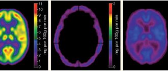
Atrophic changes in brain structures are detected during instrumental diagnostics. When conducting an Echo-EG (echoencephalography) study, in 80% of cases, compactions are observed in the periventricular (near the ventricles) region, in 20% of cases the picture is complemented by the expansion of the ventricles and spaces in which the cerebrospinal fluid is located (ducts, subarachnoid space).
Often (about 18% of cases) ischemia that affects the brain in a child occurs without obvious abnormalities in the structure of the brain matter.
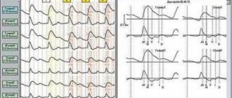
An EEG study (electroencephalography) in premature infants in 73% of cases shows the inconsistent nature of the bioelectrical activity of the brain with a predominance of slow, low-amplitude waves, periodically alternating with short-term regular waves.
In 23% of children, various pathological patterns (sample patterns) of the EEG and waves of an epileptic nature in a general oscillatory rhythm are detected. Rarely (3% of cases) the EEG does not show significant deviations from the norm in children with ischemia occurring in the brain.
Degrees of pathology in newborns
There are 3 degrees of ischemic damage to brain structures. Ischemia of the 1st degree in brain tissue in newborns is manifested by hyperactivity, sleep disturbance, loss of appetite, causeless, frequent crying, and increased muscle tone.
Symptoms of grade 2 ischemia in the brain tissue of a newborn include convulsive syndrome, increased intracranial pressure with accompanying symptoms.
There is protrusion of the fontanel, involuntary throwing back of the head, frequent crying, uncontrolled movements of the eyeballs, hydrocephalus (an abnormal increase in the diameter of the head).
Reflexes are sluggish, breathing and heart rate periodically slow down.
Stage 3 ischemia is characterized by severe depression of central nervous system functions, up to acute pulmonary failure, absence of reflexes, and coma.
Reasons for development
Ischemia in brain tissue develops as a result of oxygen starvation; all cases of pathology are associated with impaired blood flow caused by blockage or severe narrowing of the vascular lumen. Main reasons:
- Somatic diseases of infectious etiology in the mother.
- Bad habits of the mother (smoking, alcohol abuse).
- Birth injuries sustained by the fetus.
- Malfunctions of the endocrine system in the mother.
- Unfavorable course of pregnancy (toxicosis, threatened miscarriage, premature, complicated birth).
Infants who suffered from diseases during the perinatal period are susceptible to the development of ischemic lesions: pulmonary and heart failure, parasitic infestations, brain tumors, blood clotting disorders, and other pathologies of the hemostatic system.
Diagnostics
At the first stage, immediately after birth, a visual examination of the newborn is performed, an assessment of respiratory and cardiac activity, a check of reflexes, and a determination of the neurological status. To confirm the diagnosis of cerebral ischemia in an infant, the following methods are used:
- Anamnesis collection. Condition of the fetus during pregnancy and childbirth. Data from the mother’s somatic and obstetric-gynecological history, as well as features of the course of pregnancy and childbirth are taken into account.
- Neurological status of the child in dynamics. Assessment of muscle-postural tone and reflexes (Infanib scale).
- Neurosonography.
- Dopplerography of blood vessels.
- Echoencephalography.
- CT, MRI.
Instrumental studies reflect the nature and localization of organic lesions of brain structures, as well as the dynamics of the development of disorders (progress or regression).
If hypoxic or ischemic damage to areas of the brain is suspected, an electroencephalographic study is performed repeatedly, at approximately 40, 44 weeks from the moment of conception, at 6 and 12 months.
Electroencephalography with topographic mapping and visualization allows us to judge the bioelectric activity of the brain of infants. EEG study shows:
- Irritation (irritation) of cortical structures, which leads to dysfunction of the cortex. Often occurs against the background of deteriorating blood supply to areas of the brain.
- Polymorphic polyrhythm (multiplicity). Several parallel basic brain rhythms, similar in amplitude.
- Diffuse vibrations that exceed normal amplitude values.
Pathological changes often indicate a decrease in the threshold of convulsive readiness and foreshadow epileptic seizures. The quantitative content of nitric oxide in the blood indirectly indicates the contractility of the smooth muscles of the vascular wall. It is a mediator of vasodilation and regulates the expansion of the vascular lumen.
Nitric oxide is involved in the transmission of nerve impulses, improving communication between neurons.
With an increase in its concentration, the ability of the muscles to relax increases, thereby reducing the likelihood of a significant narrowing of the vascular lumen and blockage of the vessel by a blood clot.
The body accelerates the process of producing nitric oxide in the event of hypoxia or damage to the endothelium of the vascular walls.
In children with ischemic brain damage, levels of nitric oxide metabolites in the blood are elevated. The blood test also shows the enzymatic status of lymphocytes, the concentration of xanthines and hypoxanthines, and parameters of coagulation hemostasis. Usually there is a reduction in prothrombin time, increased levels of fibrinogen and soluble fibrin monomer complexes.
Treatment methods
Timely diagnosis of disorders and treatment will help to avoid serious consequences of cerebral ischemia in newborns. Complex neurorehabilitation includes:
- Drug therapy using drugs with neuroprotective effects.
- Massotherapy.
- Passive therapeutic exercises.
- Applications of ozokerite in the extremities.
- Dry immersion. Without the use of an aquatic environment, conditions of partial weightlessness are created, similar to those in which the fetus lives during intrauterine development. An effective rehabilitation measure that allows you to reduce neurological symptoms and stabilize some hemodynamic parameters.
- Physiotherapy (laser therapy, magnetic therapy).
- Music therapy.
Therapy with pharmaceutical agents is aimed at eliminating convulsive syndrome, eliminating the consequences of hypoxia and cerebral edema. Correction of psychomotor functions will be performed using drugs:
- Vitamins of group B1, B6.
- Medicines based on L-carnitine (Elkar, Levocarnitine). They normalize metabolic processes at the cellular level and have an antihypoxic effect.
- Neuroprotectors based on amino acids and neuropeptides (Actovegin). They improve the relationship between neurons, stimulate reparative (restorative) processes in the central nervous system.
- Angioprotectors. They improve the condition of vascular walls, increase the tone of smooth muscles, and prevent the penetration of calcium ions through cell membranes.
- Nootropic (Glycine, Phenotropil, Gliatilin). Increases the resistance of brain structures to hypoxia. Accelerate the utilization of glucose, stimulate the exchange of nucleic acids, accelerate the synthesis of proteins, ATP, RNA.
- Anticonvulsants.
- Muscle relaxants. Restores normal muscle tone.
For mild forms of pathology, it is recommended to do professional massage, therapeutic exercises, physiotherapeutic and water procedures, and rehabilitation treatment can be carried out without the use of any pharmaceuticals.
Possible consequences of the disease
Common (78% of cases) consequences of cerebral ischemia in newborns are disorders of auditory and visual afferentation (the continuous flow of nerve impulses from the sensory organs to the nervous system).
Frequent complications of ischemic brain damage in newborns: cerebral palsy, epilepsy, ischemic stroke, which lead to disability and death (7-28% of cases).
Against the background of oxygen starvation, dementia, sensorineural deafness and cortical blindness can develop.
Prevention
To prevent the disease, the expectant mother needs to lead a healthy, active lifestyle, give up bad habits, organize a complete, proper diet with a sufficient amount of proteins, fats, carbohydrates, vitamins and microelements. During pregnancy, you should regularly visit an obstetrician-gynecologist and, as prescribed, undergo a diagnostic ultrasound examination in order to identify abnormalities in the development of the fetus at an early stage.
Ischemia in brain tissue in newborns is a dangerous pathology that can cause disability and death of the child. The prognosis for minor brain lesions is favorable. Diagnosis in the early perinatal period and correct therapy help improve the baby’s condition and recovery.
Source: https://golovmozg.ru/zabolevaniya/simptomy-i-lechenie-ishemii-golovnogo-mozga-u-rebenka
Methods for diagnosing the disease
The following methods are used to diagnose cerebral ischemia:
- Magnetic resonance imaging;
- neurosonogram;
If obvious symptoms of a serious illness are detected, magnetic resonance imaging is performed, as well as electroencephalography, which reveals hidden seizures and other abnormalities in the functioning of the brain.
- Doppler encephalogram;
- CT scan;
- general blood and stool test;
- blood chemistry.
Based on the results of the research, an experienced doctor prescribes appropriate treatment and effective rehabilitation methods.
The consequences of cerebral ischemia in newborns depend on the severity of the pathology, its location, the number of damaged cells, and other things. Correct therapy and timely detection of the disease play a huge role. So, in half of the cases, with 3 degrees of local anemia, damage to the central nervous system and even death occurs. With grade 2 hyperemia, the consequences are much more favorable; they have a direct relationship with the development of the nervous system. Thus, complete restoration of brain cells and the entire body is possible under the influence of neuronal maturation.
Treatment of 1 severity of ischemia, in most cases, gives the desired result. Even in premature babies, the signs of the disease disappear by the age of 2–3 years, if all the doctor’s recommendations are followed and thanks to proper treatment.
Symptoms
You can suspect the presence of cerebral ischemia in a newborn already in the first days of his life. But its symptoms may manifest differently for everyone. The main symptoms of the pathology include:
- Increased excitability (the child cries for no apparent reason, his sleep is restless, he often shudders, you can notice tremors in different parts of the body).
- Depressed state of the central nervous system (muscle hypotonia, strabismus or asymmetry of the facial muscles, weakened swallowing reflex, impaired motor activity).
- Signs of hydrocephalus (excessive increase in the volume of the skull).
- Cramps.
- Coma.
Neurosurgeon, MD, tells us more about the pathology. Fayad Akhmedovich Farhad:
Doctors distinguish 3 degrees of cerebral ischemia in infants:
- Mild degree. During the first week of a newborn's life, a depressed state is observed or, conversely, the newborn is too excited. In this case, treatment begins in the maternity hospital, and after the baby it is necessary to regularly show it to a neurologist.
- Grade 2 ischemia is characterized by the presence of seizures and other neurological disorders. The newborn needs to be treated in a hospital setting.
- A severe degree of pathology is accompanied by serious disorders in the newborn’s body. He is placed in the intensive care unit. After grade 3 cerebral ischemia, the baby will require long-term rehabilitation. The child often develops various abnormalities in the functioning of the nervous system, the baby lags behind in development, is susceptible to seizures, and hearing or vision impairment occurs.
Treatment methods
The main ways to eliminate ischemia are:
- in mild cases - physiotherapeutic procedures (massage, swimming, physical therapy, exercise on a fitball, electrophoresis);
The goal of treatment is to restore normal blood circulation in the brain tissue, prevent pathological changes and eliminate the consequences of ischemia
- for moderate hyperemia – drug treatment;
- in severe cases - surgical intervention and resuscitation measures (ventilation).
Please note that in the second and third stages of the disease, they resort to drug therapy, the prescription of anticonvulsants (Diazepam, Phenobarbital), nootropic, diuretic and vasoconstrictor drugs. In mild stages of ischemia, massage is limited to increase muscle tone and blood circulation throughout the body. It is carried out in courses of 10 sessions 4 times a year.
During treatment, you must strictly follow all the doctor’s instructions, carefully monitor the child’s health and the effect of treatment on his body.
Treatment of cerebral ischemia.
Massotherapy
Treatment is aimed at normalizing and restoring the structure and function of the newborn’s brain cells and preventing the development of complications of cerebral ischemia. At grade 1, no medication is required. You can limit yourself to a massage; be sure to create psycho-emotional peace for the child. If the disease degree is 2 or higher, specialized treatment prescribed by a doctor is required.
If convulsions occur - anticonvulsants, with increased spasticity - muscle relaxants, in order to relieve cerebral edema - diuretics. Treatment in each case is selected individually by the attending physician, according to indications, taking into account the child’s weight. Massage, for older children - exercise therapy, classes with a speech therapist, animal-assisted therapy - all these are the most important methods of rehabilitation of children with cerebral ischemia.
Preventive actions
To avoid fetal illness during pregnancy, the following actions must be taken:
- perform physical exercises;
- adhere to proper nutrition;
- to refuse from bad habits;
- often be in nature;
- undergo a gynecological examination;
- register for pregnancy in a timely manner.
It is important to know that local anemia of degrees 2 and 3 has the following consequences for the child:
- epilepsy;
- cerebral palsy;
- mental retardation;
- headache;
- psychoemotional disorders.
If the first signs of a possible disease are detected, you need to immediately show the baby to a neonatologist, carefully monitor its behavior, and carry out preventive measures.
Be vigilant, take care of the health of your children!
Causes of hypoxic and ischemic lesions of the central nervous system
Hypoxic and ischemic lesions of the central nervous system are brain lesions as a result of chronic or acute fetal hypoxia, which are combined with secondary ischemia. They can occur both during pregnancy and during childbirth, in the postperinatal period.
Among the main causes of hypoxic and ischemic lesions of the central nervous system of newborns are:
- Delayed fetal development
- Thromboembolism, disturbances of uterine and fetoplacental blood flow;
- Threat of interruption, uterine bleeding;
- Bradycardia in the fetus;
- Asphyxia and acute fetal hypoxia;
- Severe bleeding during childbirth, placental abruption;
- Umbilical cord pathologies;
- Decrease in blood pressure in a newborn - below 30 mm Hg. st;
- Thromboembolic complications after childbirth - sepsis, disseminated intravascular coagulation syndrome, polycythemia;
- Congenital heart disease in a baby with persistent hypoxemia;
- Embolism;
- Increased intracranial pressure.
What is this pathology?
Cerebral ischemia in newborns is not an independent pathology, since its formation is simultaneously preceded by several pathological processes in the body. Thus, during the period of intrauterine development of the fetus, an acute oxygen deficiency may occur, as a result of which metabolic reactions in the brain are disrupted, which subsequently leads to its functional changes. These changes become the main cause of inhibition of neuronal activity, as well as the formation of local areas of necrosis.
The severity of the child’s condition and the risk of complications depend on the degree of hypoxia.
Diseases can manifest themselves throughout pregnancy or occur during childbirth. At the same time, early disruption of nervous regulation carries more serious complications for the development of the baby than later ones.
Symptoms of hypoxic-ischemic damage to the central nervous system
Damage to the central nervous system in newborns is diagnosed in the first minutes of a baby’s life, and the symptoms depend on the severity and depth of the pathology.
I degree
In mild cases of HIE, the condition remains stable; the child’s Apgar score is no less than 6-7 points; cyanosis and decreased muscle tone are noticeable. Neurological manifestations of the first degree of hypoxic damage to the central nervous system:
- High neuro-reflex excitability;
- Sleep disorders, anxiety;
- Trembling of the limbs, chin;
- Possible regurgitation;
- Reflexes can be either increased or decreased.
The described symptoms usually disappear during the first week of life, the child becomes calmer, begins to gain weight, and severe neurological disorders do not develop.
II degree
With moderate brain hypoxia, signs of brain depression are more obvious, which is expressed in deeper disorders of brain function. Typically, the second degree of HIE accompanies combined forms of hypoxia, which is diagnosed both during the intrauterine growth stage and at the time of birth. In this case, muffled fetal heart sounds, increased rhythm or arrhythmia are recorded; the newborn scores no more than 5 points on the Apgar scale. Neurological symptoms include:
- Suppression of reflex activity, including sucking;
- A decrease or increase in muscle tone, spontaneous motor activity may not appear in the first days of life;
- Severe blueness of the skin;
- An increase in intracranial pressure;
- Autonomic dysfunction - respiratory arrest, increased heart rate or bradycardia, disturbances in intestinal motility and thermoregulation, tendency to constipation or diarrhea, regurgitation, slow weight gain.
intracranial hypertension accompanying severe forms of HIE
As intracranial pressure increases, the baby's anxiety increases, excessive sensitivity of the skin appears, sleep is disturbed, tremor of the chin, arms and legs increases, bulging of the fontanelles becomes noticeable, horizontal nystagmus and oculomotor disorders are characteristic. Signs of intracranial hypertension may include seizures.
By the end of the first week of life, the condition of a newborn with the second degree of HIE gradually stabilizes against the background of intensive treatment, but neurological changes do not disappear completely. Under unfavorable circumstances, the condition may worsen with brain depression, decreased muscle tone and motor activity, exhaustion of reflexes, and coma.
III degree
Perinatal damage to the central nervous system of severe hypoxic-ischemic origin usually develops with severe gestosis in the second half of pregnancy, accompanied by high hypertension in the pregnant woman, impaired renal function, and edema. Against this background, a newborn is already born with signs of malnutrition, intrauterine hypoxia, and developmental delay. The abnormal course of labor only aggravates the existing hypoxic damage to the central nervous system.
In the third degree of HIE, the newborn has signs of severe circulatory disorders, there is no breathing, tone and reflexes are sharply reduced. Without urgent cardiopulmonary resuscitation and restoration of vital functions, such an infant will not survive.
During the first hours after birth, a sharp depression of the brain occurs, a coma sets in, accompanied by atony, an almost complete absence of reflexes, dilated pupils with a reduced reaction to a light stimulus or its absence.
The inevitably developing cerebral edema is manifested by generalized convulsions, respiratory and cardiac arrest. Multiple organ failure is manifested by increased pressure in the pulmonary artery system, decreased urine filtration, hypotension, necrosis of the intestinal mucosa, liver failure, electrolyte disturbances, and blood coagulation disorders (DIC).
A manifestation of severe ischemic damage to the central nervous system is the so-called post-asphyxial syndrome - babies are inactive, do not cry, do not respond to pain and touch, their skin is pale bluish, and a general decrease in body temperature is characteristic. Important signs of severe brain hypoxia include swallowing and sucking disorders, which make natural feeding impossible. To save the life of such patients, intensive care in intensive care is necessary, but the unstable condition still persists until the 10th day of life, and the prognosis often remains poor.
A feature of the course of all forms of HIE is considered to be an increase in neurological deficit over time, even with intensive therapy. This phenomenon reflects the progressive death of neurons that have already been damaged during a lack of oxygen, and also determines the further development of the baby.
In general, ischemic-hypoxic damage to the central nervous system can occur in different ways:
- Favorable with rapid positive dynamics;
- Favorable course with rapid regression of neurological deficit, when by the time of discharge the changes either disappear or remain minimal;
- Unfavorable course with progression of neurological symptoms;
- Disability during the first month of life;
- Latent course, when after six months motor and cognitive disorders increase.
This is interesting: Why do you need to cleanse your face?
In the clinic, it is customary to distinguish several periods of ischemic encephalopathy of newborns:
- Acute - first month.
- Restorative - within one year.
- Period of long-term consequences.
The acute period is manifested by the whole gamut of neurological disorders from barely noticeable to coma, atony, areflexia, etc. During the recovery period, the syndrome of excessive neuro-reflex excitability, convulsive syndrome, possible hydrocephalus, delayed intellectual and physical development come to the fore. As the child grows, the symptoms change, some symptoms disappear, others become more noticeable (speech disorders, for example).
Development mechanism
Violation of the metabolism of brain cells during ischemia leads to irreversible processes of destruction of nerve structures and disruption of their activity. As a rule, the mechanism of formation of the pathological process is based on a rapid decrease in the level of adenosine triphosphoric acid, which is the main energy source in the body.
Impaired cerebral circulation creates favorable conditions for the formation of ischemia
For the full functioning of neurons, a balance is required between the concentration of intracellular and extracellular ions penetrating through the membranes. Long-term oxygen deficiency creates an imbalance of transmembrane components of the brain, which subsequently dramatically changes the concentration of potassium and sodium. This leads to a decrease in membrane potentials. In parallel with these processes, there is an increase in calcium levels, which allows the release of the neurotransmitter glutamate, which overstimulates brain receptors, thereby promoting structural and functional changes in the brain.
The body's first response to perinatal oxygen deficiency is to increase blood supply to the main organs. Subsequent arterial hypotension leads to a throughput decrease in cerebral blood flow.
Pathological changes in the brain in a newborn
Thus, the following stages are distinguished in the mechanism of development of pathology:
- Intrauterine oxygen deficiency.
- Decreased oxygen supply to the brain and increased carbon dioxide concentration.
- Increased acid-base environment in the fetus.
- Swelling of intracellular components.
- Increase in brain tissue volume.
- Local decrease in cerebral circulation.
- General cerebral edema.
- Increased level of cranial pressure.
- General blood flow disorders.
- Death of brain cells.
The mechanism of ischemia formation depends on the date of birth of the child, which is associated with age-related characteristics of the structure of the brain.
Assessing the degree of complications
Ischemic disorders provoked by oxygen deficiency lead to the death of brain cells, which creates conditions for the formation of irreversible neurological disorders, the severity of which depends on the localization of structural changes.
As a rule, cells located in the pyramidal zone, cerebellar cells, ventrolateral neurons, midbrain nuclei, and stem neurons are affected.
With large-scale ischemic damage to brain cells, their complete death occurs, which leads to death.
The most common complications are decreased memory and mental performance
The consequences of ischemic disorder include the development of the following conditions:
- epilepsy;
- one-sided blindness;
- impaired mental function;
- motor function disorder.
Full assessment of complications occurs at the age of 3 years.
Diagnostic methods
Cerebral ischemia in infants is a complex diagnosis that requires a comprehensive examination in various ways. After birth, children with cerebrovascular accidents come under round-the-clock monitoring: exacerbation often occurs, deep fainting or coma occurs. The following tests and methods of examining the body help confirm the disease:
- Initial examination. Conducted by a neonatologist directly in the delivery room. Based on several indicators, he assigns Apgar scores. In children with oxygen starvation they are diagnosed at a low level. The doctor determines the presence of basic reflexes, muscle tone and attempts at spontaneous breathing.
- Analyzes. Before treatment is prescribed, the baby undergoes a general and biochemical blood test, an assessment of coagulability, and the amount of carbon dioxide.
- MRI of the brain. If the baby experiences convulsions, loss of consciousness and other signs of severe damage, a computer scan is performed. On the sections you can see areas of necrosis, the condition of blood vessels, and hematomas.
- Ultrasound of the brain. A diagnostic procedure is recommended to determine intracranial pressure and assess the condition of the cortex and medulla.
- EEG. An electroencephalogram is required if cerebral ischemia is suspected in children. A painless method helps to detect the cause of seizures, loss of consciousness, and select the optimal therapy.
Information! If necessary, ECG, X-ray and angiography are used to identify congenital pathologies of the circulatory system, tumors or aneurysms.