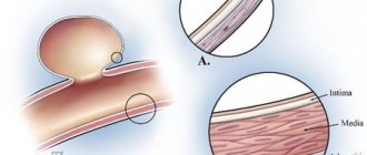
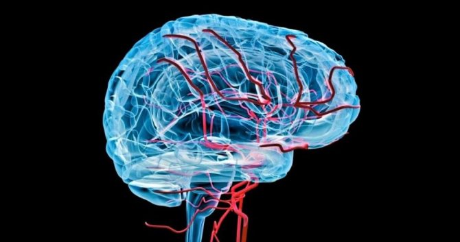
A cerebral aneurysm is an enlargement of one or more blood vessels in the brain. This condition is always associated with a high risk of death or disability of the patient if the aneurysm ruptures. In essence, an aneurysm is a protrusion of the vascular wall that occurs in one or another part of the brain. An aneurysm may be congenital or may develop during life. (ICD-10 codes: I67.0, I67.1).
Symptoms of a cerebral aneurysm
Brain aneurysms cause symptoms only when they rupture. However, intact aneurysms can also provoke the development of a clinical picture, especially when the aneurysm is large or compresses nearby nerves and tissues.
Commonly occurring signs include:
- Headache.
- Drowsiness.
- Pain as if inside or behind the eyes.
- Difficulty speaking.
- Changes in vision.
- Photosensitivity (sensitivity to light).
- Fainting (loss of consciousness).
- Disorder of consciousness.
- Painful sensations in the eyes;
- Decreased vision;
- Facial numbness;
- Hearing loss;
- Enlargement of only one pupil;
- Stiffness of the facial muscles, not all of them, but on one side;
- Seizures.
Symptoms of a ruptured aneurysm are characterized by an abrupt onset in a fairly short time. They differ in the location of the aneurysm.
Genetic failures
They include a large number of hereditary diseases, due to which the balance of protein synthesis is disrupted, affecting the elasticity of muscle fibers. These include the following diseases:
- fibromuscular dysplasia;
- Osler-Rendu syndrome;
- Marfan syndrome;
- Ehlers-Danlos syndrome;
- elastic pseudoxanthoma;
- systemic lupus erythematosus;
- sickle cell anemia;
- tuberous sclerosis.
Of course, the presence of these diseases is not an absolute sign of the presence of aneurysms, but they all increase the risk of their development under the influence of certain unfavorable conditions.
Causes of cerebral aneurysms
To date, there is no single theory explaining the formation of this vascular pathology. Most researchers believe that cerebral aneurysm is a multifactorial pathology.
Changes in the structure of the walls of blood vessels can lead to:
- atherosclerosis;
- hyalinosis;
- exposure to ionizing radiation;
- hereditary predisposition;
- inflammation of the vascular wall of a bacterial or mycotic nature;
- traumatic damage to blood vessels;
- any connective tissue diseases (they affect blood vessels, making them weak and inelastic);
- addiction to smoking, alcohol, drugs (under the influence of toxic substances, vascular tissue is actively destroyed, which is fraught with the occurrence of an aneurysm, a rapid increase in its volume and stimulation of rupture).
What are the symptoms of a cerebral aneurysm?
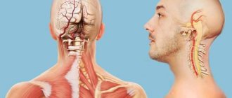
Symptoms of a cerebral aneurysm are identical in both sexes - both women and men. First of all, the dilation of the vessel makes itself felt by neurological manifestations. This is due to the fact that the vascular protrusion compresses areas of the brain.
The most common symptom of a cerebral aneurysm is cephalalgia, which varies in intensity, location and character. A migraine-type headache with pronounced sensations in one half of the head, occipital pain, discomfort in the neck, pain near the eyeballs.
Often patients do not even know that it is a brain aneurysm, attributing the symptoms to fatigue and overwork.
The symptoms of a cerebral aneurysm depend on its location. In the presence of an aneurysm, intracranial pressure increases, a diffuse headache, a feeling of pressure in the eye sockets, and nausea appear. In addition to these manifestations, headache, along with signs of compression, provokes the following symptoms of cerebral aneurysm:
- diplopia – a visual disorder in which there is a split in the horizontal plane, impaired movement of the eyeball;
- oculomotor disorders - constriction of the pupil on one side, decreased reaction to a light stimulus;
- loss of visual fields;
- partial paralysis of the facial nerve, ptosis (drooping) of the eyelid;
- pain on one side of the face with impaired sensitivity;
- speech disorders;
- paralysis, weakness in one half of the body, impaired sensitivity, inhibition of voluntary movements;
- bulbar syndrome is a whole complex of manifestations of damage to the cranial nerves (impaired speech, problems with swallowing food, drooling, nasal voice).
Danger of aneurysm
The presence of any aneurysm is associated with a high risk of intracranial bleeding. Rupture of a vascular wall defect is one of the causes of hemorrhagic stroke and subarachnoid hemorrhage. The clinical picture depends not on the type of aneurysm, but on its location, the amount of blood loss, and the involvement of brain tissue and meninges.
At the time of aneurysm rupture, a sharp, high-intensity headache and vomiting without relief most often occur. Possible loss of consciousness. Subsequently, the level of consciousness is restored or a cerebral coma develops.
If you consult a doctor in time, hemorrhage can be prevented. To do this, you need to follow all the recommendations: take prescribed medications, eat right, don’t overexert yourself, and undergo regular examinations.
Diagnostic methods
Diagnosis of an aneurysm is carried out using several methods:
Angiography
A popular method for diagnosing an aneurysm is angiography, which makes it possible to qualitatively assess the condition of the vessels, determine the shape, degree and location of the pathology. Angiography is an X-ray examination using contrast agents. Before it is carried out, a special illuminating drug is injected into the femoral artery with a catheter, which helps to identify the aneurysm and its condition. Angiography is used to diagnose aneurysms in children.
Angiography is not carried out in the only way; there is:
- Digital angiography. The data is subsequently processed by computer, resulting in high-quality images. The dosage of the contrast agent is reduced and it is injected into a vein.
- Color angiography allows you to conduct research quickly, since one image reflects the entire process.
- 3D angiography reconstructs the projection of the image in a volumetric format.
CT scan
The method is painless. During the examination, X-rays are taken, then the data is processed by a computer. The program helps to identify problems in the vascular system of the brain, identify areas of dilation, blockage and narrowing of blood vessels, see ruptures and hemorrhages.
Magnetic resonance imaging
The examination is abbreviated as MRI. This diagnosis allows you to obtain a three-dimensional image of the arteries of the brain using electromagnetic waves. The picture on the computer screen gives a clear idea of the condition of the veins and arteries.
Cerebrospinal fluid puncture
It is used when the doctor suspects that an aneurysm has ruptured. In this case, the analysis is taken by puncturing the spinal trunk with a special needle. The fluid is examined for the presence of blood, since after hemorrhage it enters the spinal cavity.
Classification of the disease
The classification is distributed according to the type of various parameters.
Size. The formation has a diameter of less than 3 mm – more than 25 mm.
Form. Formations can vary in shape: spindle-shaped (expands the wall of the vessel itself), saccular (a bag of blood, attached to the artery), lateral (on the wall of the vessel).
Number of cameras. The seal can be multi-chamber or single-chamber.
By location. The formation can occur on several different vessels.
Arterial aneurysm
The greatest danger is the protrusion of large arteries, as they feed brain tissue. In most cases, the bulge is formed as a result of a defect in the inner and outer linings of the vessel. Most often, the unpaired basilar and internal carotid arteries, as well as their branches, are affected.
Aneurysm of the vein of Galen
Aneurysm of the vein of Galen is rare. However, a third of arteriovenous malformations in young children and newborns are due to this anomaly. This formation is twice as common in boys.
The prognosis for this disease is unfavorable - death occurs in 90% of cases in infancy and the neonatal period. Embolization maintains a high mortality rate of up to 78%. There are no symptoms in half of the sick children.
Who is at risk?
Cerebral aneurysm can occur at any age. This disease is more common in adults than in children and is slightly more common in women than men. People with certain inherited diseases are at higher risk.
The risk of cerebral rupture and hemorrhage exists with all types of cerebral aneurysms. There are about 10 reported aneurysm ruptures per year for every 100,000 people, which is about 27,000 people per year in the United States). Most often, aneurysm affects people aged 30 to 60 years.
Other factors that can contribute to aneurysm rupture are hypertension, alcohol abuse, drug addiction (especially cocaine use), and smoking. In addition, the condition and size of the aneurysm also influence the risk of rupture.
Fusiform aneurysm
The third option is a spindle-shaped cerebral aneurysm. This is deformation on all sides in one area. This form suggests that one of the causes is a blood clot. Impaired motility of the vascular walls may also be the cause. The exact reasons can only be established with more detailed research. Individual characteristics of the state of health, features of the current situation - everything that is called the background of the disease and the clinical picture must be taken into account. Regardless of what form an aneurysm is found, it can either grow and the risk of hemorrhage increases, or remain dormant. The mere fact of having a disease is not a reason for radical intervention. It is important to monitor the course and take timely measures to prevent the development of pathological processes. It is desirable that therapy be aimed at this, which is problematic without early diagnosis.
The later the pathology is discovered, the worse it is. People rarely take care of their health at the level necessary to prevent the growth of different types of aneurysms in different parts of the brain. You need doctor's orders, giving up habits, changing your daily diet, volitional effort, conscious regulation, and developing healthy habits. This does not mean that the person himself is to blame. Ecology, nervous tension - there is no escape from these negative factors. Few people these days can boast of ideal health between the ages of 30 and 60. Depending on their size, aneurysms are divided into small, medium and giant. Small ones are those whose diameter does not exceed 11 mm. Medium ones are aneurysms with a diameter from 11 mm to 25. Giant ones are those whose diameter is more than 25 mm. Other, more detailed classifications are also used. This classification is universal and general. The diameter of the aneurysm, discovered as a result of examinations, allows us to understand how dangerous the situation is.
Useful to know: What is brain cancer, causes, characteristic signs and treatment
to contents ^
Aneurysm rupture
When an aneurysm ruptures, a sharp and very severe headache occurs. The patient may describe it as the worst headache ever experienced.
In addition, rupture of a cerebral aneurysm may be accompanied by:
- loss of consciousness
- blurred vision or diplopia (double vision)
- vomiting
- nausea
- photophobia
- stiff neck
- drooping eyelid
- convulsions
An unruptured aneurysm does not manifest itself in any way until, as it grows, compression of nearby nerves occurs. In this case, various symptoms may appear, including blurred vision, eye pain, paralysis or numbness of the face.
Classification of aneurysms
Based on the external signs of formation, the following types of cerebral aneurysm are distinguished:
- Saccular - looks like a round sac that is attached to the artery. Inside this sac is filled with blood. It can also be compared to a berry hanging on a branch.
- Lateral – looks like a tumor. Located in the wall of the vessel.
- Fusiform - can form in the place where the vessel expands.
The most common type of brain aneurysm diagnosed is the saccular type.
Depending on their origin, cerebral aneurysms can be:
- True - appear due to the fact that the wall of the vessel begins to protrude unnaturally to one side.
- False - located near the artery and not associated with the blood vessel. Such an aneurysm begins to fill with blood through a small hole in the wall of the vessel.
- Exfoliating - the cavity of such a formation is located inside the hollow wall of the vessel. Blood can get inside through a hole located in the vessel.
Elena Malysheva and her assistants talk about the types of manifestations, nature and symptoms of the disease in their TV show about health:
Aneurysms can be single or multiple, single-chamber or multi-chamber, congenital or acquired.
Diagnostics
When asymptomatic, cerebral aneurysms usually become incidental diagnostic findings discovered during examination of a patient for another reason. When clinical symptoms appear, cerebral aneurysm is diagnosed on the basis of existing neurological symptoms, as well as data from instrumental studies, which include: radiography of the skull; computer or magnetic resonance imaging of the brain; X-ray or magnetic resonance angiography.
The final diagnosis of aneurysm of the cerebral arteries, determination of its location, size and shape are possible only with the help of angiography, which is performed even in the acute period of a stroke. In some cases, contrast-enhanced computed tomography of the head can be informative.
Basic diagnostic methods:
Angiography. This is an x-ray of the brain vessels, in which contrast agents are used.
CT (computed tomography). This method is considered the best. It is painless, quick, non-invasive, helps to find the lesion, and in case of rupture, determine the size of the hemorrhage.
CT angiography. It differs from CT in that a contrast agent is injected.
MRI (magnetic resonance imaging). MRI uses a strong magnetic field and radio waves to capture images of the brain.
Cerebrospinal fluid analysis. It is carried out if there is a suspicion that the aneurysm has ruptured. The patient is given a local anesthetic.
Various vascular deformations
The mechanisms of the appearance of cerebral aneurysms are not fully understood, research continues. It is not known how to stop the process and eliminate all factors. One person is doing well, despite the fact that aneurysmal disease was discovered, while the other is not. The situation is getting worse, and the reason for this may be ordinary stress, nervous overstrain, which cannot be avoided in the modern world. The pressure will increase, and the expansion of the blood vessels in the brain may become more significant. How to protect a person from negative changes, how to protect and restore an abnormal, deformed area of blood vessels? There are no answers yet. It is known today that the following deviations from the norm are to blame:
- defects in the muscular layer of blood vessels in a certain area - the walls do not narrow and do not respond to signals from the nervous system;
- damage to the middle layer of the walls of blood vessels - the elastic membrane, which becomes denser over the years, and in some areas there are gaps for the penetration of useful substances, is destroyed;
- hyperplasia of the layer of the vessel walls, called intima, which is responsible for the penetration of useful substances;
- hyperplasia of atheroma of the arterial trunk - an increase in the number of fatty plaques and scar tissue, acceleration of degeneration in atheroma, which makes the area of the vessels fragile, weak, unable to further transport blood;
- damage to collagen fibers, which ensure the stability of the shape of blood vessels in a normal state, are a strong structure, inelastic, and should prevent stretching of the walls of blood vessels from their deformation
- thinning of the walls while maintaining such characteristics as strength and inflexibility, which together increases the risk of hemorrhage.
Useful to know: Focal changes in the brain substance of a dyscirculatory nature
Scientists have studied in detail the changes in the walls of blood vessels during an aneurysm in all their diversity. However, an effective medicine that could heal as if by magic has not yet been invented. Learning to save lives when a cerebral aneurysm ruptures was a priority task before.
to contents ^
Treatment
The leading method of treating aneurysm is surgery. It will remove the formation itself and restore the integrity of the blood vessels.
Surgery is the only effective treatment for cerebral aneurysm. If the size of the defect is more than 7 mm, then surgical treatment is mandatory. Emergency surgery is required for patients with a ruptured aneurysm. The following types of surgical intervention are possible:
Direct microsurgical intervention (brain trepanation and direct surgical removal of the seal)
Endovascular operations (high-tech method, allows you to remove an aneurysm without craniotomy)
Drug correction (to prevent aneurysm rupture)
The type of surgical intervention largely depends on the severity of the patient’s condition; in difficult situations, it is impossible to do without craniotomy.
Treatment of cerebral aneurysm
The only way to get rid of such a formation is to perform a surgical operation.
However, they resort to it only in extreme cases, because any surgical intervention on the brain has enormous risks.
The operation can damage healthy blood vessels. In addition, attacks often occur after it, and the aneurysm itself may reappear.
In any case, after a cerebral aneurysm is detected, the patient is prescribed an individual treatment plan.
If the aneurysm is small, doctors prefer watchful waiting, believing that the risk of having an aneurysm is equal to the risk associated with surgery.
At the same time, the tumor is constantly monitored in order to respond in time and begin the necessary treatment. To do this, it is necessary to undergo diagnostics annually and monitor your well-being.
Such simple tips will help the patient not to miss important symptoms that indicate a ruptured aneurysm.
If a large aneurysm is detected or its rapid growth, a mandatory operation is performed. It can only be carried out by a highly qualified specialist.
Methods of conducting operations
There are 2 ways to perform the operation:
- Open intervention and removal, clipping of a cerebral aneurysm, strengthening the vessel wall or bypassing it.
- Endovascular embolization of a patient's cerebral aneurysm.
Aneurysm clipping
Clipping involves installing a special clip on the aneurysm. This is done in order to completely exclude the affected area of the vessel from the general blood flow.
Clipping is also used to remove blood that collects in the tumor.
While strengthening the walls, the affected area is wrapped with special surgical gauze, which leads to the formation of a capsule from the surrounding connective tissue. During bypass surgery, the surgeon will tighten the affected vessel, redirecting blood flow.
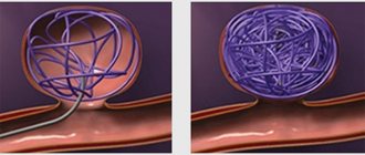
Endovascular embolization is performed without opening the skull.
To do this, a balloon or microspiral is inserted through the femoral artery into the damaged area, which leads to the removal of the tumor from the general circulatory system.
Today, this particular technique is considered more progressive than clipping.
Disadvantages of clipping
Although this method has some disadvantages:
- High price.
- Inability to remove existing blood clots.
- Risk of developing hypoxia.
See what the endovascular embolization procedure looks like. Doctors use natural access to the brain through an artery located in the thigh:
- In some cases, vasospasm is possible.
- In approximately 20% of all cases, the microspiral can change shape, as a result of which blood flow is restored and the patient requires repeated intervention.
Sometimes the patient may be prescribed conservative treatment. It consists of receiving:
- Painkillers.
- Anticonvulsant drugs.
- Antiemetics.
- Drugs that can bring blood pressure back to normal.
- Calcium channel blockers, which are needed to prevent spasms.
Prevention
In this regard, experts highlight a number of recommendations that can prevent the development of pathology:
- Eliminate bad habits: smoking, drinking alcohol and drugs.
- It is necessary to treat arterial hypertension and constantly monitor blood pressure levels.
- Nutrition should be rational with reduced consumption of table salt. Anything fatty, salty, smoked, or with a lot of herbs and spices should be excluded from foods.
- Regular exercise, primarily cardio training, allows you to maintain a high level of health.
- If you have diabetes mellitus and other somatic diseases, it is necessary to monitor their course and follow the prescription of your doctor.
Related posts:
- Erection disorders in men When the first symptoms of erectile dysfunction appear in a male…
- Soft tissue abscess A skin abscess is an intradermal inflammatory process caused by bacterial flora, most often...