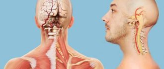
Home » Ultrasound of the head and neck »
Echo EG is used to examine the functioning of the brain using ultrasound echography. For this purpose, sound waves of a certain frequency are used. They are reflected from any tissue of the head, such as the meninges, blood, skull bones, and so on. Foreign bodies, tumors, and all kinds of pathological formations get into the reflective structures. Many people are interested in how an echoencephalogram can help a doctor make the correct diagnosis and prescribe therapeutic treatment.
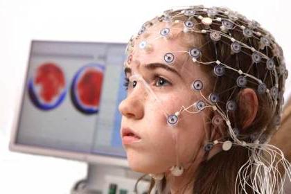
Echoencephalography of the brain
After deformation of a special plate with which the research probe is equipped, an ultrasonic signal is generated. Moreover, the plate is capable of simultaneously sending and receiving a signal in reflected form.
Each tissue has a different surface that perceives ultrasonic waves. For example, the skin has its own reflected signal; a cyst has a completely different appearance. Thus, an image of the brain and its structure appears on the display.
Echoencephalography is performed in different ways:
- M-method
- Ultrasound scanning
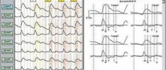
When the M-method is used, a graph is shown on the monitor screen where peaks are clearly visible, showing internal formations in the cranial cavity. When an ultrasound examination of the brain is performed, the internal structure is shown flat on the screen.
Echo
Echo is a 2008 American horror film, a remake of the 2004 Filipino horror film. This film was directed by Yam Laranas. The plot of the film revolves around Bobby, who was released early from prison. He ended up in prison for unintentional murder. Returning home, he learns that his mother has died and moves into her apartment.
Bobby hears strange noises in his mother's apartment. He can't find the source of the sounds. The main character learns that in the last days of her life, his mother locked herself in the apartment and did not come out. All the neighbors called her crazy. She died locked in a closet in her own house. The house manager tells all this to the main character. Clearing out and slowly settling into the apartment, Bobby simultaneously gets a job as an auto mechanic and tries to improve his relationship with his ex-girlfriend. But his neighboring neighbors do not allow him to live in peace. Eternal swearing from behind the wall does not allow you to rest. The neighbor in the stairwell, a policeman, constantly makes scandals with his wife, beating her along the way. Bobby tries to stop them a couple of times, but no one opens the door for him. Fearing for the little girl, the daughter of neighbors, one day in the midst of a strong family quarrel behind the wall, Bobby calls the police to the neighbors.
The film Echo touches on a very serious topic: domestic violence and its impunity. Not being involved in violence against someone does not absolve you from responsibility. This is exactly what the filmmakers were trying to convey. After all, while the policeman outside the door was beating his wife and daughter to a semi-conscious state, all the neighbors pretended not to hear anything, closed the doors more tightly, and drew the curtains. The film conveys a simple and important truth: not being involved in violence does not save you from its consequences; inaction is also evil.
The film Echo is worth watching not only for fans of the horror genre. In addition to the elements of a horror film, namely: inexplicable noises, the sounds of a piano, the depressing atmosphere of the apartment as a whole, there is a strong moral message to the viewer. The topic is raised not only of domestic violence, but also of prejudice towards former prisoners. It is very difficult for the main character to find a job. Friends and neighbors turned their backs on him. Although none of them asked what circumstances pushed Bobby to such a terrible act as murder.
The narration in the film is very slow and smooth, which is not typical for horror films. But the rest of the film is filled with classic horror movie techniques. Incomprehensible shadows, frightening sounds, rustling sounds, screams. All this creates the atmosphere inherent in horror films, for which this genre is loved. True horror connoisseurs should watch Echo online for free in good quality.
When is echonecephaloscopy prescribed?
For young children, when the fontanelle has not yet become overgrown, all areas of the brain are examined using echoencephalography.
An echoEG is performed on an adult to detect all kinds of formations associated with pathological phenomena:
- tumors
- abscesses
- intracranial hematomas
- head injuries
- hydrocephalus
In addition, echoencephalography is performed for a more accurate diagnosis when there are certain diseases:
- neck bruises
- stroke
- blood flow disorders
- concussion
- ischemia
For children, Echo EG of the brain is prescribed:
- In case of defective physical development.
- If there is a sleep disorder.
- Muscle hypertonicity is pronounced.
- Enuresis was diagnosed.
- Stuttering.
What do Echo-EG indicators mean?
There are 3 signal complexes with which a conclusion is made:
- Elementary. The sensor receives these signals extremely quickly. Their formation occurs as a result of the ultrasound wave being reflected from the skin, cranial bones and muscles.
- Median. The formation of signals occurs as the waves come into contact with structures located between the hemispheres.
- Finite. The formation of signals occurs as a result of the contact of the wave with the dura mater.
Then the indicators are deciphered . The conclusion of the decoding of the Echo-EG of the brain is normal:
- Between the start and end signal, the echo signal has average values. Equal M-echo distances between hemispheres are required.
- The value of the median complex should not increase. If there are any deviations from the norm, then this indicates
presence of high intracranial pressure.
- It is unacceptable for the M-signal ripple to exceed 30%. If these indicators are high (up to 60%), then this indicates that the person is prone to the appearance of hypertensive pathology.
- Normally, between the final and initial signals there should be small pulses of the same amplitude, and there should be an equal number of them.
- The midsellar impulse should fluctuate around 3.9–4.1. A lower value indicates high intracranial pressure.
Preparation for echoencephalography
Echoencephaloscopy of the brain is performed for both children and adults without prior preparation. You don’t need to follow a specific diet, you don’t need to specifically drink a glass of water.
Echoencephalography is done regardless of age. Pregnant women and mothers who are breastfeeding can undergo this examination.
The exception is those cases when there are open wounds on the head. To conduct an examination of the brain matter, in these cases it is better to use MRI.
When an encephalography is performed on a baby, parents help conduct the examination. They hold the baby's head in a certain position for several minutes.
Research methodology
Echoencephaloscopy is usually performed lying down, but sometimes, for example in children, it is performed in a sitting position. This research method is very often used for emergency diagnostics. That is why the developers created such devices to be compact, lightweight and easily transportable.
Echoencephaloscopy can be performed in a doctor’s office; it is often done in an ambulance that arrives on call. Echoencephalography can be carried out directly on the street; the device does not require a centralized power supply; it can be powered by built-in batteries. Studies of children and adults are performed within 10 minutes, several modes are used.
Transmission
This method uses two sensors. They are installed on the head on two opposite sides.
One sensor sends a signal, the other receives it. Thus, the “midline” is determined. In most cases, this line completely coincides with the anatomical line. However, if there are injuries or accumulation of blood in the skull, this dependence will be disrupted.
What parameters are normal?
The obtained indicators are compared with normal data. The main thing the doctor pays attention to is M-echo. Normally it should be the same on both sides. Pathologies are indicated by a shift in the direction opposite to the present volumetric neoplasm.
In a healthy person, the M-echo displacement is up to 2 mm. The median structures must be located along a strictly median plane. There should not be any pathological formations of a malignant or benign nature.
Emission
This technology requires only one sensor. It is installed at special points that do not interfere with the passage of ultrasound.
The sensor position is slightly shifted. This makes it possible to obtain a better informative image.
Slowly moving the sensor over the scalp, an echoencephalography is obtained in the form of a slice of the brain. Moreover, the cut line corresponds to the movement of the sensor.
This method is considered not particularly accurate if it concerns pathological lesions of small size. The fact is that moving the ultrasonic probe very often shows inaccurate results.
That is why, when it is necessary to conduct a primary diagnosis of the brain, the doctor prescribes not echoencephalography, but MRI.
How the received data is decrypted
Echoencephalography technology is based on several components. Doctors called them complexes.
The first includes areas located near the sensor, muscles, tissues, and skull bones.
The second refers to the contact of ultrasonic waves with the hemispheres of the brain.
The last, third signal is the dura mater of the marrow, located on the opposite side of the sensor.
Decoding the examination results takes the above-described complexes as a basis. Additional bursts are very rarely used.
Decoding the EchoEg results begins with an M-echo assessment. This parameter is located in the middle between a pair of complexes. In this case, the distance from the selected point to these complexes should be the same. A slight displacement is allowed, but not more than five millimeters.
The pulsation limits created by the M-echo should not go beyond 50%. In the case when this indicator is above 50%, the doctor can diagnose the development of hypertension.
At the number 4 or mark 39, the mid-sellar index is determined. If the indicators are less than the specified figures, we can talk about a violation of intracranial pressure. This indicator is not determined in children; only adults undergo such examination.
The interpretation of the EchoEg results is carried out by a neurologist. After comparing the research data and the client’s complaints, it is possible to determine an accurate diagnosis and prescribe the correct treatment.
Features of the examination
As a rule, during the examination the person lies down (in rare cases, sits). The duration of this procedure is approximately 10–15 minutes.
Today, one-dimensional ultrasound of the brain can be done at home, and even in an ambulance, but only if the device is equipped with a battery.
Echo-EG is carried out in 2 different modes:
Emission mode (when 1 sensor is used). It should be installed in places that will allow ultrasound to easily pass through the cranial bones directly to the brain. You will need to move the sensor in order for the image to be more informative.
- Transmission mode. 2 sensors are used at once. They are installed on both sides of the head, but on the same axis. As a rule, the line on which the sensors are located coincides with the midline of the head.
To obtain two-dimensional echoencephalography, it is necessary to move the sensors around the perimeter of the head. The effectiveness of this procedure in identifying small formations is quite low.
In the case when an initial study of the brain is carried out, designed to identify minor disorders, it is recommended to resort to MRI.
What features does echoencephalography of the brain have?
The doctor’s experience is very important when conducting an examination. The correct understanding of the results obtained depends on his professionalism.
After undergoing echoencephalography, the patient must be examined by a neurologist. Only he can prescribe the correct therapeutic treatment.
Echoencephalography of the brain is considered the most modern and completely safe way to study the brain.
Using EchoEG, you can make an accurate diagnosis and determine the location of tumor formation.
Such research is carried out very quickly, its cost is affordable to every citizen of the Russian Federation.