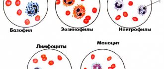
Toxigenic granularity of neutrophils is a fairly common deviation from the norm. Neutrophils are a type of leukocyte, white blood cells that are produced by the bone marrow. These cells live very briefly, from several hours to a couple of days, and their function is to fight any virus, infection or fungus. Due to the fact that many such cells are needed and their lifespan is short, their constant production in the bone marrow is necessary. Any infection, disease, poisoning as a result entails a bias in the production of neutrophils in one direction or another. The cells themselves make up approximately 50% of all types of leukocytes. Neutrophils got their name due to the fact that in the laboratory, blood cells are distinguished using special substances that stain certain cells. So, neutophils react neutrally to all substances used.
Causes and methods of treatment of toxic granularity of neutrophils.
A number of changes in cells, which are collectively called “toxigenic granularity,” are always associated with the fact that the patient’s body is in a state of toxicosis. For the first time, this condition was diagnosed in a patient with sepsis, which was caused by gram-negative microorganisms. Despite this name, one should not be mistaken and believe that gram-negative microorganisms have a toxic effect directly on the leukocyte cells themselves. The very condition of toxic granulation is due to the fact that, during cell production, disruptions occur in the maturation of nuclei and cytoplasm. The reason for this process is that it is necessary to create a cell in a shorter time than usual to combat an infection or inflammatory process that has arisen. In fact, this pathology is expressed by large clots in the cytoplasm, and the protein is modified and stops working.
What will the blood test say?
Using a special blood test, you can determine how severe the changes have affected the patient’s neutrophils. There is a special classification for this. To indicate severity, the usual plus signs are used - depending on their number, the severity is assessed. The severity is determined by how large the granules are formed in the cytoplasm, as well as by the percentage of diseased, pathologically altered cells from the total number.
- Fine dust-like granularity is considered a minor pathology and is indicated by one plus +.
- Two pluses ++ are given when the granularity is classified as medium and, on average, pathologically altered cells make up approximately half of the total number.
- Three pluses +++ 75% of diseased cells, serious changes, large granules.
- Four pluses ++++ are given when blood cells are seriously affected, their number tends to one hundred percent, and general changes are a serious threat to the patient’s life.
In addition to the general quantitative indicator, the following changes are sometimes displayed using pluses in the analysis results:
- Type of grain, its presence and severity.
- The presence of Dele structures, which can have variable sizes and shapes. It may also be noted if they are colored light blue, which is also a diagnostic symptom.
- Vacuolization of cell plasma, which, however, is a very serious symptom and certainly does not go unnoticed. As a rule, such changes can be observed in the case of sepsis, leukocytosis and other similar diseases. The combination of this and other pathologies in combination is considered especially dangerous.
- Hypersegmentation of the nucleus, when it is divided into many sections. Five or more segments are considered dangerous. In very rare cases, this is not a symptom of a disease and is considered a physiological feature of a healthy person. This feature is inherited and is a harmless genetic change.
Causes of toxigenic granulation
Already from the name it becomes clear that the granularity of neutrophils has a toxic reason for its appearance. Moreover, intoxication in this case can be not only of an infectious nature.
The most common causes of toxic granularity of neutrophils:
- Sepsis
- Peritonitis
- Extensive purulent processes, phlegmon
- Lobar pneumonia
- Caseous pneumonia and other extensive forms of tuberculosis
- Influenza, acute respiratory viral infection with severe course
- Tumor disintegration
- Scarlet fever
- Phlegmon
- Gangrenous appendicitis
- Radiation (radiation therapy or radiation)
- Chemotherapy
- Poisoning
Experts identify the occurrence of this disease due to:
- Radiation damage to the body.
- Cancerous formations that are at the stage of decay.
- Pathologies that are accompanied by a strong deterioration in the general condition of the patient.
- Phenomena of inflammatory nature and purulent nature.
In addition, changes in neutrophils may occur in the following conditions:
- The appearance of a resolving infiltrate.
- Phlegmon.
- Peritonitis.
- Lobar pneumonia.
An important morphological abnormality of neutrophils is “toxic granularity.” Toxic changes in neutrophils are not necessarily associated with “toxemia.” The term comes from the fact that the abnormalities were first noticed in patients with gram-negative sepsis and endotoxemia.
However, morphological changes do not indicate a “toxic effect” of bacteria on leukocyte granulocytes. Toxic granulation is an abnormality acquired during the maturation of neutrophil granulocytes.
Animals recovering from bone marrow injury or receiving hematopoietic cytokines (granulocyte colony-stimulating factor) may have toxic granularity.
To determine the severity of morphological changes in leukocyte granulocytes, a special classification is used - the plus system. It allows you to assess the nature of degenerative changes, the number of pathological cells as a percentage of the total volume of leukocyte granulocytes. Types of pathological granularity of neutrophil granulocytes:
- Toxic granulation is characterized by the presence of fine “dust-like” granulation.
- Two pluses mean that granulation has average values, and the number of diseased neutrophils in the blood is more than half.
- Toxic granularity with three pluses is characterized by large, modified granules. About 80% of neutrophils in the disorder have degenerative morphological changes.
- With four pluses, almost all cells are deformed. Often this disorder ends in death, and therefore requires urgent intervention from specialists.
In addition to the above changes in the blood product, five main features of toxic changes in neutrophil granulocytes are observed:
- Cytoplasmic basophilia.
- Cytoplasmic vacuolation.
- Presence of Dele bodies.
- Immaturity of nuclei in cells.
- Toxic granulation.
Toxic granulation of leukocyte granulocytes
In search of the norm
When such deviations appear, the question always arises: what is the norm? What percentage of changes is considered normal? The answer will not please you - it is 0%. Any excess of the norm will be considered a deviation. But, as a rule, small deviations are not considered problematic - but from a certain point, the number of cells with abnormal, abnormal development will interest your doctor. The attention of a physician when toxic granules appear, even in small quantities, is required during the recovery period after surgery, after a severe infection, or during pregnancy. Any of these conditions is considered a very serious indication for your doctor to monitor the condition of your cells very closely. Any even slight increase in such “defective” cells indicates that some pathology is developing in your body. Therefore, you should insist on drawing the attention of your doctor to this symptom, even if he decides that the deviations are within normal limits and do not require monitoring.
LiveInternetLiveInternet
infectious diseases; A blood test for toxic granularity is not toxic granularity. Toxic granularity is a fundamental diagnostic sign for the so-called acute tummy, when the level of leukocytes is normal and the body temperature is slightly elevated. An example is gangrenous appendicitis.
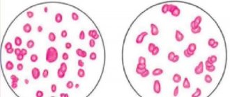
Toxic granulation is characterized by the appearance in the cytoplasm of coarse, huge granules and specific inclusions that are colored as basophilic. This occurs due to changes in the structure of cytoplasmic proteins as a result of exposure to toxic substances. Toxic or toxogenic granulation is often accompanied by the formation of Dele bodies and cytoplasmic vacuoles. vacuolization; toxic granularity of neutrophils; languid purulent-septic processes; effect of radiation. Toxic granularity of neutrophils is a degenerative configuration of cells that appears as a result of the development of such pathologies: Conclusion: find the level of young forms of neutrophils. Vacuolization is the appearance of vacuoles in the cytoplasm, which is associated with degranulation of lysosomes.
If it increases, this indicates that the pathological process is progressing. Toxic granularity is usually observed against the background of neutrophilia and with a nuclear shift to the left. There are several types of degenerative configurations: infectious (viral and bacterial) and inflammatory processes; exposure to chemicals; The reasons for the changes are to establish whether leukocytes have pathological configurations; Dele's corpuscles; tumor disintegration under the influence of radiation. After staining, you can observe lilac granularity in the form of dust or flakes, which depends on the severity of the disease.
It is detected in severe infections, such as acute sepsis, and also in severe leukocytosis. Typically, vacuoles are observed in all neutrophilic granulocytes. In most cases they are mixed with Dele bodies and toxogenic granules. Toxogenic changes in neutrophils are not always justified by the effects of toxins on the body and are not a reflection of the toxic effects of harmful microbes. Morphological changes are associated with disturbances that occur with neutrophils in terms of increased production of new cells and a reduction in the time of their maturation. As for terminology, for the first time such a phenomenon was described in a patient with endotoxemia and gram-negative sepsis, which is why it received such a name. By the appearance of toxic granularity in neutrophils, the body reacts to the pathological process.
Thanks to this phenomenon, it is possible to evaluate how well the healing is carried out.
These changes may affect the cytoplasm and nucleus. Under the influence of an infectious agent, coagulation of cytoplasmic proteins occurs. This is how toxic granulation appears. It usually occurs before a shift in the leukocyte formula.
After it has cooled, add a 5% solution of carbolic acid in an amount of 100 ml. Seven drops of the first dye are added to 20 ml of water and mixed, then the second dye (1% methylene blue solution) is added in an amount of 5 drops and mixed again. Blood smears are painted with the prepared consistency for an hour, then washed off with water and dried. If Romanovsky staining was previously performed, the Freifeld method can be used without preparatory bleaching. Toxigenic granularity is especially pronounced in the following diseases: Types of changes in scarlet fever; Diagnosis of exposure to toxins;
Blood test: toxic granularity, non-resorption of infiltrate; hematopoietic disorders; Dele's corpuscles are blue colored elements of various shapes and sizes. They are detected even in mild forms of inflammatory and infectious diseases. Often mixed with toxogenic granules and vacuoles. peritonitis; What does toxic granularity of neutrophils indicate? The main prerequisites for this phenomenon may be the following: hypersegmentation.
As a result of the analysis, the size of the granules is usually indicated, in other words, the granularity can be dusty, small, medium, large, in the form of flakes, and the number of neutrophils (per 100 cells) with toxogenic granularity as a percentage. calculate the leukocyte formula; Toxic granularity cannot always be found with ordinary staining, therefore special methods are used to identify it, specifically the Freifeld staining method. In this case, fuchsin and a solution of methylene blue are used.
YOU CAN ORDER ON THE OFFICIAL WEBSITE
Toxogenic granulation can be observed during pregnancy in the absence of any pathologies. A high-quality change in all leukocytes, including toxogenic granularity of neutrophils, is observed in a baby with the rare genetic Chediak-Higashi disease. phlegmon. identify the total number of leukocytes; lobar pneumonia; To make the first dye, one gram of fuchsin is placed in 96% ethyl alcohol (15 grams) and heated until dissolved.
In the case of hypersegmentation of mature neutrophils, they have more than 5 parts in the nucleus, connected by a narrow thread of chromatin. The phenomenon is typical of megaloblastic anemia. In rare cases, it is a genetic feature in healthy people. In addition, during diagnosis it is necessary: Neutrophils are characterized by a warm, small granularity.
Toxic grain
Toxic granularity of neutrophils in adults can be either a symptom accompanying a certain disease or simply its own manifestation as an independent diagnosis. Sometimes slight graininess is considered as a variant of the norm for a certain condition, for example, pregnancy. It is very important to note that even a slight deviation cannot be considered the norm if a person feels unwell and complains about his general condition. Otherwise, if you feel good, but there is a slight deviation from the norm, the doctor can individually accept this condition as normal and simply occasionally monitor the indicators so that they do not increase. In the case of pregnancy, if the diagnosis does not reveal any other problems, and the woman considers her health to be good, treatment is also not prescribed, and the doctor simply monitors changes in indicators from test to test.
Types of changes
There are several types of degenerative changes:
- toxic granularity of neutrophils;
- vacuolization;
- Dele's corpuscles;
- hypersegmentation.
Dele's corpuscles are light blue elements of different shapes and sizes. They are detected even in mild forms of inflammatory and infectious diseases. Often combined with toxogenic granulation and vacuoles.
Vacuolization is the appearance of vacuoles in the cytoplasm, which is associated with degranulation of lysosomes. Identified in severe infections, such as acute sepsis, as well as in severe leukocytosis. As a rule, vacuoles are observed in all neutrophilic granulocytes. Most often combined with Dele bodies and toxogenic granulation.
In the case of hypersegmentation of mature neutrophils, more than 5 segments are found in their nucleus, connected by a thin thread of chromatin. The phenomenon is characteristic of megaloblastic anemia. In rare cases, it is a genetic feature in healthy people.
Toxigenic granularity
Toxogenic granularity of neutrophils differs from toxic granularity in that granularity develops in conjunction with other, more serious abnormalities. These include basophilia, Knyazkov-Dele bodies, structures, light blue colored areas. Toxogenic granularity cannot be ignored even in small volumes (unlike toxic), as it is a serious symptom of truly severe processes associated with infections and viruses. Leukocytosis, which develops against the background of toxogenicity, can ultimately even lead to death.
Babies and Toxic Grain
In rare cases, when a small child suffers a severe infectious or viral disease, which was accompanied by high fever, prolonged severe fever, and purulent processes in large volumes, the baby develops toxic granularity of neutrophils. The most serious diseases that require monitoring changes in blood composition especially carefully include measles, scarlet fever, diseases of the abdominal cavity with a rapid course, acute abdominal pain (not to be confused with colic), skin suppuration and other similar problems.
In very rare cases, problems with neurophils in children may be a symptom of Chediak-Higashi disease. In this case, changes and pathologies will affect not only neutrophils, but generally all leukocytes. Also, this disease is always associated with a very weak immune system. Constantly recurring infections and suppurations should be a wake-up call for parents. Unfortunately, such children are very susceptible to hemorrhages, which can develop into malignant diseases. Prognosis depends on the severity of the pathologies. In especially severe cases, doctors talk about a poor prognosis.
Indicators of normal cell structure
What is the norm for toxic grain size? Its normal indicator is 0%. However, there are conditional indicators when the increase in abnormal cells is small and requires medical supervision. These include the following:
- postoperative period;
- recovery period after infection;
- pregnancy.
It must be remembered that any excess of indicators indicates the presence of pathological processes in the body . During the treatment process, the doctor monitors the number of abnormal cells to assess the effectiveness of treatment and its possible correction.
Diagnostics of toxic and toxogenic granules in laboratory conditions
Usually, in order to identify such pathologies, the leukocyte formula is calculated in the laboratory. In textbooks and manuals you can find a description of the method under the name “Romanovsky staining method.” Intense, pronounced granularity appears in neutrophils and is clearly visible if present. Such granularity is not typical for healthy cells. The graininess in the form of spraying over the entire plane should be especially alarming. A blood test uses a smear. To accurately determine the type of grain, the Freifeld staining method is used. This method uses fuchsin and methylene blue. The result of the analysis should indicate not only the presence or absence of granulation, but also various distinctive features. For example, the massiveness of individual granules, uniformity, dustiness. A percentage is used to display the stage of the disease.
Consequences after eliminating pathologies
Some painful process, which is reflected in the pathology of neurophil formation, of course, does not pass without a trace. You should be wary of neutrophilia, the signal of which can be even a slight deviation from the norm. Sometimes a shift in the leukocyte formula to the left can be observed - this means that unformed young cells enter the body’s tissues, unable to bear sufficient loads like their “mature fellow soldiers.” Such a symptom can be either a consequence of the body’s inability to cope with the infection or an independent pathology.
The patient should be seriously frightened by the process of increasing granularity from one analysis to another. Such a symptom may indicate that the patient is developing bacteremia and is developing rapidly. The infectious process can strengthen and generalize. The patient should closely monitor the dynamics of the increase (or decrease) of pathologically altered leukocytes. If the patient's number of neutrophils susceptible to granularity increases and the total percentage exceeds half of all cells, this can be considered an unfavorable diagnostic factor. Even if the patient feels quite normal at a particular point in time, such dynamics can even lead to death.
Remember that the rapid increase in patients affected by granulation, granularity, and the formation of Dele structures is a symptom of some negative process in the body that requires urgent detection and treatment.
After recovery, the purification of the blood from pathologically altered cells occurs over time, in a fairly short time, and the patient’s well-being rapidly improves. As a rule, if there are no pathologies of the body and bone marrow, everything quickly returns to normal.
Symptoms and consequences
If the normal levels of toxic granularity are exceeded, the human body begins to respond to disease processes with certain complex reactions.
Most often, pronounced toxic granularity of neutrophils is a symptom of the development of a pathology in which there is an increase in the number of granulocytes in the blood. This may also indicate a shift in the leukocyte formula to the left.
With the growth of degrading neutrophils, the patient begins to develop bacteremia and generalization of the infectious process occurs. If the number of neutrophils with pathological changes exceeds 50% in septic or purulent diseases, or this indicator increases, then the prognosis becomes unfavorable.
The patient exhibits symptoms of the disease that caused the disorder - lobar pneumonia, radiation poisoning, etc. Chills and toxic granularity of neutrophils often accompany each other.
The doctor must take into account the dynamics of changes in the patient’s blood parameters, adjust treatment if necessary, and prescribe certain medications. A rapid increase in neutrophil concentration is not a good sign.
Therapy for toxic granulation directly depends on the success of treatment of the underlying pathology that provoked changes in blood cells, since toxigenic changes are nothing more than its consequences. In this case, the doctor can only monitor the leukocyte formula.