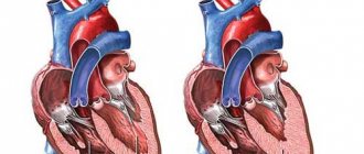
There are a number of diseases affecting the cardiovascular system. A condition in which disruptions in the metabolism of myocardial tissue are observed is called myocardial dystrophy (MCD). As a result, the walls of the organ become thinner, and heart function suffers. The pathology is secondary and develops against the background of the underlying disease.
Stages of pathology development
Cardiac muscle dystrophy is not associated with inflammatory or degenerative tissue changes. It develops as follows:
- Metabolic imbalance causes a defect in the nervous regulation of the myocardium, the production of adrenaline increases, which leads to increased heart rate. Systematic tachycardia contributes to the “wear and tear” of tissues, due to which the walls of the heart are depleted.
- Weak tissues are not able to absorb the required amount of oxygen, the ischemic effect leads to the fact that the myocardium suffers from hypoxia.
- Oxygen deficiency leads to an increase in calcium levels in the blood. An increased content of this element complicates tissue respiration. Harmful substances that deform cells begin to be produced. Deformed lysosomes produce enzymes that adversely affect the structure of cardiomyocytes.
- Lipid metabolism suffers and is disrupted. As a result, free radicals accumulate in the tissues and can deform the myocardium.
The changes that have occurred lead to a sharp shortage of viable cardiomyocytes.
When diagnosing myocardial dystrophy, symptoms and treatment will directly depend on the causes that provoked the pathology. The disease is classified according to the same causal principle.
Treatment
It is necessary first of all to treat the underlying disease that caused myocardial dystrophy. In many cases, etiotropic therapy helps restore the normal functional state of the heart muscle. Agents that improve myocardial metabolism, in particular mildronate, are widely used. Drugs such as riboxin, cocarboxylase, ATP are currently not recommended for use due to the lack of evidence of their effectiveness. In myocardial dystrophy, lipid peroxidation of cell membranes is activated, which is one of the mechanisms for the formation of functional and structural disorders, therefore antioxidants that suppress these processes (vitamin E) are included in the treatment complex. When supraventricular rhythm disturbances occur, calcium antagonists (verapamil) are prescribed. They also help eliminate changes in calcium metabolism in myocardial cells, leading to impaired contractility of muscle fibers. Beta blockers may also be prescribed. They help cope with rhythm disturbances and eliminate the harmful effects of excess catecholamines on the myocardium. In order to strengthen intracellular membranes and prevent the entry of aggressive enzymes from lysosomes into the cell, membrane stabilizers, for example, Essentiale, can be prescribed. Improving the supply of oxygen to the myocardium is achieved through the use of hyperbaric oxygenation, taking oxygen cocktails, and prescribing Mexidol. To eliminate electrolyte imbalances, a diet rich in potassium is used, as well as medications containing this trace element (panangin). If circulatory failure develops, appropriate therapy is carried out.
Classification of myocardial dystrophy
The classification, which is based on provoking factors, looks like this:
- Dyshormonal myocardial dystrophy. This variant of pathology always develops against the background of one or another hormonal imbalance. Menopausal changes in the body of women, age-related decreases in testosterone production in older men, pathologies of the thyroid gland, and other endocrine disorders cause metabolic disruptions in the body. As a result, the heart does not receive enough nutrients in full, and myocardial dystrophy develops.
- Dysmetabolic myocardial dystrophy. This form develops due to metabolic disorders caused by poor nutrition. A deficiency of certain elements in the diet leads to anemia, vitamin deficiencies, and, as a consequence, metabolic disorders. Protein deficiency, diabetes, and other important components can be included in this group. For example, an adult who has chosen veganism, or a woman who is trying to lose weight and is constantly following a strict diet, should remember that a lack of nutrients is fraught with serious heart problems.
- Myocardial dystrophy of mixed origin is a condition that occurs as a result of other established causes, with the exception of dyshormonal and dysmetabolic disorders. Mixed or complex genesis includes the consequences of intoxication, infectious diseases, and neuromuscular pathologies.
A separate group includes myocardial dystrophy of unspecified origin, i.e. a condition the cause of which could not be determined after a comprehensive examination and the use of all known diagnostic methods.
The above classification in cardiological practice can be significantly expanded. That is, working with a specific patient, a cardiologist can more accurately identify the cause that provoked cardiac dystrophy. Practitioners distinguish:
- Tonsillogenic MCD. If a child or adult often suffers from sore throats or is diagnosed with chronic tonsillitis, as a result of constant inflammation of the tonsils, the activity of some parts of the brain is activated, which are responsible for the increased production of adrenaline and norepinephrine, causing the myocardium to contract in an enhanced manner. Increased load causes overstrain of muscle fibers, and myocardial dystrophy develops.
- Anemic MCD. With anemia, the heart muscle, like other tissues, suffers from hypoxia. The fact is that it is hemoglobin that transports oxygen to tissues, and with its deficiency, hypoxic phenomena begin to develop. The myocardium tries to compensate for the oxygen deficiency and contracts faster than usual. Systematic increased load leads to myocardial dystrophy.
- MKD sports overloads. If an athlete constantly exceeds his biological capabilities and trains intensely, the heart is forced to beat faster than usual. A natural consequence is that increased load leads to myocardial dystrophy.
- Alcoholic MKD. The cause of the pathology is alcohol abuse. Do not think that problems with myocardial activity occur only in people who drink heavily. If the patient has a weakened immune system, is subject to frequent stress or has other chronic diseases, the alcohol factor will be important even with small but regular intakes of alcohol.
- Toxic MCD. This form of the disease is provoked not only by toxic and narcotic substances. This also fully includes medications, such as glucocorticoid hormones and some chemotherapy drugs.
- Menopausal MCD. Occurs against the background of estrogen deficiency.
- MCD for thyroid diseases (hypothyroidism and hyperthyroidism). The development of myocardial dystrophy is provoked by a deficiency or excess of thyroid hormones.
- Neuroendocrine MCD. The cause of this form of pathology is considered to be chronic stress. The myocardium, forced to work under conditions of chronic stress, contracts in an intensified mode, mobilizing its “emergency” capabilities and experiencing constant tension.
Cardio website
Other cardiac pathologies
If the patient has myocardial dystrophy, it will be difficult to assign a code according to ICD-10 in this case. Since the last revision, a separate code is no longer allocated for this pathology. However, there is a code that is used from the section on cardiomyopathy.
Myocardial dystrophy is understood as a specific heart disease that is not inflammatory in nature. It is characterized by metabolic disorders that occur in myocytes. At the same time, the contractility of the heart muscle changes, and heart failure appears.
Classification of diseases ICD-10 is a normative document that contains codes for diseases systematized by sections. This is an international development that facilitates the collection and storage of information to create statistics for all countries. ICD-10 was created by the World Health Organization. The document is revised every 10 years and additions are always made to it. If previously there was a separate code for myocardial dystrophy, now it is not assigned to this disease according to ICD-10. However, doctors may give the number I.42, which implies cardiomyopathy. Despite the similarity of names, cardiomyopathy and myocardial dystrophy are considered different phenomena, since the first involves a wider range of different cardiac pathologies that are not associated with heart disease and the vascular system. Therefore, these terms cannot be used as synonyms. But for myocardial dystrophy there is still one code according to ICD-10, which assumes only cardiomyopathy with metabolic disorders and nutritional problems.
In general, section I42 assumes cardiomyopathy, but excludes conditions that are complicated by pregnancy and postpartum ischemic disease. Number I42.0 suggests a dilated type, and 42.1 is a hypertrophic obstructive pathology. Number 42.2 refers to other forms of hypertrophic pathology. Code 42.3 is endomyocardial disease, and 42.4 is fibroelastosis of the endocardial type. If the patient has other restrictive cardiac pathologies, then the number is set to 42.5. If cardiomyopathy is of alcoholic origin, then code 42.6 is assumed. If the disease is caused by the influence of drugs or other external factors, then the number 42.7 is set. Other cardiomyopathies have the number 42.8, and if the forms of the pathology are not specified, then the code is 42.9.
Definition of obliterating atherosclerosis of the vessels of the lower extremities in ICD 10
There are several forms of myocardial dystrophy. Depending on the type of dystrophic processes in the heart, the following are known:
- restrictive;
- dilatational;
- hypertrophic.
Depending on the severity of the disease, the following forms are distinguished:
- 1. Compensation. Hemodynamics are still maintained at the level required for the body.
- 2. Subcompensation. The mechanisms that are responsible for hemodynamics are strained, even with moderate physical activity. There is already pronounced dystrophy of the heart muscle.
- 3. Decompensation. With little physical activity, deviations in hemodynamics are noticeable. The contractile capabilities of the myocardium are sharply weakened.
Depending on the pathogenesis, primary and secondary forms are distinguished. The primary one assumes that the cause of the disease has not been established. Secondary means that myocardial dystrophy appeared as a complication against the background of another disease.
Depending on the disease that caused dystrophy of the heart muscle, the following forms are distinguished:
- 1. Dishormonal. Appears due to disturbances in the synthesis of sex hormones. A person often gets tired, has problems sleeping, is thirsty, and loses weight. A stabbing or aching pain is felt in the area of the heart.
- 2. Tonsillogenic. It is a complication after tonsillitis. Endurance deteriorates, aching pain in the heart area, and arrhythmia appear.
- 3. Alcoholic. Develops as a consequence of prolonged alcohol intoxication. The membranes of cardiac tissue cells are destroyed due to exposure to ethanol. Potassium and fatty acid reserves decrease. This leads to shortness of breath and arrhythmia. Pain in the heart area almost does not appear.
- 4. Anemic. Often appears in pregnant women. At later stages, toxicosis also appears.
- 5. Diabetic. Appears in patients with type 1 diabetes mellitus. In this case, diabetic disorders can be detected in the coronary vessels, and angina pectoris develops.
Causes and symptoms
The occurrence of myocardial dystrophy as a primary disease is quite rare. Usually this pathology is secondary. All causes of myocardial dystrophy can be divided into 2 groups. The first includes all the heart ones. For example, this could be cardiomyopathy or myocarditis. The second group includes non-cardiac causes. These are problems with metabolic processes, anemia, intoxication of the body, diseases caused by infections, and exposure to adverse environmental factors (radiation, weightlessness, overheating, etc.).
Because of this, the myocardial muscles do not receive enough nutrients and oxygen and experience intoxication. Gradually the cells die and are replaced by connective tissue. Scars appear. As a result, the main functions of the myocardium are disrupted - contraction, conductivity, excitability and automaticity. Because of this, all cells of the human body suffer.
At the initial stage of the disease, symptoms do not appear, so the disease does not make itself felt.
But if the disease is not treated, heart failure gradually develops, which can even lead to death. Therefore, as soon as the first signs of illness appear, you need to urgently go to the hospital. The reason for this is the following:
- shortness of breath with minor physical exertion;
- strong increase in heart rate at low loads;
- weakness, a person gets tired quickly;
- pain and discomfort in the chest area;
- cough in the evening and at night with a large amount of sputum.
Symptoms of the disease, depending on its form and causes, may be different. If they are detected, you should immediately consult a doctor for diagnosis and selection of appropriate therapy.
Myocardial dystrophy is a specific non-inflammatory disease. With this disease, metabolic disorders are observed due to the action of various factors. Since the last revision, there is no separate code for this syndrome in ICD-10, so the code must be selected from section I42, which describes the types of cardiomyopathy.
Features in children and adolescents
Myocardial dystrophy has no age limit. The most common age group in which the pathology occurs are people over 40 years of age, but MCD develops in both newborns and children - preschoolers and schoolchildren.
Features of pediatric MCD include difficulties in therapy. In children, the disease is more difficult to treat, since the growing body needs a more resilient heart, and due to dystrophy, the myocardium cannot meet high demands.
In a newborn, MCD develops as a result of intrauterine infections or perinatal encephalopathies, and the syndrome of maladaptation of the heart and blood vessels can also be the cause. A teenager can receive a diagnosis of MCD due to the following reasons - infections, frequent sore throats, anemia, myocarditis, intense physical training, metabolic disorders. Sometimes the disease is caused by some acute process that was previously diagnosed - rheumatism and similar pathologies.
Causes
Among the causes of the development of myocardial dystrophy, cardiac, extracardiac and external factors can be distinguished. Let's take a closer look at them.
Cardiac factors
This includes all pathologies of the cardiovascular system that led to the development of MCD, these are:
- Heart disease.
- Circulatory disorders.
- Pulmonary heart.
- Arterial hypertension.
- Myocarditis and others.
Extracardiac factors
The most extensive group, which includes numerous chronic and congenital pathologies:
- Chronic anemia.
- Metabolic diseases – hypothyroidism and others.
- Hormonal pathologies - menopause, problems during puberty.
- Chronic gastrointestinal diseases - malabsorption disorders, pancreatitis.
- Tumor diseases.
- Vitamin deficiencies, lack of nutritional components caused by chronic pathologies.
- Congenital pathologies.
External factors
This includes all provoking factors associated with defects in behavior, lifestyle and load distribution. This:
- "Sports" heart.
- Neuroses, chronic stress, depression.
- High physical activity.
- Toxic, alcoholic, medicinal effects.
Causes of myocardial dystrophy
Myocardial dystrophy of the heart muscle occurs due to severe stress on the heart during improper and intense training. Also, an unbalanced diet, short rest, and constant interruption of sleep can trigger the appearance of MCD. Other common factors in the development of myocardial dystrophy include:
- infectious diseases in the inflammatory phase;
- chronic tonsillitis;
- intoxication of the body (poisoning, alcohol, cigarettes, drugs);
- overweight;
- myxedema;
- avitaminosis;
- diabetes;
- thyrotoxicosis;
- anemia;
- radiation;
- muscular dystrophy;
- genital inflammation;
- collagenosis;
- the appearance of menopause in women;
- overheating;
- lack of potassium in the body;
- Cushing's syndrome;
- starvation;
- prolonged stay on mono-diets;
- deposition of salts in the heart muscle.
Diagnostic approaches
Diagnosis of cardiac pathologies is varied. A patient examination always begins with a visual examination of the patient and an analysis of his complaints. During the examination, the doctor performs percussion (tapping with fingers) and auscultation (listening with a phonendoscope). Percussion allows you to determine the boundaries of the heart muscle, which expand during myocardial dystrophy. However, the percussion method cannot be called indicative in comparison with more modern diagnostic methods - ECHO-CG, MRI and others.
Auscultation helps to identify the presence of a heart murmur, muffled first tone, impaired ri src="https://davlenienorm.com/wp-content/uploads/2018/10/miocardiodistrofiya-distrofiya-miocarda-klassifikatsiya-diagnostika-lechenie-6.jpg" class=”aligncenter” width=”530″ height=”310″[/img]
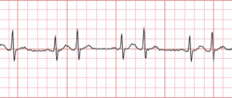
The basis of any cardiac examination is always the ECG. This is an accessible and very informative diagnostic method, which allows not only to identify pathologies in the functioning of the heart, but also to determine the further scenario for examining the patient.
Chest X-ray, MRI, stress tests are prescribed by a cardiologist to clarify the diagnosis after an ECG.
Additionally, laboratory tests are carried out, in particular, a biochemical blood test, tests for thyroid hormones, and a general blood test are important.
Other complex diagnostic procedures - scintigraphy, coronary angiography, ultrasound of the heart are prescribed strictly individually, after a recommendation from a cardiologist is given for this.
Diagnosis of the disease
An objective examination is the first stage of diagnosis. During admission, an irregular pulse and systolic murmur are detected. Tone 1 weakens at the top. The ECG reveals arrhythmia, the phenomenon of myocardial repolarization, and a decrease in its ability to contract.
Stress tests give a negative result. “Gallop rhythm” appears after phonocardiography. An echocardiogram is used to diagnose enlargement of the atria and ventricles and detect structural changes in tissue. There are no traces of organic pathology.
After taking an x-ray, it is possible to see that the damage to the myocardium is quite deep, and there are signs of myopathy. A biopsy is a last resort method when all other diagnostic methods do not provide an accurate result. Scintigraphy makes it possible to identify areas of focal changes. Differential diagnosis should be carried out with myocarditis, cardiosclerosis and coronary heart disease.
Stages and symptoms
During myocardial dystrophy, there are 3 stages.
Stage 1 of the disease is called the compensation stage. It is characterized by the destruction of cardiomyocytes in certain areas of the heart muscle. To eliminate the defect that has arisen, the cells surrounding the damaged area enlarge and proliferate. As a result, the volume of the myocardium becomes larger. If you begin treatment of the pathology at this stage, the myocardium is able to fully recover.
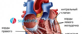
Heart with myocardial dystrophy
Symptomatic manifestations of the compensation stage are rapid fatigue, inability to work for a long time, pressing pain, shortness of breath with minimal increases in load.
Stage 2 is called the subcompensation stage. Areas of non-viable cardiomyocytes become larger, merging with each other.
At the same time, healthy cardiomyocytes begin to work harder, trying to compensate for deficiencies, and increase in volume even more. Contractile function is impaired due to too much volume of the myocardium, and the volume of blood that the heart can pump out decreases.
A characteristic symptomatic sign of stage 2 is shortness of breath, which occurs both during and after exercise. Later, the heart rhythm is disturbed - tachycardia or bradycardia occurs. Sometimes already at this stage the patient develops swelling of the lower extremities. With adequate treatment, good results can be achieved and normal contractile activity can be restored.
The last, stage 3 of myocardial dystrophy is a state of decompensation. The patient's myocardial structure is disturbed; even on an x-ray, an expansion of its boundaries is noticeable. A large and weak heart muscle is not able to contract well. Blood circulation in the body is disrupted, irreversible changes occur, as a result of which the patient’s death is possible.
Symptoms of stage 3 are as follows: severe pallor of the skin, weakness, constant shortness of breath, serious disruptions in the functioning of internal organs, arrhythmias, and congestion in the lungs.
Symptoms of MCD
Signs of myocardial dystrophy can most often be noticed only during intense physical activity. At rest, the disease is passive. Symptoms of MCD can very often be confused with other diseases, so myocardial dystrophy is quite difficult to identify and diagnose at the primary stage. The main signs of MCD disease:
- spasmodic or aching chest pain;
- fast fatiguability;
- impotence;
- state of depression;
- shortness of breath when moving;
- weakness;
- cardiopalmus;
- pain in the heart area;
- interrupted functioning of the heart muscle;
- arrhythmia;
- systolic murmur at the apex of the heart;
- slowing heart rate.
In chronic cases, cardiac myocardial dystrophy, treatment, symptoms - all this becomes complex. The patient experiences severe shortness of breath at rest, an enlarged liver, and the appearance of edema.
Treatment and prognosis
A cardiologist treats myocardial dystrophy. For patients with stage 1 or 2 of the disease, it is important to select adequate drug therapy and adjust the regimen. They do not need to be hospitalized. It is very important to establish the cause of heart problems; its elimination is the primary goal of therapy.
Medicines are selected by a doctor and taken strictly as prescribed. Self-medication for myocardial dystrophy is unacceptable, since lost time is fraught with complications. Cardiologists use several drug groups to eliminate symptoms, these are:
- Beta blockers. “Bisoprolol”, “Metoprolol”, “Anaprilin”, “Bicard” normalize the heart rhythm and reduce the heart rate.
- Drugs that improve metabolism - Mildronate, Thiotriazolin, Riboxin. Helps improve myocardial nutrition and restore damaged cardiomyocytes.
- “Curantil”, “Dipyridamole” are drugs that improve microcirculation.
- “Panangin”, “Asparkam” - replenish the deficiency of potassium and magnesium.
- Vitamin preparations - vitamin C, B vitamins.
Treatment is not limited to this small list of medications. Depending on the causative factors, the doctor may prescribe an additional drug to patients - an antihypertensive, hormonal, enzyme, antiplatelet agent, etc. You should understand how important it is to timely select not only drug therapy, but also to adjust the patient’s general regimen.
Important: If medical recommendations for lifestyle correction are not followed, drug therapy does not produce results.
Nutrition
It is of great importance for heart health. If you like salty, spicy, sweet, everything “delicious”, get ready that over the years you will develop excess weight, problems with blood pressure and other “joys”. If, on the contrary, you eat according to your own established principles, for example, you give preference to plant foods, excluding meat, you go on diets for a long time and use dubious food additives - problems also cannot be avoided.
Therefore, the conclusion is simple - nutrition for myocardial dystrophy should be complete, balanced, satisfying all the needs of unhealthy cardiomyocytes.
This is achieved through the following principles:
- Limitations of table salt. The recommended amount is 3 grams per day.
- Double the amount of vitamins. The diet is supplemented by fresh vegetables and fruits, which should appear on the menu up to 6 servings per day.
- Proper drinking regime. Drinking is plain water, fruit drinks without sugar. Soda, sugary drinks and boxed juices are prohibited. The volume of drinking per day, including first courses, is no more than 1.2 liters.
- Reducing the overall calorie content of the diet by eliminating fatty foods, smoked meats, fatty broths and sausages.
You can take the Mediterranean diet as a basis. By the way, people who adhere to it almost do not suffer from heart disease.
Prevention of myocardial dystrophy is not only a proper diet. The correct regimen plus the exclusion of all causative factors described above is the best preventive algorithm.
To Army? Need to think
Parents whose child suffered from myocardial dystrophy in childhood or whose teenager now suffers from it are always concerned about the question, what about the army? It would seem that the heart is sick, it is definitely impossible to serve.
The issue will be resolved individually, based on a detailed examination of the young man. If abnormalities in the functioning of the heart are detected, the service will be excluded. If the diagnosis was made in early childhood and was completely removed with age, then service is possible.
In conclusion, we would like to remind you that a dangerous or non-dangerous prognosis of a disease always depends on the patient’s reasonable pedantry - how correctly you follow the regimen, take prescribed medications, and take care of yourself. Take care of yourself.
Did you like the article? Save it!
Still have questions? Ask them in the comments! Cardiologist Mariam Harutyunyan will answer them.
Ivan Grekhov
Graduated from the Ural State Medical University with a degree in General Medicine. General practitioner

