Causes of left atrial hypertrophy and treatment for an enlarged heart
Often this pathology occurs due to genetic defects (hereditary predisposition). Some of the most common triggers are high blood pressure and obesity.
Other reasons that can enlarge the left half include the following factors:
- Mitral valve stenosis and insufficiency. These diseases often provoke inflammation of the left atrium;
- Hypertrophic cardiomyopathy - thickening of the ventricles (hereditary pathology);
- Aortic stenosis is an abnormal narrowing of the aorta;
- Acute and chronic lung diseases. Any infection and inflammation of the respiratory system can lead to hypertrophy;
- Stress.
Enlarged heart - can it return to normal?
An enlarged heart, depending on the cause, may return to normal size if prompt treatment is given. In many cases, however, this does not occur, in which case the goal of therapy is to quickly determine the cause and provide treatment to stabilize the situation and prevent further development of cardiomegaly.
Reversible causes include:
- Early stages of hypertensive heart disease caused by high blood pressure.
- Early stages of significant congestive heart disease.
- The early stages of a heart attack and sudden weakness of the heart muscle, known as cardiomyopathies, which may be associated with pregnancy, severe stress or a viral infection.
The right side of the heart has a remarkable ability to recover from acute stress, so treating the underlying cause can promote full recovery.
Video: Cardiomegaly, stretching of all cavities of the heart, rhythm disturbance of DCM
Enlarged heart: left ventricular hypertrophy on x-ray
The reasons are similar to the disorder in the corresponding atrium:
- high blood pressure (hypertension);
- aortic valve stenosis;
- cardiomyopathy;
- excessive physical activity;
- obesity.
The risk increases with muscular dystrophies and Fabry disease.
The disorder develops gradually. In the early stages of left ventricular enlargement, there are no symptoms, then shortness of breath and chest pain occur. A person gets tired quickly, suffers from dizziness, rapid heartbeat, and may faint.
Forecast
It depends on what exactly caused the enlargement of the heart:
- With arterial hypertension, the prognosis is favorable. If you take the medications prescribed by your doctor on time, your heart will soon return to normal and will no longer enlarge.
- In case of ventricular septal defect – relatively favorable. If the operation is not performed in time, there is a risk of developing aortic valve insufficiency, severe arrhythmias, left ventricular dysfunction and sudden death. If the patient is operated on, the heart will no longer bother him.
- For dilated cardiomyopathy – unfavorable. Full recovery occurs only after transplantation. However, it is not always possible to find a donor for a heart transplant. In addition, the risk of postoperative complications is high.
- In hypertrophic cardiomyopathy – relatively unfavorable. With an asymptomatic course of the disease, patients die before the disease is detected. With proper therapy, the risk of death is reduced.
- Metabolic cardiomyopathy has a favorable prognosis. When metabolism is established, complete recovery occurs.
- With aortic stenosis without treatment, life expectancy ranges from 1 to 4 years from the onset of symptoms. If the operation is performed in a timely manner, the prognosis is relatively favorable.
- If mitral stenosis is left untreated, 50% of patients die within 5 years of the first symptoms appearing. After surgery, the prognosis is relatively favorable.
- In case of Ebstein's anomaly, it is relatively favorable. The risk of sudden death is 3–4%.
- For myocarditis – favorable. Complete recovery occurs after 4–8 weeks in 90% of cases, after a year – in 10% of cases.
- For exudative pericarditis – favorable. All operated patients recover.
- In case of amyloidosis – unfavorable. The maximum life expectancy is 5 years from the date of diagnosis.
Right atrial hypertrophy
This part of the organ is highly dependent on the functioning of the lungs, so pathologies of the respiratory system lead to changes in the right atrium and its ventricle.
The most common reasons:
- Lung diseases;
- Tricuspid valve stenosis;
- Tricuspid regurgitation (tricuspid valve insufficiency);
- Pulmonary embolism;
- Congenital heart defects.
The disorder is indicated by the same symptoms as when the right side of the organ is affected: fatigue, chest pain, difficulty breathing, rapid pulse.
Causes of pathology
From this article you will learn: why the heart can become enlarged, and whether a large heart is always a sign of illness.
What symptoms appear if the heart is enlarged due to disease. Treatment of pathology. Both the entire heart and its individual chambers can enlarge. This may be a symptom of cardiovascular system defects, inflammatory processes, or a consequence of excessive stress on the myocardium.
The problem is dealt with by a cardiologist and a cardiac surgeon.
Some diseases that cause heart enlargement can be completely cured with medication or surgery, but there are others that can only be completely cured by organ transplantation.
There are two types of enlargement of the entire heart or its individual chambers:
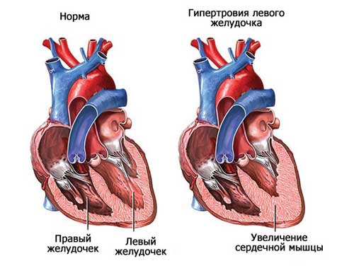
- Hypertrophy. This is thickening of the walls. Occurs due to the growth of the myocardium (muscular membrane). The left ventricle is most susceptible to this, since it bears the greatest load. Hypertrophy does not always require treatment. Click on photo to enlarge
- Dilation. This is a “stretching” of the chambers of the organ - an increase in their cavity.
This may be excessive stress on the heart muscle or heart or vascular defects.
Relatively safe causes of cardiac muscle growth
- high intensity physical activity;
- difficult pregnancy and childbirth.
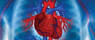
The most important organ that ensures the vital functions of the entire body consists of 4 chambers: two ventricles and two atria. There are two sections of the heart - right and left, each of which includes an atrium and a ventricle.
Normally, the boundaries of the heart change throughout life. People who play sports experience an increase in its size, which is considered a completely natural process and does not cause any cause for concern.
The weight of a man's heart is 332 grams, a woman's - 253 grams. An enlarged heart is observed when the myocardium grows and (or) expands its cavity.
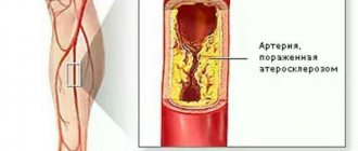
Most often, the organ enlarges to the left, which is observed in many diseases: hypertension, stagnation of blood in the systemic circulation, heart defects. Enlargement of all parts of the organ is dangerous.
This condition is called “bull’s heart”, and only surgical intervention can improve a person’s quality of life.
The reasons influencing the enlargement of the organ are:
- Hypertonic disease. An increase in pressure leads to changes in the cardiovascular system: vascular tone increases, the thickness of the muscle layer increases, and the systemic circulation suffers.
- Coronary heart disease: angina pectoris, myocardial infarction. Oxygen starvation of the organ tissues occurs with the death of their cells and replacement with connective tissue, which leads to an increase in the size of its left section.
- Rheumatic heart disease. It is a consequence of tonsillitis (frequent tonsillitis). Rheumatic disease is manifested by an inflammatory process occurring in the tissues of the organ. As a result, the valves suffer and defects form.
- Myocarditis.
- Renal dysfunction.
- Alcohol abuse. The most common example is alcoholic cardiomyopathy.
- Smoking.
- Acute pericarditis (inflammation of the serous membrane).
- Congenital heart defects.
Right ventricular hypertrophy
It is worth noting that this pathology is very rare. Its appearance can be triggered by only four reasons:
- pulmonary hypertension;
- tetralogy of Fallot;
- pulmonary valve stenosis;
- ventricular septal defect.
As in other cases, the initial stage does not manifest itself in any way. As it progresses, difficulty breathing occurs, accompanied by chest pain. Dizziness can result in loss of consciousness. Swelling of the legs is also observed.
In many ways, the symptoms of hypertrophy are similar to angina pectoris and coronary heart disease. This is why it is necessary to consult a doctor. Only a cardiologist can, after a thorough examination and receive the research results, make an accurate diagnosis.
Causes of an enlarged heart in an adult, symptoms and diagnostic measures
Correct mode
An enlarged heart is not always pathological, and therefore the question often arises: how to behave in the event of such a disorder? Obviously, a lot depends on the reason. Professional athletes or those who are passionate about one sports discipline always develop a pathology called athlete's heart syndrome.
Athletes require a greater range of blood delivery, which is obviously ensured by the greater compression force of the heart. The effect of this additional work is a physiological enlargement of the heart, without any risk to health.
If the additional work of the heart is caused by any obstacle in the delivery of blood, then a pathological enlargement of the myocardium occurs.
To make a correct diagnosis of an enlarged heart, and then prescribe appropriate treatment methods, the specialist resorts to a number of tests and examinations:
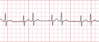
- ECG, or Holter monitoring, i.e. recording an ECG for 24 hours, as well as an ECG under stress, which allows you to detect arrhythmia.
- An echocardiogram, which detects the presence of cardiac obstructions.
- RX. To assess the cardiac shadow.
- Nuclear magnetic resonance using gadolinium contrast agent. To highlight fibrous areas of the myocardium.
- Cardiac catheterization with insertion of a microchamber to assess possible eruptions from the ventricle into the aorta.
Expansion of the boundaries of the heart is called cardiomegaly. Enlargement of the heart chambers is often caused by the accumulation of metabolic products in the heart muscle, which means that true cardiomegaly develops.
The exact etiology of the problem is established after diagnosis.
Important! The pathology detected in a newborn is very dangerous, since about 35% of children with it die in the first three months of life, and 20% develop chronic left ventricular failure.
Symptoms
Attention! Often there are cases of asymptomatic progression, then the pathology is discovered by chance during a routine examination.
Certain congenital defects cause the death of a child, so it is important to diagnose them as soon as possible (from the first days of life to six months) in order to carry out cardiac surgical treatment. This is done by cardiologists and cardiac surgeons.
Diagnostics
- Chest X-ray - the image clearly shows the shadow of the expansion of the heart, and blood stagnation is detected.
- Electrocardiography (ECG).
- Echocardiography (EchoCG) determines the physical parameters of the heart muscle, including the size of the chambers, the presence of necrosis and ischemia of the heart.
- Ultrasound of the heart muscle.
- Computed tomography (CT).
- Magnetic resonance imaging (MRI).
- Immunological and biochemical blood test, which determines the level of hemoglobin, bilirubin, urea, protein and hormones.
Important! The effectiveness of treatment directly depends on the correct diagnosis and cause of the disease. Therefore before. Before treating pathology, the doctor carefully studies the results of tests and instrumental studies.
Treatment directly depends on the causes of the disease. All activities are primarily aimed at organizing a healthy lifestyle for the patient and eliminating the cause of the disease. The patient is recommended a special diet that excludes fatty, salty and spicy foods, and giving up bad habits. The doctor prescribes special exercises.
Surgical intervention is prescribed only in emergency cases when the patient's life is at risk. The most dangerous and advanced form is considered to be “bull heart”, in this case only a transplant can help.
If disturbances occur against the background of valve pathology, then prosthetics are performed. In case of severe heart rhythm disturbances, a pacemaker is installed under the skin to normalize it.
Important! For prevention and additional therapy, traditional medicine is used.
Symptoms
In this section, you will learn in detail about what happens with the pathologies listed above, what symptoms they are accompanied by, and their causes.
The size of the heart can be determined using the following methods:
- Percussion (tapping the surface of the chest with fingers). Allows you to determine the boundaries of the organ during the initial examination.
- EchoCG (ultrasound of the heart). It helps not only to find out the size of the heart, but also to determine the reason for its enlargement.
- Chest X-ray. Allows you to detect heart enlargement during a routine examination.
Further diagnostics may include an ECG, Holter monitoring, and various blood tests.
Enlarged heart in children
Suspecting a similar condition in a child, the pediatrician will send him for x-rays, biopsy of cardiac tissue, echocardiography and ECG. Such measures will help to timely identify possible pathology and begin treatment.
Treatment methods for hypertrophied myocardium consist of establishing the normal functioning of the ventricles and heart valves, improving tissue nutrition and vascular permeability.
Reducing the volume of fluid that circulates throughout the body will also reduce the load on the myocardium and stabilize the patient’s condition.
People with hypertrophied myocardial syndrome must be registered with a cardiologist and systematically undergo drug therapy. Regular examination of the heart is indicated to monitor the situation over time.
cardiograf.com
As a rule, an enlarged heart is detected by chance during a routine examination and chest x-ray. The diagnosis of an enlarged heart, or cardiomegaly, shocks parents. Cardiomegaly can be primary or secondary. Secondary cardiomegaly develops against the background of infectious diseases, toxic lesions, and also with respiratory failure.
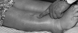
An enlarged heart in a child is accompanied by a slowdown in weight gain and overall development, tissue swelling, frequent colds, shortness of breath, heart rhythm disturbances, pale or bluish skin.
Treatment
It consists in eliminating the underlying disease, one of the symptoms of which is a large heart.
| Arterial hypertension | Taking antihypertensive drugs (beta-blockers, ACE inhibitors, angiotensin 2 receptor antagonists, etc.) |
| Ventricular septal defect | Surgical correction. |
| Dilated cardiomyopathy | Complex. May include taking ACE inhibitors, diuretics, beta-blockers, aldosterone inhibitors, digoxin, indirect anticoagulants. If arrhythmias occur, it is possible to install a defibrillator-cardioverter. But treatment does not completely eliminate the risk of death. The only way to completely cure the disease is organ transplantation. |
| Hypertrophic cardiomyopathy | Limiting physical activity. Prescription of ACE inhibitors and angiotensin 2 receptor antagonists, as well as beta-blockers and antiarrhythmic drugs. To eliminate the risk of fatal arrhythmias, a defibrillator-cardioverter is installed. |
| Metabolic cardiomyopathy | Rejection of bad habits. Taking medications to restore metabolism. These can be hormonal medications, enzymes, antienzymes, metabolites, cofactors, vitamins, amino acids, etc. |
| Aortic stenosis | Valve replacement. |
| Mitral stenosis | Valve replacement. |
| Ebstein's anomaly | In asymptomatic cases, treatment is not carried out. If signs appear, plastic reconstruction or prosthetic replacement of the tricuspid valve is performed. |
| Myocarditis | Antiviral, antibacterial, anti-inflammatory. Avoid physical activity until complete recovery. Symptomatic treatment of complications (ACE inhibitors, antiarrhythmics, etc.) |
| Exudative pericarditis | Subtotal pericardectomy – cutting of the pericardial sac. |
| Amyloidosis | Melphalan, Prednisolone, Thalomid, Dexamethasone, Lenalidomide are prescribed. But even a heart transplant cannot completely cure the disease. |
Hypertension and cardiomyopathy
Symptoms of heart enlargement are most often observed with arterial hypertension. Millions of people suffer from this disease. Hypertension is more often detected in people in adulthood and old age. An increase in heart size always occurs with this pathology. At the initial stage of the disease, an increase in the apical impulse is noted. As the disease progresses, the left ventricular cavity expands and becomes larger. This helps to shift the boundaries of the heart down and to the left. Further, all other sections (both atria and the right ventricle) increase in size. It is important that the severity of changes in organ size depends on the course of hypertension. If it progresses very quickly, then cardiomegaly also develops transiently.
Cardiomyopathy is a common cause of this disease. Cardiomyopathy of the hypertrophic type leads to an increase in the volume of all departments, but the ventricles change to a greater extent. The size of the organ increases slightly. Viral infection is of no small importance in the development of this pathology. Idiopathic cardiomyopathy is of greatest importance. In this situation, it is not always possible to determine the cause of changes in cardiac function. In some cases, the etiological factors are alcoholism and viral infection.
Return to contents
Diagnostics
As medical statistics show, diagnosing bovine heart disease is often very difficult. The patient may complain of feeling unwell, and the symptoms may indicate the presence of other ailments. Experts use the following diagnostic methods:
- echocardiogram;
- palpation and auscultation;
- radiography;
- CT scan;
- blood chemistry;
- caterization;
- biopsy.
Timely consultation with a doctor, high-quality diagnosis, and a properly designed treatment regimen can quickly restore health and prevent the development of complications.
Key points
- Enlarged heart, or “bull heart,” is a serious disease that, if left untreated, can be complicated by heart failure.
- The occurrence of an enlarged heart is most often associated with other diseases such as genetic disorders, hypertension, and heart defects.
- Treatment of an enlarged heart is carried out according to indications. In some cases, medication is sufficient, but there may be a need for surgical intervention.
- After the operation, there must be a recovery period, which, depending on the severity of the disease, can be two or more weeks.
- Cardiomegaly is dangerous due to its complications, since in severe cases sudden death, valve insufficiency, etc. can occur.
Video: Hypertrophic cardiomyopathy. Big heart disease
Sources
2. “What Is an Enlarged Heart (Cardiomegaly)?” WebMD. 2019-01-30.
Among the common heart pathologies, an enlarged heart is not so common. The reasons for this in an adult may be different. This pathological condition is not considered as a separate disease, since it develops as a consequence of an existing disease of the cardiovascular system.
A big heart
For more than two centuries, cardiologists around the world have been fighting heart pathologies, using new treatment methods. Most diseases are acquired, and if examined regularly, the risk of severe consequences is significantly reduced.
One of the striking symptoms of pathologies of this profile is an enlarged heart. To people who don’t know what it is, the name will seem scary, but if you notice the signs of the disease in time and begin treatment, the consequences will not be dangerous.
What causes a greatly enlarged heart?
For adequate functioning, the size of the heart must be within normal limits. Only under this condition does the organ fully pump blood. If its parts are hypertrophied, functional ability decreases. In cardiology, when the organ is greatly enlarged, the term cardiomegaly is used. Doctors call it bull's heart.
Causes:
- aortic valve insufficiency,
- birth defects,
- hypertonic disease.
Attention! Cardiomegaly develops slowly with frequent beer consumption. That's why doctors call it beer heart.
When drinking 3 liters of alcohol per day, the heart muscle is overloaded. The organ has to pump much more fluid. And then compensatory capabilities are activated - the heart is forced to double in size. His chambers are expanding.
Although the size of the organ increases during illness, the volume of working mass decreases sharply. Cardiocyte muscle cells are replaced by dense connective tissue.
Attention! With bovine heart, the muscle is unable to stretch. Therefore, it does not have enough power for a full contraction. As a result, the myocardium becomes flabby and wears out. There is a danger of developing heart failure.
At the beginning of the disease, the organ copes with the load, and its owner does not experience any difficulties. However, there comes a period when the weakened heart muscle is not able to push out a normal volume of blood with each contraction. This means that fluid stagnates in the legs, lungs, and liver. In medical terms, heart failure begins to develop. Signs include shortness of breath, swelling of the legs, accumulation of fluid in the abdomen (ascites). The liver increases in size, and various heart rhythm disturbances occur.
Most beer drinkers get fat because the “drink” is very high in calories. Obesity further overloads the pumping function.
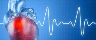
Acute enlargement of the heart: causes of the disease, main symptoms, treatment and prevention
An acute manifestation of volemic overload, characterized by overstretching of the heart muscle, which occurs against the background of a sharp increase in the volume of circulating blood due to incorrect calculation of the dose and rate of infusion or transfusion.
Causes
Fluid overload in people who do not have chronic diseases almost never occurs, since the heart independently copes with the increased volume of circulating blood, and the excretion systems quickly cope with removing excess water from the body.
The development of this condition in a healthy person is possible only when the permissible volume of infusion is very much exceeded, which in practice is detected very rarely.
There are several predisposing factors for hypervolemic syndrome, including coronary enlargement, which can provoke the development of pathology.
Kidney failure caused by a decrease in the functional abilities of the urinary system, leading to a slow removal of excess water and metabolic products from the body.
As a result of such processes, a positive fluid balance is formed, in which the volume of incoming H2O significantly exceeds the amount of urine.
In this case, the severity of the patient’s condition directly depends on the severity of fluid imbalance.
In chronic heart failure, the heart is forced to function at the limit of its residual resources. As a result of pathological processes, it is unable to fully pump even a normal volume of blood.
With a sharp filling of the vessels with additional fluid, an excessive increase in preload occurs, an acute expansion of the cardiac chambers develops, which provokes an even more pronounced decrease in their functionality.
Some respiratory diseases, such as bronchiectasis, vasculitis, bronchial asthma, chronic obstructive pulmonary disease, cause increased pressure in the pulmonary circulation.
As a result of such disturbances, the load on the right sections increases, which provokes their expansion, potentiates a mechanical disturbance in the activity of the left ventricle, against which acute or chronic cor pulmonale is formed.
The further process of development of changes does not differ from that in chronic heart failure.
Symptoms
At the beginning of the development of pathological disease, the disease is manifested by a rapid increase in systolic blood pressure, while diastolic blood pressure increases less pronouncedly.
As a result, the difference between systolic and diastolic pressure increases. The pathological pulse is characterized by weak filling and tension, tachycardia is detected. Patients experience severe headache and difficulty breathing.
Sometimes bronchospasm is detected with the development of an attack of cardiac asthma.
As the pathology progresses, the symptoms intensify.
Signs of peripheral circulatory failure appear, such as cyanosis of the nasolabial triangle, earlobes, fingertips and hypoxia, characterized by diffuse cyanosis, inclusion of auxiliary muscles in the breathing process, flaring of the wings of the nose and the appearance of psychomotor agitation. There is a decrease in blood pressure due to a decrease in the strength of heart contractions. With the development of pathology, tachycardia persists and has a compensatory nature.
During the terminal phase, all manifestations of the disease worsen significantly, and white or pinkish foam begins to emerge from the mouth, pronounced bluishness of the skin is noted, the heart rate and arterial pressure decrease to critical levels.
Diagnostics
The diagnosis is made on the basis of existing clinical symptoms. To confirm the diagnosis, the diagnostic algorithm may include a physical examination, general and biochemical blood tests, as well as ultrasound and electrocardiogram of the heart.
Treatment
Treatment of acute volume overload is aimed at normalizing vital signs. Treatment is considered effective if it reduces the heart rate to normal levels. The heart rhythm should be restored, the symptoms of pulmonary edema eliminated.
As restorative methods, general routine measures, medication and resuscitation are used. At the initial stage, intravenous administration of the infusion solution should be stopped. The patient is placed in a sitting position, which allows blood to be partially deposited in the extremities and reduce cardiac stress.
A tourniquet may be applied briefly to three of the four limbs.
Drug support consists of prescribing loop diuretics and oxygen inhalation. If cardiac activity is weakened, the administration of inotropic drugs such as dopamine, dobutamine, levosimendan and cardiac glycosides will be required. In case of pulmonary edema, administration of morphine or promedol will be required.
Diagnosis and treatment
Diagnosis of the syndrome requires studying the patient's medical history. Chronic diseases, reasons for recent hospitalizations, medical records, test data, etc. are studied. This includes the appointment of a hardware examination using special equipment.
Examination methods:
- Listening and heart rate monitoring.
- Blood test for biochemistry.
- Biopsy examination.
- Chest X-ray. It is on x-rays that the boundaries of the expansion of the contours of the organ are clearly visible.
- ECHO KG, because the examination shows when the heart muscle has signs of necrosis or ischemic pathology.
- Ultrasonography.
- A CT scan or MRI will show an increase in the volume of the organ.
- Coronary angiography.
- It is necessary to study hormonal levels, work schedule, attitude to sports and the general condition of the body, since often a harmful lifestyle affects the course of the disease.
Treatment
Patients are prescribed:
- Diuretic medications to remove fluid retention.
- Antithrombotic drugs that affect blood clotting, acting against the formation of plaques and blood clots in blood vessels.
- “Heart” tablets (captopril, for example).
- Valve replacement if there is pathology.
- Installation of a pacemaker.
- Antihypertensive drugs for hypertension.
- Hormonal drugs.
Surgical intervention is prescribed in emergency cases, when serious consequences are predicted, in particular, the diagnosis of “bull’s heart”. To correct the situation, a transplant is needed, the operation is expensive and requires a donor organ.
As part of the treatment complex, the cardiologist prescribes compliance with the correct diet, work and rest. Together with drug treatment, this tactic gives a good result.
Correct mode
The patient needs:
- Minimize salt and sugar in your diet.
- Avoid fatty, fried and spicy foods.
- Quit smoking and alcohol.
- Walk for 30 minutes a day.
- Measure blood pressure regularly.
- Perform special gymnastics.
Only timely diagnosis increases the chances of effective treatment. Every budget clinic offers methods for detecting the syndrome. This makes it possible to conduct the necessary examinations free of charge and at any time.
Treatment methods
It is difficult to treat an enlarged heart. Typically, cardiomegaly is a consequence of some disease, if it does not develop after drinking alcohol. The main therapy is to slow progression.
Treatment regimen for cardiomegaly:
- Antihypertensive medications stabilize blood flow and help shrink the organ.
- diuretics,
- anticoagulants to prevent thrombosis,
- beta blockers that regulate rhythm.
Surgical methods are used - installing a pacemaker or implanting valves. Coronary artery bypass grafting is widely used.
All efforts of doctors will be minimized if the patient does not stop drinking alcohol. In addition, a person should follow a low-fat diet. Complex treatment uses physical activity to improve blood circulation.