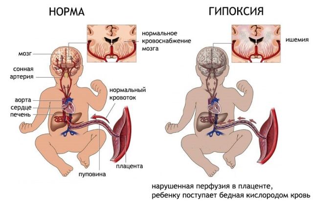
Oxygen starvation of the fetus, resulting from impaired blood circulation in the placenta, leads to serious illnesses and deviations in the development of the child. One of the disorders that occurs for this reason is a subependymal cyst of the brain, which is diagnosed in newborns due to prolonged circulatory disorders, hypoxia and lack of nutrients.
A subependymal or cerebral cyst of the brain is a benign hollow formation in the ventricles of the brain, filled with fluid, formed due to hypoxia and insufficient blood supply. Since hypoxia is often diagnosed during pregnancy, the risk of formation is very high.
An intracerebral cyst in a newborn child is a natural protective response of the body to atrophy of brain tissue.
To prevent the expansion of necrosis of brain tissue, the body replaces the damaged areas, creating a natural barrier in the form of a cystic cavity. A small cyst does not pose a threat to the normal functioning of the brain.
But if fetal hypoxia is not treated during pregnancy, the cyst begins to increase in volume, leading to such dangerous manifestations as convulsions, deterioration in well-being and health, and psycho-emotional disorders.
Causes and risk factors
Fetal hypoxia is the main cause of subependymal cyst formation and has a significant impact on the development of the infant.
The main risk factors for the manifestation of a cyst are the diagnosis of moderate or acute fetal hypoxia in a pregnant woman. There are three factors for the appearance of a subependymal cyst:
- Cerebral ischemia . The most common cause of any cyst formation. Ischemia occurs due to impaired blood supply to brain tissue. In the place where the brain matter dies, a void appears, which the body fills with cerebrospinal fluid. The small size of the cyst does not lead to serious disorders; it only requires drug treatment and monitoring over time. If it grows and puts pressure on other parts of the brain, the child experiences convulsions, regurgitation, protrusion of the fontanel, and inhibition of physical and mental development.
- Hemorrhage in the head of a newborn is the second reason for the appearance of a neoplasm. The most severe deviations in the development of the child will be if hemorrhage occurs during uterine development of the fetus, and not during passage through the birth canal. The main causes of hemorrhage are the presence of infections, acute fetal hypoxia and birth trauma.
- Acute or moderate fetal hypoxia is a disruption of the functioning of the pregnant woman’s body and placenta. The main causes of hypoxia include anemia, toxicosis in the third trimester, multiple pregnancy, Rh conflict between the child and mother, polyhydramnios, placental insufficiency, and infectious diseases.
Why is a posterior fossa cyst dangerous?
Tarlov cysts are sacs filled with cerebrospinal fluid that are located in the area of the nerve roots of the spine, at its base. Contents What is Classification Causes Clinical picture Diagnosis Treatment Complications Prognosis Prevention In most cases they are asymptomatic, but sometimes they occur...
Cysts in the brain are a consequence of pathological processes. By disrupting the integrity of brain structures, they can cause neurological abnormalities or even death. Contents What is Classification Causes Clinical picture Diagnosis Treatment Complications Prognosis Prevention What is an arachnoid cyst The human brain has…
A brain cyst is essentially a space-occupying formation in the brain. The cavity has a spherical shape containing liquid. It is located in areas where there is dead nerve tissue.
It can appear anywhere in the brain, both singular and plural. Just don’t forget that it is not a tumor formation!
The main reason for the appearance of cysts in newborns is intrauterine pathology of the central nervous system, or as a result of injury after childbirth.
In infants, a cyst can form due to poor cerebral circulation, which can lead to necrosis of nerve tissue. A cyst may also appear after an injury or an inflammatory disease such as meningitis or encephalitis. It can also form after a cerebral hemorrhage.
All this can lead brain tissue to degeneration and necrosis, which results in the formation of a cavity in this area, which gradually begins to fill with fluid and compresses nearby tissue. Neurological symptoms appear, the baby lags behind in growth and development.
Classification
There are three main types of brain cysts in children:
- Arachnoid cyst.
- Subependymal cyst.
- Choroid plexus cysts.
An arachnoid cyst looks like a sinus, which can have a variety of shapes and sizes. Can be located anywhere in the brain. The cause of its appearance is inflammatory processes, such as trauma, meningitis, hemorrhage. It is characterized by rapid growth, which causes compression of tissues close to it, and the progression of severe consequences.
Subependymal cyst is one of the severe forms of these formations. The child needs constant and careful monitoring. This cyst is formed due to insufficiently correct functioning of the cerebral circulation in the place where the ventricles of the brain are located. This pathology can cause severe ischemia and tissue necrosis.
Choroid plexus cysts begin their development even when the unborn child is inside the mother. This pathology occurs as a result of a herpes viral infection. If a cyst was discovered during pregnancy, then there is no particular danger to the child, since after some time these cysts subside on their own. But if this type of cyst was discovered in a child much later, then there is a risk of a severe course of the disease.
The symptoms of this disease largely depend on the area of the brain in which the cystic cavity has formed.
If it is located in the back of the head, it can cause damage to the visual center, and visual impairment can also occur: its sharpness decreases, double vision occurs, and a feeling of a foggy veil before the eyes.
A cyst in the cerebellar tissue leads to gait disturbance, deterioration of movements and dizziness.
If the location of the cyst is the sella turcica (next to the pituitary gland), then the baby develops complications with the endocrine system: he lags behind in his physical and sexual growth.
In addition, there is a risk of seizures, paresis or paralysis of the limbs, and decreased hearing acuity.
It is worth noting that if the cyst begins to grow in volume, then the intracranial pressure will increase due to the fact that the volume of the cranium has a constant size, but the number of tissues slowly increases. High intracranial pressure is manifested by the following symptoms: dizziness, a feeling of fullness and pulsation in the head, nausea, lethargy, vomiting, increased drowsiness.
In infants in the first year of life, cysts are diagnosed using ultrasound examination (neurosonography). It is necessary to examine the baby before one year if there is a suspicion of a cyst, since the fontanel is not yet completely overgrown, and the bones of the skull are not in a closed form. It is during this period that excellent research results can be obtained.
With the help of MRI and CT examinations, a fairly clear and informative picture is obtained about the location, shape and size of the cyst cavity.
Localization in the brain and symptoms
A subependymal cyst is localized at the site of impaired blood supply to brain tissue, most often in the ventricular region. Symptoms of the disease depend on the location and size of the cyst. The presence of a pathogenic focus in a certain area of the brain leads to disruption of the body functions for which it is responsible.
A cyst in the occipital region affects the functioning of the visual system, visual acuity decreases, double vision, etc. A cyst in the temporal lobe leads to hearing impairment, in the cerebellum – gait and coordination of movements are impaired, in the pituitary gland – hormonal imbalances.
The small size of the cyst may not manifest itself in any way on the child’s behavior. But larger ones lead to convulsions and strokes, which can result in paralysis of the body.
The main symptoms observed in the presence of a cerebral cyst in the head of a newborn child:
- sleep disturbance;
- anxious behavior;
- breast refusal;
- poor weight gain or loss;
- long, causeless crying;
- hyper- or hypotonicity of muscles;
- tremor of the chin, twitching of the limbs;
- hearing impairment;
- blurred vision, sometimes complete loss;
- regurgitation “fountain”;
- loss of consciousness, coma;
- pulsation and swelling of the fontanelle;
- convulsions.
The consequences of growth and lack of treatment of the cyst lead to mental and physical retardation in the development of the child.
Other locations of cysts in infants
Ovarian cyst in infant
This pathology occurs quite often in newborn girls, is considered a functional tumor and does not belong to malignant neoplasms, and also has a tendency to resolve on its own, without requiring surgical intervention. Ovarian cysts are treated with various medical methods. The difference is considered to be multiple cysts (polycystic ovary syndrome), which negatively affect the child’s hormonal levels or tend to transform into a malignant tumor that develops rapidly and has aggressive growth.
Malignant neoplasms of the ovaries in infants are extremely rare.
Spermatic cord cyst in infant
A spermatic cord cyst is an accumulation of fluid due to non-fusion of the vaginal process of the peritoneum (in the membranes of the spermatic cord). In terms of functionality, this type of cyst is similar to testicular hydrocele; it also seems that the treatment of this neoplasm with the treatment of hydrocele is surgical intervention.
During intrauterine development, the fetal testicle descends into the scrotum through the inguinal canal along with the growth of the peritoneum. This process normally resolves before the birth of the child, but if the processes of its spontaneous removal are disrupted, a cystic neoplasm of the spermatic cord is formed, which during diagnosis is often confused with an inguinal hernia, which has similar symptoms - enlargement of the scrotum and swelling in the groin area. If these signs appear in a newborn, parents should urgently contact a pediatric urologist or surgeon.
Testicular cyst in a baby
Testicular cysts in a newborn are benign tumors that look like a cavitary neoplasm with fluid in the area of the epididymis. Cysts have a smooth, soft and well-defined structure. It is necessary to differentiate this neoplasm from hydrocele, hernias, and varicocele.
The diagnosis is clarified using ultrasound and other instrumental studies, examination and medical history. The size of the testicular cyst does not exceed 1-2 cm and can cause discomfort and urination problems for the infant. Treatment of the cyst is carried out by surgical intervention after a year of observation due to the likelihood of spontaneous resorption of the tumor. Untreated spermatic cord cysts in adulthood can cause obstructive forms of infertility, erectile dysfunction and impotence.
Kidney cysts are asymptomatic and do not affect kidney function. A cystic neoplasm is determined using an ultrasound examination of the kidneys, which allows you to accurately determine the location of the cyst and the characteristics of its blood supply.
There are several types of kidney cysts in newborns:
- unilateral cysts resulting from the development of concomitant kidney diseases;
- cortical cysts (when diagnosing this type of cyst on one kidney, a tumor is often detected on the second kidney).
In addition to ultrasound examination, to diagnose cysts, newborns undergo a duplex scan of the kidneys, which makes it possible to determine the malignancy of the process.
Treatment of kidney cysts is carried out through drug treatment, and there are also cases of spontaneous resorption in the first year of a child’s life.
Spleen cyst in infant
A splenic cyst in a newborn is defined as a cavity in the parenchyma of an organ filled with fluid. However, surgical removal of this type of cyst is not recommended - there is a high probability of organ loss, so treatment is carried out using medications.
The causes of the development of splenic cysts are determined by congenital disorders of embryogenesis. Sometimes false cysts develop, which resolve on their own and do not require treatment.
Cyst on the tongue of an infant
A cyst on the tongue of a newborn is determined by abnormalities in the development of the thyroglossal duct, and is quite common.
The clinical picture depends on the size of the tumor and its location on the tongue:
- small cysts are defined as a tumor on the tongue without clinical manifestations;
- a large cyst located in front often interferes with eating, so it must be removed.
In the vast majority of cases, a cyst on the tongue of a newborn resolves on its own in the first months of the child’s life. When the cyst progresses, the treatment method depends on the structural features and location of the cyst.
The main method of surgical intervention for a cyst on the tongue is dissection of the cystic neoplasm.
Cyst in a newborn's mouth
A cyst in a newborn in the oral cavity is a genetic pathology associated with various infectious processes in the body. Depending on the location, tongue cysts, palatal and gingival cysts with their own histogenesis are distinguished.
Diagnosis, determination of the cause of the cyst and decisions on treatment methods are made by the dentist. For this, various diagnostic methods are used (x-ray or ultrasound of the oral cavity), which make it possible to determine the localization of the tumor. It is important to know that 90% of these cysts resolve in the first year of life, so medical and surgical treatment for up to a year is used when absolutely necessary.
Palatal cyst in infant
Cysts on the palate of newborns (Epstein's pearls) are not considered a pathological phenomenon and are observed in almost all babies in the first weeks of life and disappear on their own after the first month of the child's life.
They are formed from epithelial inclusions located along the line of fusion of the palatal plates and appear as yellowish or white tubercles in the area of the palatal suture. Palatal cysts do not require treatment.
Cyst on the gum of a baby
Gingival cysts in infants are formed in utero from the ectodermal ligament (dental plate) that forms the basis for the formation of teeth, both primary and permanent. The remains of the plate are considered to be the cause of small gum tumors and cysts. Neoplasms localized directly on the gum are called Bohn's node, and cysts developing on the process of the alveolar ridge are called gingival cysts.
These cysts look like small balls of white or yellowish color; they are absolutely painless and do not cause inconvenience or discomfort to the baby. They resolve on their own in the first weeks of a child’s life or disappear completely when baby teeth appear.
Diagnostic criteria
Using neurosonography (ultrasound) in newborns, it is easy to find cysts and other abnormalities in the brain. The fontanel, as a rule, does not close until a year, so this procedure is the fastest, easiest and most reliable.
Neurosonography must be done for premature babies, during severe pregnancy and protracted labor. If the doctor discovers a small subependymal cyst, then the brain tissue returns to normal on its own; you just need to be under the supervision of a pediatric neurologist.
If the cyst exceeds the minimum values, then additional diagnostic measures, MRI, etc. are required. Diagnosis using these methods should be carried out twice a year.
Repeated examination of the cyst and recording its growth can lead to serious disturbances in the development of the child and a threat to his life.
Diagnosing a multi-chamber subependymal cyst is very problematic, since the symptoms can be easily confused with other disorders.
MRI of a subependymal cyst in a newborn child
Diagnostics
The main method for diagnosing subependymal cysts is ultrasound neurosonography. This procedure allows you to achieve an accurate result. Moreover, the manipulation is absolutely safe and painless for the child.
Ultrasound examination of the brain is prescribed for infants whose fontanelles have not yet ossified. The planned procedure should be carried out when the baby is one month old. However, if there are pathologies during pregnancy or after a difficult birth, manipulation is prescribed earlier.
Additional methods are sometimes used to clarify the diagnosis. Computed tomography or magnetic resonance imaging allows you to accurately study the characteristics and location of the tumor. Due to exposure to radiation, CT scans are done no more than twice a year.
Qualitative approach to therapy
Treatment of a cerebral cyst is aimed at preventing its growth. To do this, you need to find and eliminate the main cause that provokes oxygen starvation. During certain life stages of a child's growth, medical care will vary.
First aid
After birth, a neonatologist performs resuscitation care.
Removes fluid from the respiratory tract, stimulates artificial respiration; in severe cases, an oxygen mask is used to supply pure oxygen.
The first three days after birth...
A neurologist conducts a daily examination and prescribes a restorative course of medication and rehabilitation therapy. Massage, physical therapy and sedatives are prescribed.
In childhood, the doctor observes the dynamics of the child’s development rate.
Medicines are used to stimulate speech functions and normalize the psycho-emotional state. At this stage, you may need the help of specialists such as a speech therapist and a psychologist.
...and beyond
When a child reaches adolescence, vitamin medications are prescribed to stimulate brain function and normalize metabolism. It is also necessary to take hormone replacement medications.
All physiotherapeutic and medicinal methods are prescribed only after the results of the examination and identification of deviations in the child’s development.
Treatment methods
If a child is diagnosed with a small cyst, and it does not cause changes in his behavior, then only observation and control by a specialist is required. At the initial stages of development, the neoplasm is treated conservatively with several groups of drugs:
- antiviral and antibacterial;
- immunomodulatory;
- nootropics (Picamilon, Pantogam) to supply the body with glucose and sufficient oxygen;
- medications Longidaza and Karipain to improve blood supply.
The course of treatment is 3–4 months, the regimen is repeated twice a year. Once the cause is eliminated, the tumor resolves on its own.
If the cyst enlarges, surgical treatment is required. In practice, two types of intervention are used: palliative or radical. The first method involves removing the contents of the tumor while preserving the walls of the bladder, which poses a risk of relapse. There are two types of such an operation:
- Shunting. A channel is created in the brain to drain cystic fluid. A significant drawback of the method is the possibility of infection.
- Endoscopic surgery. The contents of the tumor are removed through a puncture in the head. The method is not applicable to all cysts due to the inaccessibility of some areas of the brain to the endoscope.
Radical removal of a cystic bladder is an operation accompanied by craniotomy, as a result of which the formation is removed along with the walls. The procedure is traumatic and is accompanied by a long rehabilitation period.
Surgery is not necessary for all types of cysts:
- subependymal requires regular monitoring and examination, removal is necessary only when neurological symptoms progress;
- new formation of the intermediate sail is the least dangerous anomaly, requiring only monitoring of the baby;
- a choroid plexus cyst, as a rule, disappears on its own in the first year of a child’s life;
- the need to remove a retrocerebellar cyst depends on its size;
- arachnoid and dermoid are necessarily removed.
The basis of treatment is working with the cause that provoked the occurrence of cystic formation: hypoxia and its consequences are eliminated.
Treatment of the disease takes place in several stages:
- The first minutes of birth. In case of hypoxia that occurs during labor, immediate initiation of resuscitation measures is required to reduce the risk of cyst formation. When resuscitating newborns, fluid is removed from the oropharynx, trachea and nasopharynx, artificial respiration is performed, and devices for respiratory support can be used: an oxygen tent, oxygen masks, nasal cannulas.
- The first three days. During this period, the newborn’s condition is monitored by a neurologist, who prescribes medications, a massage course and physiotherapy, which help eliminate oxygen starvation and improve the child’s condition. The following medications can be prescribed to children: injections of vitamin B12, Cortexin, Diacarb, Asparkam.
- Childhood. Drugs are prescribed that improve metabolic processes in the brain, promote the formation of skills, and increase cognitive activity - nootropics, for example Pantogam. Vitamin therapy is also indicated.
If the cyst is actively growing and is large, surgical treatment is indicated: fluid is removed from the cyst using an endoscope. In severe cases, open brain surgery may be required.
Dr. Komarovsky, a famous pediatrician, claims that cystic formations identified during neurosonography in infants do not pose any danger and do not require treatment.
Consequences and prognosis of the disease
The formation of small cysts does not have a serious impact on the quality of life, psychological and mental development of the child. Problems appear if it increases in volume. This may be affected by the presence of infections and hypoxia.
As the cyst enlarges, symptoms such as irritability, bronchopulmonary diseases, heart defects, developmental delays, and anemia begin to appear.
The first year of a child’s development does not show any serious deviations. Parents begin to notice violations at 2-3 years of age. The child has hyperexcitability, delayed speech development, and poor coordination. More serious manifestations include coma and death.
What are pseudocysts?
In articles on medical topics, you can often find the expression cerebral pseudocyst in newborns. Unlike cysts, it is not a pathological phenomenon and has a different mechanism of formation. After the birth of a child, cerebrospinal fluid enters the choroid plexuses of the ventricles of the brain, forming pseudocysts. They are small, round, do not grow, and are involved in the production of cerebrospinal fluid necessary for the normal functioning of the brain.
Subependymal pseudocyst is usually determined only using instrumental examination methods. As a rule, it does not give any clinical symptoms. Pseudocysts resolve on their own in children.
Features of the pathology
A cyst of subepinedymal localization is distinguished by the fact that it is mainly benign histologically, but there is a possibility of rapid growth and malignancy. Therefore, all children with a history of intrauterine hypoxia or birth trauma require careful dynamic monitoring of the condition and size of this formation. They are at risk.
Based on the growth pattern and histological structure, brain tumors are conventionally divided into benign and malignant. But with rapid growth, they are all malignant. After all, tumors develop in the confined space of the cranium, put pressure on the surrounding brain structures, block the circulation of cerebrospinal fluid and venous outflow, causing a clear clinical picture of malignancy.
Preventive measures
In order to prevent the appearance of a cyst in the head of a newborn, it is necessary to establish the cause of its appearance. During pregnancy this is quite problematic.
It is necessary to do a genetic analysis, including amniotic fluid, but this analysis is done only in case of serious abnormalities in the development of the fetus.
Therefore, a woman needs to lead a healthy lifestyle, both before conception and during pregnancy, monitor her diet, and protect herself from intoxication and inflammatory processes.
Surgical treatment is performed very rarely, so the diagnosis of a subependymal cyst is not as scary as many people think. All that is necessary is observation by a neurologist for timely treatment of the disease and prevention of serious deviations in the development of the child.
Prevention of pathology development
A cyst is a consequence of disturbances in the functioning of the brain. To eliminate it, it is important to discover the cause of the anomaly. During the period of fetal ripening, this is not possible. However, in some cases genetic analysis is used for diagnostic purposes. Amniotic fluid is used as the main material. It is advisable to carry out such manipulations only in cases of suspected serious abnormalities in the fetus.
A woman should monitor her health before and during pregnancy. Everything must be done to prevent intoxication or inflammation.
Surgery is prescribed for a small patient only as a last resort. This diagnosis is not as scary as it might seem to parents at first glance. It is necessary to register with a neurologist, who will prevent the development of serious deviations in the general health and development of the baby.
To protect against subependymal cystic cavity, hypoxia must be prevented. To do this, a pregnant woman must eat properly and nutritiously in order to saturate her body and fetus with a sufficient amount of vitamins and minerals.
It is equally important to quit smoking and drinking alcohol. Any infectious pathology, even a cold, must be treated in a timely manner and only under the supervision of a specialist. Expectant mothers also need to avoid contact with various toxic substances.
If all these recommendations are followed, the risk of a pathological defect in a newborn is significant. In addition, such prevention will help avoid other complications during pregnancy.
Subependymal cysts in infants are a widespread formation. Small formations usually resolve on their own and do not cause any abnormalities, while large neoplasms are dangerous with serious neurological consequences and even death.
Therefore, before giving birth, preventive recommendations should be carefully followed, and if the child has a disease, do not delay treatment.
0Share0Share0Share0Share0Share0Share