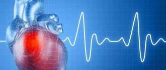
Atrial septal defect - description, causes, symptoms (signs), diagnosis, treatment.
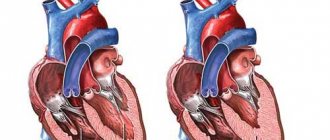
Atrial septal defect (ASD) is a congenital heart defect with communication between the atria. Statistical data: 7.8% of all congenital heart disease detected in infancy, and 30% in adults;
VSD of the ostium secundum type - 70%, ostium primum - 15%, sinus venosus - 15%; Lutembashe syndrome accounts for 0.4% of all cases of ASD, the combination of ASD with mitral valve prolapse - 10–20%; the predominant gender is female (2:1–3:1). Code according to the international classification of diseases ICD-10:
- Q21.1 Atrial septal defect
Etiology: factors that form congenital heart disease (see Tetralogy of Fallot).
Pathogenesis • The magnitude and direction of shunt depend on the size of the defect and the relative compliance of the ventricles • In adults, the right ventricle is more compliant than the left, as a result of which shunt occurs from the left atrium to the right • A small shunt leads to a moderate volume overload of the right heart, and pressure in the pulmonary artery remains normal • The severity of pulmonary hypertension may be insignificant even with a large shunt • Only in rare cases does severe pulmonary hypertension develop, leading to right ventricular failure and right-to-left shunt • Unlike VSD, with ASD the shunt is smaller and affects only the right side of the heart.
Variants of ASD • Ostium secundum (secondary defects) are localized in the area of the oval fossa, are often multiple, accompany many syndromes: Holt-Oram syndrome (ASD of the ostium secundum type in combination with digital hypoplasia), Lutembashe syndrome (combination of ASD with mitral valve stenosis), combination of ASD with mitral valve prolapse, etc. • Ostium primum (primary defects) are usually large in size, localized in the lower part of the septum, at the attachment point of the mitral and tricuspid valves, interatrial and interventricular septa. They are part of an open AV canal and are often combined with abnormal drainage of the pulmonary veins, splitting of the anterior mitral valve leaflet, mitral regurgitation and Down syndrome • Defects of the sinus venosus type are localized near the mouth of the superior vena cava and the sinus node, often combined with sick sinus syndrome, AV - nodal rhythm and abnormal drainage of the pulmonary veins.
Clinical picture
Complaints: shortness of breath, palpitations, fatigue during physical activity, retarded physical development, frequent infections, paradoxical embolisms.
Objectively • Pallor of the skin • Harrison's furrows - displacement of areas of the chest as a result of chronic shortness of breath • Splitting of the first tone with a pronounced component of the tricuspid valve • Pronounced fixed splitting of the second tone (pronounced - due to - due to prolongation of the time of blood ejection from the right ventricle; fixed - due to - due to the fact that the dependence of venous return on the phases of breathing is leveled by discharge from the left atrium) • An ejection click and a soft systolic murmur of relative stenosis of the pulmonary artery in the second intercostal space to the left of the sternum • Due to an increase in blood flow through the tricuspid valve, a low-frequency diastolic murmur sometimes occurs above the xiphoid process of the sternum.
Instrumental diagnostics
• ECG. Signs of hypertrophy and overload of the left sections, and with pulmonary hypertension - also of the right. With ostium primum, there is a sharp deviation of the EOS to the left due to the displacement of the hypoplastic branch of the left leg of the His bundle forward. Various variants of sick sinus syndrome, AV block. With a defect such as sinus venosus - lower atrial rhythm or AV rhythm - junction.
• Jugular venography: equal amplitude of A and V waves.
• X-ray examination of the chest organs. Strengthening the pulmonary pattern. Expansion and lack of structure of the roots of the lungs, bulging of the right atrium arch and upward displacement of the right cardiovasal angle. Fluoroscopy reveals increased pulsation of the roots of the lungs (a rather specific sign). The “Turkish saber” symptom with concomitant anomalous drainage of the right pulmonary veins into the superior vena cava.
• EchoCG. Hypertrophy and dilatation of the left sections, and with pulmonary hypertension - also of the right. Visualization of ASD in Doppler and B-mode. Differentiation from an open foramen ovale (the anatomical closure of the latter occurs no later than 2 years of life) is the inconsistency of visualization of the discharge in color Doppler mapping and the presence of a leaflet in the cavity of the left atrium. Diagnosis of associated anomalies (abnormal drainage of the pulmonary veins, valvular defects, etc.). The degree of discharge and the ratio of pulmonary minute blood flow to systemic blood flow (Qp/Qs) are determined. Adults undergo transesophageal echocardiography. With intravenous contrasting of the right parts of the heart, there is a negative contrast effect (displacement of the contrast agent by a stream of blood from the left atrium).
• Radionuclide angiocardiography (first pass method or equilibrium): registration of pathological discharge and its quantitative assessment, diagnosis of concomitant abnormal drainage of the pulmonary veins and ventricular dysfunction.
• Probing of the cardiac cavities •• Indicated for suspected pulmonary hypertension, before open-heart surgery and with conflicting clinical data •• If the catheter can be passed from the right atrium to the left, then this in itself cannot be a sign of an atrial septal defect: sometimes the catheter it is possible to carry out through the open foramen ovale •• Tests are carried out with aminophylline and oxygen inhalation to determine the prognosis regarding the reversibility of pulmonary hypertension •• The ratio of pulmonary minute blood flow to systemic blood flow (Qp/Qs) is calculated - a reference indicator of the amount of discharge.
• Right atriography, angiopulmonography: flow of contrast from the right atrium to the left; identification of concomitant abnormal pulmonary venous drainage.
Drug therapy. In uncomplicated ASDs of the ostium secundum type, infective endocarditis prophylaxis is usually not carried out. For ASDs of the ostium primum type, large defects of the sinus venosus type, and a combination of ASDs with mitral valve defects, antibiotics are prescribed before and for 6 months after uncomplicated surgical correction. For right ventricular failure, diuretics are prescribed.
Surgery
Indications: Qp/Qs ratio is 1.5 or more, defects of the ostium primum type, large defects of the ostium secundum type, concomitant hemodynamically significant anomalies (abnormal drainage of the pulmonary veins, mitral stenosis, etc.).
Contraindications: severe concomitant pathology that threatens the patient’s life; end-stage circulatory failure, irreversible pulmonary hypertension, the ratio of total pulmonary vascular resistance to peripheral vascular resistance is 0.9 or more.
Methods of surgical treatment. Endovascular correction with a button or two-patch Sideris device or an Amplatz device is feasible for central defects no larger than 2 cm in size. In the absence of experience in endovascular treatment, small defects are sutured under artificial circulation. In other cases, ASD repair with a synthetic or autopericardial patch under artificial circulation is recommended.
Specific postoperative complications • Sick sinus syndrome (after correction of sinus venosus type defects) • AV block (after correction of ostium primum type defects) • With mitral regurgitation that existed before surgery, symptoms may worsen after correction of ASD • Atrial fibrillation that occurred before surgery , as a rule, persists after it.
Forecast. In early childhood the course is benign. In rare cases, severe circulatory disorders can lead to death in the first months of life. Spontaneous closure of the defect is possible before the age of 5. The average life expectancy without treatment is 40 years. 5–15% of patients die before age 30. 10 - year survival rate - 90%, 20 - year - 88%, 30 - year - 67%; 40 years old - 44%, 50 years old - 25%, 60 years old - 13%, 70 years old - 7%. More than 75% of patients with large defects die from other causes. For uncomplicated defects of the ostium secundum type, perioperative mortality is less than 1%, it is slightly higher for defects of the ostium primum type, the latter also require mitral valve replacement or repair.
Abbreviations. Qp/Qs is the ratio of the pulmonary minute volume of blood flow to the systemic one.
ICD-10 • Q21.1 Atrial septal defect
Source: gipocrat.ru
Possible complications
Atrial septal defect can cause serious complications if not treated promptly. After all, the heart is constantly under stress. As a result, it becomes depleted and various disturbances arise in it.
One common consequence is Eisenmenger syndrome. When it occurs, pulmonary hypertension occurs over a long period and is characterized by improper blood discharge. Another complication may be infectious endocarditis, which is an inflammatory process in the inner lining and valves of the heart. The development of this pathology is provoked by the penetration of infection.
Often patients are struck by a stroke. It occurs due to the fact that a blood clot or any other microembolus passes through a defective hole and blocks the vessels supplying the brain.
In addition to these diseases, ASD can lead to the following disorders:
- Failure of heart rhythm.
- Pulmonary hypertension.
- Ischemic heart disease.
- Rheumatism.
- Pneumonia.
- Acute heart failure.
- Disorders of the digestive organs.
The risk of death with an atrial septal defect is quite high if the pathology is not treated at all. With therapy, only half of patients live to be 50 years old, and many are considered disabled.

Mechanisms of development of anomalies and causes
Aneurysm of the interatrial septum in newborns
The etiology of this anomaly is not fully understood today, despite the fact that aneurysm of the interatrial septum (as the term “interatrial septum” is abbreviated) has been known for a long time.
Studying the mechanism of development and causes of the anomalous phenomenon, doctors have identified several theories. Aneurysm of the interatrial septum in a newborn causes disagreement among scientists. One group argues that it is associated with a genetic factor, that is, it is a hereditary pathology, and the other - that abnormal disorders occurred during intrauterine development and could be caused by infectious diseases of the expectant mother.
Regarding the mechanism of development of atrial septal aneurysm in children, doctors describe another quite probable process. During fetal development, this septum contains the oval window, which closes soon after the baby is born. Presumably, under the influence of various factors destabilizing the process, a weak spot (thinned, insufficiently dense) remains at the site of this window, which, under the pressure of the blood flow, begins to stretch and forms an abnormal protrusion, i.e., an aneurysm. Closing the window too late can also cause an abnormal structure of the septum, which contributes to the formation of an aneurysm.
In adults, abnormal protrusion develops as a result of a previous myocardial infarction. The dangerous influence of the development of atherosclerosis, arterial hypertension, and smoking cannot be ruled out.
Based on what caused the formation of the anomalous phenomenon, the appropriate code for the aneurysm of the interatrial septum is selected according to ICD 10. For example, congenital anomalies are in group Q21, and the consequence of a heart attack is I23.1.
Epidemiology
Statistics show that men over 40 years of age are more susceptible to the disease. However, no one is immune from pathology, even small children, who may have a congenital cardiac aneurysm.
In the vast majority of cases, an aneurysm is diagnosed in the area of the anterolateral wall and apex of the left ventricle of the heart. Aneurysm of the right ventricle, right atrium, posterior wall of the left ventricle, interventricular septum and aorta of the heart is considered a rarer diagnosis.
The most common and dangerous cause of the development of weakness of the heart muscle is a previous myocardial infarction (according to various sources, from 90 to 95% of all cases of the disease). It is associated with 5 to 15% of cases of left ventricular aneurysm. If we take the total number of cases of interventricular aneurysm and left ventricular pathology, then they constitute about 15-25% of the total number of patients.
[8], [9], [10], [11], [12], [13], [14], [15], [16]
How dangerous is the anomalous phenomenon?
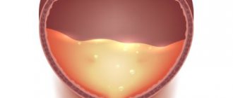
Aneurysm of the interatrial septum
Knowing that over time, the weakened part of the interatrial septum will thin out even more, and the abnormal protrusion will increase in size, those patients who have been diagnosed with this disease begin to fear its rupture. Other types of aneurysms really threaten human life if their integrity is violated. In the case of an aneurysm of the interatrial septum, doctors say that everything is not so dangerous. The rupture will not seriously affect the work of the myocardium, much less lead to its stop. The fact is that the pressure of the blood flow in this particular segment of the organ is not so strong as to lead to fatal consequences. The only thing that forms at the site of the rupture is a defect, but patients live happily with it for many years.
With all this, the anomaly cannot be called harmless. The main problem is that an aneurysm located in the interatrial septum can lead to such a dangerous phenomenon as an embolic stroke. This is due to the fact that blood clots form in the bulging “bag”. If it ruptures, the particle can travel through the bloodstream to the brain, blocking a blood vessel and causing a stroke. Also, when an aneurysm ruptures, the blood clot threatens to enter not only the brain, but also other organs, provoking, for example, a renal infarction.
Clinical picture of a dangerous condition
At the very beginning of its development, the anomaly is not accompanied by any signs and does not manifest itself. Further, its symptoms are most often associated with age:
- from 1 year to 3 years: attention should be paid to the appearance of some delay in the physical development of the baby; he may not have time to gain the required weight, or be too susceptible to viral infections;
- from 4 to 7 years: the child cannot withstand physical activity, complains of weakness, chest pain, and is stunted. Pallor of the skin and arrhythmias are observed;
- after 7 years: children of this age are also lagging behind in physical development; there may be a delay in the development of the reproductive system, chest pain. When listening, the doctor hears characteristic abnormalities: soft systolic murmurs.
- sudden chest pain;
- feeling of discomfort;
- increased weakness;
- inability to cope with any physical activity.
Symptoms of a cardiac aneurysm
The fact that a cardiac aneurysm can have different sizes, locations, and causes of pathology causes significant differences in the manifestation of the disease in different people. However, in order to catch the disease at the very beginning, without waiting for the aneurysm to grow to critical sizes (a decrease in muscle resistance even in a small area of 1 cm is clinically significant), you need to know and pay attention to at least those symptoms that are typical any type of cardiac aneurysm.
The first signs by which a cardiac aneurysm of any location is determined include:
The symptoms of a cardiac aneurysm may be superimposed on various manifestations of other existing pathologies of the cardiovascular and respiratory systems, which significantly complicates the diagnosis of the disease. And the symptoms themselves, depending on the size of the aneurysm, can be expressed to varying degrees. With a small or congenital aneurysm of the heart, the disease may proceed for a long time without any suspicious symptoms and appear much later.
Diagnostic procedures and treatment of abnormalities
Diagnosis of atrial septal aneurysm
To identify an anomaly, an ultrasound of the heart and an electrocardiogram are sufficient; a CT scan may also be needed. Simple diagnostics make it possible to identify an abnormal phenomenon immediately after the birth of a baby, and many women only learn about such an abnormality in their body during pregnancy through an ultrasound scan.
Only the attending physician can make a conclusion about whether it is necessary to treat an aneurysm of the interatrial septum and what kind of treatment, based on the diagnostic results. If a protrusion is detected that does not exceed 10 mm, then the patient can only be shown dynamic observation. If the anomaly exceeds the permissible norm, then maintenance drug therapy can be carried out (these can be drugs that lower blood pressure, thin the blood, improve metabolism).
If there is a threat of rupture of the aneurysm of the bladder or the development of pulmonary hypertension, a decision may be made to perform surgical intervention.
Source: medsosud.ru
Aneurysm of the interatrial septum
Aneurysm of the interatrial septum is a fairly common pathology that occurs among children and adults. We are talking about the curvature of that same septum (protrusion) to one side. It is classified as a minor anomaly of the heart and is not considered too dangerous. In most cases, patients diagnosed with such a pathology are simply registered with a cardiologist, without responding to any treatment. But sometimes therapy is still required.
What is the essence of the disease
If we talk about aneurysm of the interatrial septum in children, then in this case it is congenital. At the time when the fetus is at the stage of intrauterine development, there is a small hole (window) in the interatrial septum. After the baby is born it closes. These are the normal indicators. But sometimes it happens that after closing the window, the thinnest part of the partition is formed in this area. Under the influence of blood flow, the latter begins to stretch and undergo curvature.
As for the reasons that could provoke the development of atrial aneurysm in a newborn, they have not been precisely studied. Factors that increase the likelihood of pathology include a hereditary predisposition to heart disease, insufficient supply of vitamins to the fetus during its development, exposure to negative external factors on the fetus, and infectious diseases that developed in a woman during pregnancy.
Considering the fact that in the case under consideration the anomaly does not manifest itself in any way and does not affect the functioning of the heart and its pumping function, then specific treatment is also often not required. The child will simply be registered with a cardiologist, systematically undergoing examination and examination. The doctor, assessing the patient’s condition, will be able to give recommendations that will need to be followed in the future.
Important! The decision on the advisability of treatment is made in each case separately. Everything will depend on the results of the ultrasound, which can be used to judge the size of the aneurysm. If they do not exceed 10 mm, then such a pathology is considered practically safe. If the indicators of septal bulge are greater, then the doctor will give separate recommendations for such a patient.
Information
- Minutes of meetings of the Expert Council of the RCHR of the Ministry of Health of the Republic of Kazakhstan, 2015 List of used literature: 1) Belov Yu. V. Guide to vascular surgery with an atlas of operative techniques.
 Moscow." De Novo. - 2000. p.53 55. 2) Belov Yu.V., Stepanenko A.B., Gens A.P. and others. Technologies for surgical treatment of aneurysms of the thoracic and thoracoabdominal aorta. // Annals of the Russian Scientific Center for Chemistry of the Russian Academy of Medical Sciences. - 2001. - No. 10. p. 22-29. 3) Belov Yu.V., Khamitov F.F. Diagnosis of aneurysms of the thoracoabdominal aorta. // Thoracic and cardiovascular surgery. 2001. - No. 3. - p.74. 4) Burakovsky V.I., Bockeria JI. A. Guide to cardiovascular surgery. Moscow. - 1989. p. 27 - 28. 5) Pokrovsky A.V. Diseases of the aorta and its branches. M., - 1979. p. 199-234. 6) Pokrovsky A.V. Dissecting aortic aneurysms. Diseases of the heart and blood vessels, ed. E.I. Chazova. Moscow.: "Medicine". - 1992. - vol. 3. - p. 308-309. 7) Hiratzka LF, Bakris GL, Beckman JA, et al. 2010 ACCF/AHA/AATS/ACR/ASA/SCA/SCAI/SIR/STS/SVM Guidelines for the Diagnosis and Management of Patients With Thoracic Aortic Disease: Executive Summary. J Am Coll Cardiol.2010;55(14):1509-1544. doi:10.1016/j.jacc.2010.02.010. Peter Danyi, MD; John A. Elefteriades, MD; Ion S. Jovin, MD Medical Therapy of Thoracic Aortic Aneurysms Are We There Yet? Contemporary Reviews in Cardiovascular Medicine Circulation. 2011; 124: 1469-1476doi: 10.1161/CIRCULATIONAHA.110.006486 9) Prateek K. Gupta, Himani Gupta and Ali Khoynezhad Hypertensive Emergency in Aortic Dissection and Thoracic Aortic Aneurysm – A Review of Management/Pharmaceuticals 2009, 2, 66-76; doi:10.3390/ph2030066
 Moscow." De Novo. - 2000. p.53 55. 2) Belov Yu.V., Stepanenko A.B., Gens A.P. and others. Technologies for surgical treatment of aneurysms of the thoracic and thoracoabdominal aorta. // Annals of the Russian Scientific Center for Chemistry of the Russian Academy of Medical Sciences. - 2001. - No. 10. p. 22-29. 3) Belov Yu.V., Khamitov F.F. Diagnosis of aneurysms of the thoracoabdominal aorta. // Thoracic and cardiovascular surgery. 2001. - No. 3. - p.74. 4) Burakovsky V.I., Bockeria JI. A. Guide to cardiovascular surgery. Moscow. - 1989. p. 27 - 28. 5) Pokrovsky A.V. Diseases of the aorta and its branches. M., - 1979. p. 199-234. 6) Pokrovsky A.V. Dissecting aortic aneurysms. Diseases of the heart and blood vessels, ed. E.I. Chazova. Moscow.: "Medicine". - 1992. - vol. 3. - p. 308-309. 7) Hiratzka LF, Bakris GL, Beckman JA, et al. 2010 ACCF/AHA/AATS/ACR/ASA/SCA/SCAI/SIR/STS/SVM Guidelines for the Diagnosis and Management of Patients With Thoracic Aortic Disease: Executive Summary. J Am Coll Cardiol.2010;55(14):1509-1544. doi:10.1016/j.jacc.2010.02.010. Peter Danyi, MD; John A. Elefteriades, MD; Ion S. Jovin, MD Medical Therapy of Thoracic Aortic Aneurysms Are We There Yet? Contemporary Reviews in Cardiovascular Medicine Circulation. 2011; 124: 1469-1476doi: 10.1161/CIRCULATIONAHA.110.006486 9) Prateek K. Gupta, Himani Gupta and Ali Khoynezhad Hypertensive Emergency in Aortic Dissection and Thoracic Aortic Aneurysm – A Review of Management/Pharmaceuticals 2009, 2, 66-76; doi:10.3390/ph2030066
 List of protocol developers:
Conflict of interest: none.
Reviewers: Konysov Marat Nuryshevich – Doctor of Medical Sciences, KGP at the Atyrau City Hospital, chief physician.
Conditions for review of the protocol: review of the protocol 3 years after its publication and from the date of its entry into force or in the presence of new methods with a level of evidence.
All iLive content is reviewed by medical experts to ensure it is as accurate and factual as possible.
We have strict sourcing guidelines and only link to reputable sites, academic research institutions and, where possible, proven medical studies. Please note that the numbers in parentheses ([1], [2], etc.) are clickable links to such studies.
If you believe that any of our content is inaccurate, out of date, or otherwise questionable, please select it and press Ctrl + Enter.
It is not for nothing that doctors classify pathologies of the heart, which is a kind of engine of the whole organism, as the most dangerous to human life. Previously considered diseases of older people, they have an unfortunate tendency to reduce the age of patients. Some pathologies with a fairly high percentage of deaths, such as cardiac aneurysm, can develop in both adults and newborns. And this is already a signal to learn as much as possible about this pathology in order to prevent its development if possible.
[1], [2], [3], [4], [5], [6], [7]