Classification of hemangiomas
Depending on the structure, simple, cavernous and combined hemangioma are distinguished, and simple is cellular and capillary:
- Cellular develops in the prenatal period, such a hemangioma in newborns is immediately visible, it contains many angioblasts - immature precursors of normal choroid cells, and over time it can significantly increase in size.
- Capillary hemangioma consists of small vessels and appears in the first few days of a child’s life; it is located in the superficial layer of the skin.
- Cavernous hemangioma is located under the skin and consists of cavities filled with blood. The skin over it is unchanged or has a bluish tint, but as the formation grows it may acquire a bluish or purple color.
- A combined tumor combines the features of capillary and cavernous; its symptoms depend on the predominance of one type or another.
Both capillary and cavernous angioma have three stages of development:
- active growth: lasts for the first 6 – 8 months, while the tumor becomes clearly visible;
- growth stabilization: lasts until the age of 1.5 years;
- involution: reduction in the size of the neoplasm, fading of color, up to the complete disappearance of signs of the disease; this period takes up to 7 years.
Stages and degrees
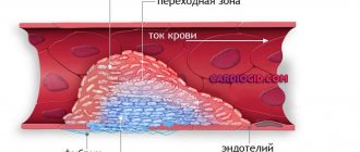
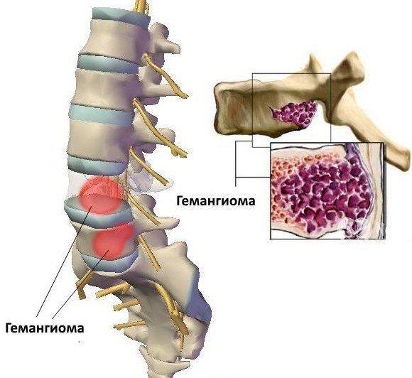
Vertebral hemangioma is detected on MRI within the first minutes from the beginning of the diagnostic examination process. As this formation progresses, 3 main stages of its development are distinguished.
Proliferation
The proliferation stage is the period of the most active growth of vascular tissue, when the tumor gains mass and covers most of the affected vertebra. At this stage, a slow growth of the hemangioma is possible with an increase in its volume up to 1 mm per year, or a more active growth of this neoplasm.
In the latter case, the risk of severe complications associated with impaired blood supply to the tissues of the spinal column, as well as a compression fracture of the spine, increases. A hemangioma detected at the proliferation stage must be removed as soon as possible.
Stabilization
The stabilization stage of hemangioma growth is characterized by the complete cessation of the pathological process of further growth of the tumor body. The patient may still not feel any symptoms of the disease, or he may experience the first signs of discomfort in the area of the vertebra, which is surrounded by an excess amount of vascular tissue.
At the stabilization stage, benign hemangiomas respond well to surgical treatment with minimal risk of complications.
Spontaneous regression
The stage of spontaneous regression is characterized by the beginning of the process of degenerative changes in the structure of the tumor. At this stage, the vascular tissue of the vertebral hemangioma begins to resolve with a gradual decrease in its volume.
The reason for the regression of a foreign tumor of this type may be associated with the activation of a defense mechanism or increased activity of the body’s immune system. Despite the partial reduction in tumor volume, its complete disappearance without surgery is unlikely.
Symptoms and diagnosis of hemangioma in children and adults
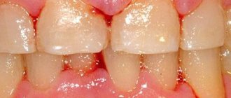
Skin hemangioma can occur in different areas, but most often it is localized on the head, in the cervical region, behind the ear, on the lip, on the back of the head, and in the area of the knee joints. Less commonly, the tumor appears on the body and limbs. In rare cases, newborns are diagnosed with liver hemangioma, damage to the genital organs, kidneys, and bones.
Capillary hemangioma appears as a flat pink spot that gradually rises above the skin and turns bright red. Deeper cavernous formations, when located under the skin, can be almost invisible, and when localized in internal organs, they cause disruption of their function with corresponding symptoms.
The disease is usually recognized when examining a child. Hemangioma in adults sometimes requires more in-depth diagnostics to exclude skin cancer: ultrasound with Doppler ultrasound to assess blood flow in the tumor tissues;
- MRI to assess the location of deep angiomas and their proximity to internal organs;
- biopsy to exclude a malignant process.
Diagnostics
Methods used to diagnose the presence of hemangioma:
- Research through palpation and inspection.
- Laboratory diagnostics.
- Non-invasive informative methods. When the tumor is internally localized, ultrasound is used in combination with Doppler sonography of the hemangioma.
- MRI or CT.
- X-ray of the spine, bones, skull and other parts of the body.
- Angiography.
The course of most hemangiomas is benign, which is a favorable prognosis. Simple tumors can regress ; many types do not develop. In cases where hemangioma negatively affects neighboring organs, disrupting their functions, then the optimal treatment method is selected to restore all indicators and function of the organs.
Treatment of hemangiomas
Most of these tumors do not require medical intervention. Treatment of hemangioma is necessary only if it interferes with visual function. Tumors located on the face and head are also usually removed early, as they can cause social or psychological problems in the child in the future.
Treatment options:
- using gels containing beta-blockers or taking them in tablet form;
- injection of corticosteroids into the tumor or their use in the form of ointments;
- laser removal of hemangioma;
- cryodestruction of small capillary hemangiomas of the skin and lips;
- surgical removal of a large tumor that impairs vision or leads to disfigurement of appearance.
Traditional methods include lotions with celandine, oak bark decoction, tincture of ginseng and mumiyo. However, it is worth remembering that in most cases the tumor almost completely disappears with age.
Complications and consequences
Hemangioma never turns into a malignant neoplasm, so it is not life-threatening. But the deeper the tumor grows into the skin, the more likely complications are.
Due to poor circulation, the skin does not receive enough nutrients, so ulcers and cracks may appear, and hair often begins to grow in areas of the skin where it should not be.
Hemangioma can grow not only into muscles, but also into bones, which leads to disruption of the functioning of the musculoskeletal system. When the bone is damaged, osteoporosis develops.
The most dangerous consequences are bleeding. With rapid growth, the tumor destroys tissue and can damage organs, which leads to disruption of their functioning.
Complications with hemangioma
In rare cases, the growth of angioma is accompanied by complications:
- when located on the eyelid – disruption of normal vision development;
- when localized in the area of large arteries, venous angioma can also affect the integrity of the vessel walls;
- a tumor in infants is easily injured, ulcerated and infected;
- Possible deterioration of blood clotting due to excess production and then depletion of platelets;
- angioma of the brain or internal organs can cause internal bleeding;
- vertebral hemangioma is complicated by back pain, pathological fractures, compression of the spinal cord; if the cervical spine is affected, paralysis of all limbs and dysfunction of the pelvic organs may develop.
Causes of neoplasm
The reasons why hemangioma occurs are still not known exactly. Since hemangioma occurs in fetuses and infants, many believe that the cause is:
- the birth of twins, triplets, and in general, multiple births;
- “old” age of the mother;
- abnormalities in the baby's weight and timing (prematurity, postmaturity, low and high weight);
- severe pregnancy (edema, gestosis, eclampsia).
It is known that no matter what types of hemangiomas occur, hemangioma in premature babies is capable of increasing 2-3 times faster.
Statistics say that hemangioma on the skin (subcutaneous hemangioma) occurs in 8-9% of cases of all births, but by 7-8 years of age, most of these formations disappear on their own. Where are these formations found, and what are they?
Why choose the clinic “Mama Papa Ya”
The network of family clinics “Mama Papa Ya” offers medical services for hemangiomas to patients of any age. The clinic's branches are located in Moscow and other cities. Advantages of visiting this medical institution:
- the opportunity for children and adults to get advice from doctors of various profiles - dermatologist, cosmetologist, surgeon, neurologist, pediatrician;
- individual decision on the need to remove hemangioma;
- the use of low-traumatic methods of tumor removal;
- affordable prices for diagnostic and therapeutic procedures.
To obtain a doctor’s consultation, we invite you to call the clinic’s phone number listed on the website and make an appointment at a convenient time. You can also leave a request by filling out a special form on our website.
Reviews
Marina Petrovna
The doctor carefully examined my husband, prescribed an ECG and made a preliminary diagnosis. She gave recommendations on our situation and ordered additional examination. No comments so far. Financial agreements have been met.
Roakh Efim Borisovich
I am simply delighted with the doctor and the clinic. I haven't had fun in clinics for a long time. Everything went perfectly from a logistics point of view, strictly on time. I also received aesthetic pleasure both as a patient and as a person. I could communicate and this communication gave me great pleasure. My deepest bow to the ultrasound doctor.
Luzina Sofya Khamitovna
I really liked Dr. Vlasova. Pleasant and sweet woman, a good specialist. I received an answer to all my questions, the doctor gave me a lot of good advice. I was more than satisfied with the visit.
Evgeniya
We visited the “Mama Papa Ya” Clinic with our child. A consultation with a pediatric cardiologist was needed. I liked the clinic. Good service, doctors. There was no queue, everything was the same price.
Olga
I really liked the clinic. Helpful staff. I had an appointment with gynecologist E.A. Mikhailova. I was satisfied, there are more such doctors. Thank you!!!
Anonymous user
I removed a lump from Alina Sergeevna, the operation was great! Many thanks to her for her sensitive attention and approach to every detail.
Anonymous user
Today I was treated at the clinic, I was satisfied with the staff, as well as the gynecologist. Everyone treats patients with respect and attention. Many thanks to them and continued prosperity.
Iratyev V.V.
The Mama Papa Ya clinic in Lyubertsy is very good. The team is friendly and responsive. I recommend this clinic to all my friends. Thanks to all doctors and administrators. I wish the clinic prosperity and many adequate clients.
Belova E.M.
Today I had a mole removed on my face from dermatologist I.A. Kodareva. The doctor is very neat! Correct! Thanks a lot! Administrator Yulia Borshchevskaya is friendly and accurately fulfills her duties.
Anonymous user
I would like to express my gratitude to the staff of the clinic: Mom, Dad, and me. The clinic has a very friendly atmosphere, a very friendly and cheerful team and highly qualified specialists. Thank you very much! I wish your clinic prosperity.
Christina
I liked the first visit. They examined me carefully, prescribed additional examinations, and gave me good recommendations. I will continue treatment further; I liked the conditions at the clinic.
Anna
Good clinic, good doctor! Raisa Vasilievna can clearly and clearly explain what the problem is. If something is wrong, she speaks about everything directly, not in a veiled way, as other doctors sometimes do. I don’t regret that I ended up with her.
The first signs of the disease
A person is born with a hemangioma or it appears in the first months of life. A common location is the scalp. The face, ears, neck, and skin under the hair are affected. Less commonly, it appears on the back, stomach, and arms.
If you look at the tumor in an enlarged form, you can see overgrown pathological vessels that are intertwined into balls. The structure of the tumor is cavernous, it protrudes above the skin, so there is always a risk of damage to the surface.
Stages of disease development:
- Congenital hemangioma or its appearance.
- Increase in size.
- Stopping growth.
- Resorption of the tumor.
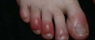
Early symptoms of the disease include the appearance of red dots, spots, and scratches on the skin, the origin of which is not related to domestic trauma. Hemangioma tends to grow, the intensity of which varies.
Symptoms
It appears in the first days and weeks after birth and occurs in three stages.
First manifestations
The primary manifestations of cutaneous and subcutaneous cavernoma depend on how deep it is located and the type of vessels that form it. A rounded elevation appears on the child’s skin with a small spot in the center of a bright red, crimson, bluish or purple color. The color depends on the depth of the cavity. The closer to the surface of the skin, the brighter the color. If the tumor is deep under the skin, the color of the skin does not change, only swelling is visible. The color of the spot also depends on the type of affected blood vessel. Arteries give a bright color, veins give a dark, bruise-like color.
The surface of the spot is smooth, less often rough, the boundaries are clear. The formation is soft and painless to the touch. The symptom of “compression” and “pouring” is characteristic - when pressed with a finger, the tumor decreases and turns pale, and then restores its shape. This occurs due to the presence of cavernous cavities communicating with each other - when pressure is applied, blood flows out and then fills the cavity again. When you scream, strain, or cough, the cavernoma enlarges, fills with blood, and becomes brighter. On a cut it looks like a sponge, filled with blood and riddled with passages and holes.
Growth phase
Lasts up to 6-9 months, sometimes longer. The hemangioma grows in breadth and depth. With diffuse growth, the formation becomes shapeless, growing in all directions and in depth, growing into the skin and subcutaneous tissue. If it is limited to a capsule, then it remains the same in appearance, increasing in size. The process is painless. Speed varies. It is impossible to predict either the intensity of growth, or the size that the formation will reach, or the age until which the growth of the cavernoma will continue. Possible complications are bleeding, ulcers on the surface, infection, thrombosis.
Rest phase
Growth is completed and the formation takes on its final shape and size. This happens by the age of 5-7 years. Local cavernoma looks like a berry or a node - convex, clearly defined and rough. Diffuse is a large spot with uncertain boundaries. Sometimes it occupies a significant area, for example, half of the face or leg.
Regression phase
A reverse development occurs - the tumor turns pale, becomes softer, gradually decreasing in size. This usually happens by age 12. At the site of a large cavernous hemangioma, scars, skin atrophy, and hyperpigmentation can form.
Vascular tumor of internal organs usually occurs without clinical manifestations.