© Author: Z. Nelly Vladimirovna, laboratory diagnostics doctor at the Research Institute of Transfusiology and Medical Biotechnology, especially for SosudInfo.ru (about the authors)
A measure of red blood cell size variation (volume distribution width) is measured by automated hematology systems and referred to as RDW. This is anisocytosis of erythrocytes (Er). A similar task of determining anisocytosis in a general blood test was solved (and is now sometimes solved, if necessary), by the Price-Jones curve, the graphical drawing of which is a labor-intensive and tedious process.
Laboratory services have now largely moved away from using the Price-Jones curve as a method. Visual assessment of the degree of anisocidosis during a morphological examination of a smear (viewing the specimen under a microscope) is often accompanied by many errors, which, in general, is never noticed with devices for measuring erythrocyte and platelet indices - analyzers practically do not make mistakes if they are correctly calibrated. The devices detect anisocytosis much more effectively, and also allow you to get rid of visual artifacts.
Physiological norms of red blood cells
It just so happens that everyone (both doctors and patients) calls red blood cells cells, although in fact this is not entirely true, but this definition has taken root, so there is no point in deviating from the generally accepted unspoken rules and changing anything. Cells and cells...
In addition, before starting to describe this erythrocyte index, I would like to answer one of the frequently asked questions - what is the diagnosis of anisocytosis in children associated with? Of course, one can understand the concern of parents who discovered an unfamiliar word in the results of their baby’s hemogram, but this medical term inherently does not differ in relation to the blood test of a child or an adult. Although, it is true, anisocytosis with a predominance of microcytes is more often observed in children, which indicates the development of iron deficiency anemia - it is most typical for childhood and adolescence. Otherwise, everything is the same, so the description of the indicator will not focus on either childhood or adulthood.
So, red blood cells are formed elements of blood, nuclear-free blood cells that have a round or slightly oval outline and the shape of biconcave discs. This form is necessary for red blood cells so that they can significantly increase the surface area for full gas exchange.
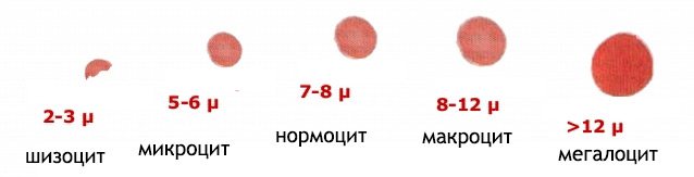
Normal values for the diameter of red blood cells do not leave the boundaries of 7 - 8 microns, and the physiological range of fluctuations in this indicator of red cells is 5.5 - 9.5 microns (graphic expression - Price-Jones curve). Average values of erythrocyte volume range from 80 to 100 femtoliters (or cubic microns) - this is the MCV norm. Cells that have these parameters are called normocytes, and the vast majority of red blood cells should belong to this category.
Calculation of erythrocyte indices included in the general blood test (hemogram): MCH (average content of the red pigment - hemoglobin, Hb), MCHC (average concentration of Hb in a red blood cell), MCV (average erythrocyte volume), as well as an indicator of heterogeneity (heterogeneity) red blood cells by volume (red blood cell anisocytosis degree - RDW) is currently assigned to hematology automatic analyzers. The normal range for RDW (or red blood cell anisocytosis) is 11.5% to 14.5%.
The cause of anisocytosis in a general blood test may be deficiency conditions (lack of vitamin B12, folic acid, iron), a malignant tumor process, complications after blood transfusions (immune reactions), impaired hematopoiesis in the bone marrow, chronic infection, cardiac pathology (development of coronary artery disease).
Treatment methods
Therapy is determined by the specific type of disease. The task is not so much to correct the level of formed cells, but to eliminate the primary process.
Here are just a few possible methods:
- Bone marrow pathologies suggest a bone marrow transplant. This is a complex procedure, and there are also problems with selecting donor material. As a symptomatic measure, transfusion of red blood cells and, if necessary, a liquid fraction (plasma) is used.
- Liver diseases require systematic use of hepatoprotective drugs. Essentiale, Karsil as basic. In difficult clinical cases, transplantation is used. It is possible in the initial stages of cirrhosis, with subcompensation, before critically dangerous bleeding has yet begun.
- Hormonal imbalance is also a possible cause. Replacement therapy using synthetic drugs is necessary.
- Insufficient concentrations of iron, vitamin B12, and folic acid require the introduction of these substances from the outside.
Treatment methods are determined by the situation. As a rule, therapy is carried out using medications in an outpatient setting. Stationary correction - according to indications.
Anisocytosis of red blood cells
Meanwhile, in various anemic conditions, depending on the type of pathology, both the diameter and volume of blood cells can vary noticeably in one direction or another - the population of erythrocytes becomes heterogeneous (the presence of such a modification of cells allows for differential diagnosis of anemia). Thus, in addition to normocytes, abnormal red blood cells of different volumes appear in the blood (heterogeneity of the red cell population):
- Unnaturally enlarged, huge, “bloated” - megaloblasts (the diameter of these cells exceeds 9.5 microns);
- Slightly fewer megaloblasts, but more normocytes - macrocytes;
- Small formed elements (liliputian cells) - microcytes.
Most hematology analyzing instruments designed to study the basic characteristics of the cellular composition of peripheral blood calculate the degree of red blood cell anisocytosis using the formula as the coefficient of variation of the mean red blood cell volume (MCV):
- RDW(%) = SD/MCV(fl) x 100%,
SD – standard deviation of volume Er, MCV – average volume Er.
With the arrival of hematological analyzing systems in the laboratory service, it finally became clear that conditions in which the indicator would be reduced simply do not exist. Well, if it is still lowered, it means that something is wrong with the automatic analyzer that performed the calculation, so the staff will have to do calibration in order to get the correct results of anisocytosis in the general blood test, and the patient will have to visit the laboratory again to don't worry in vain.
Prevention
The development of anisocytosis can be prevented by following measures to prevent the underlying disease that caused it.
Video from YouTube on the topic of the article:
Education: higher education, 2004 (State Educational Institution of Higher Professional Education “Kursk State Medical University”), specialty “General Medicine”, qualification “Doctor”. 2008-2012 – postgraduate student of the Department of Clinical Pharmacology, KSMU, Candidate of Medical Sciences (2013, specialty “pharmacology, clinical pharmacology”). 2014-2015 – professional retraining, specialty “Management in Education”, Federal State Budgetary Educational Institution of Higher Professional Education “KSU”.
The information is generalized and is provided for informational purposes. At the first signs of illness, consult a doctor. Self-medication is dangerous to health!
According to statistics, on Mondays the risk of back injuries increases by 25%, and the risk of a heart attack by 33%. Be careful.
74-year-old Australian resident James Harrison has donated blood about 1,000 times. He has a rare blood type whose antibodies help newborns with severe anemia survive. Thus, the Australian saved about two million children.
Dentists appeared relatively recently. Back in the 19th century, pulling out diseased teeth was the responsibility of an ordinary hairdresser.
In order to say even the shortest and simplest words, we use 72 muscles.
Tooth decay is the most common infectious disease in the world, which even the flu cannot compete with.
If your liver stopped working, death would occur within 24 hours.
Human blood “runs” through the vessels under enormous pressure and, if their integrity is violated, it can shoot at a distance of up to 10 meters.
Our kidneys are capable of purifying three liters of blood in one minute.
Over the course of a lifetime, the average person produces no less than two large pools of saliva.
The highest body temperature was recorded in Willie Jones (USA), who was admitted to the hospital with a temperature of 46.5°C.
The first vibrator was invented in the 19th century. It was powered by a steam engine and was intended to treat female hysteria.
In the UK there is a law according to which a surgeon can refuse to perform an operation on a patient if he smokes or is overweight. A person must give up bad habits, and then, perhaps, he will not need surgical intervention.
Millions of bacteria are born, live and die in our intestines. They can only be seen under high magnification, but if they were put together, they would fit in a regular coffee cup.
Human bones are four times stronger than concrete.
The average life expectancy of left-handers is shorter than that of right-handers.
The task of accurately establishing paternity is as ancient a problem as the search for the meaning of life. At all times, men were interested in whether they were raising their own children.
source
A doctor's eyes are irreplaceable
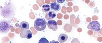
Any “smartest” analyzer, meanwhile, does not replace the work of laboratory diagnostic doctors, whose functional responsibilities include conducting hematological studies. By placing the sample in the apparatus, the laboratory technician simultaneously prepares (paints and dries) a smear intended for visual viewing (microscope + doctor’s eyes) and determining the degree of anisocytosis of erythrocytes.
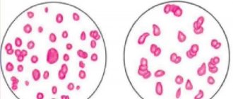
anisocytosis in the blood, red blood cells - different sizes
If in the preparation the entire field is covered with cells of approximately equal volumes (normocytes), then no notes are made about anisocytosis in the hemogram form. This indicator is reflected in a general blood test if abnormal red blood cells claim to be widespread in the smear. It is clear that if the doctor sees small cells, he indicates anisocytosis with a predominance of microcytes , if large cells, he notes macrocytosis .
However, along with micro- and macrocytosis separately, there is another option - mixed anisocytosis . Mixed-type anisocytosis is said to occur when up to 50% of the field is occupied by abnormal cells and it is not clear which of them predominates: megaloblasts, macrocytes (vitamin B12 deficiency, folic acid deficiency or pernicious anemia may be suspected), or the smear is predominantly “populated” by midgets – microcytes (then there will be an assumption about the development of iron deficiency – IDA). In such cases, the doctor reports mixed type anisocytosis with a mark on the analysis form and thereby draws the attention of the attending physician, who, in turn, will prescribe additional examination for the patient.
It should be noted that such circumstances sometimes force the laboratory diagnostics physician to return to the use of a graphical expression of these parameters (or the Price-Jones curve).
In addition, it is not for nothing that the term “degree of anisocytosis of erythrocytes” is used; it is really distinguished by degrees, highlighting:
- Minor anisocytosis - abnormal cells occupy up to 1/4 of the blood smear (up to 25%), the result can be expressed as one plus (+);
- Moderate – abnormal cells within half of the entire community included in the smear (up to 50%), or result: ++;
- Pronounced – 3/4 of all red blood cells are taken over by cells with an altered volume (up to 75%) or result: +++;
- High (acute) degree of anisocytosis - abnormal cells practically occupy the entire field (up to 100%), the result can be expressed as 4 pluses (++++).
At the same time, regardless of the complexity of the situation, you should not try to compare the Price-Jones curve, since it had to be “drawn” anyway, and the histogram produced by the analyzer, because the first reflects the value of the distribution by diameter, and the second by volume, so two methods have a number of differences and features.
Symptoms
Not only a general clinical blood test can indicate such a deviation. Several characteristic clinical signs may indicate an anomaly.
The main symptoms of anisocytosis:
- constant weakness that does not go away even after a full night's rest;
- decreased ability to work;
- constant drowsiness;
- headaches that are localized in the upper part of the head or in the back of the head;
- muscle weakness, especially after waking up;
- fast fatiguability;
- decreased concentration;
- dryness and redness of the tongue;
- change in taste preferences;
- problems with swallowing;
- impaired skin sensitivity;
- increased heart rate;
- dizziness;
- dyspnea;
- pain in the abdominal area;
- hepatosplenomegaly;
- pallor of the skin and nail plates.
In addition to the external signs characteristic of this condition, the clinic will include symptoms of the underlying disease.
Anisocytosis of erythrocytes and diagnosis of anemia
Assessing the size of erythrocytes and calculating erythrocyte indices plays an important role in the diagnosis of anemia, since it reflects the state of bone marrow hematopoiesis (erythropoiesis). The indicator of the degree of anisocytosis makes it possible to distinguish the following variants of the anemic state, that is, to identify the types of anemia:
- A shift towards macrocytes indicates rejuvenation of red blood cells and their incomplete maturation, which is usually associated with a lack of vitamin B12, folic acid or other hematopoietic factors involved in hematopoiesis. In this case, we can talk about megaloblastic or macrocytic anemia;
- A large number of small cells - microcytes (or anisocytosis with a predominance of microcytes) may indicate the direction of erythropoiesis towards micronormoblasts, which occurs when there is insufficient iron content in the body (iron deficiency anemia) or other specific components - microcytic anemia;
- Regarding the norm, everything is not so clear. A normal level of anisocytosis does not mean the absence of pathology, which, for example, occurs in the case of aplastic anemia or anemic conditions associated with chronic pathology.
It should be noted once again that the presence in the blood of a homogeneous, although noticeably changed in the direction of decreasing cell volume (microcytes) population, as a rule, leaves the RDW indicator normal, therefore in such cases the values of another index are taken into account - MCV and thus carry out differential diagnosis of microcytic anemia.
In addition, to obtain a complete picture of red blood, the anisocytosis of erythrocytes, issued by the analyzer in digital terms, is compared with the data of a histogram reflecting the frequency of occurrence of cells of different volumes.
Diet
When a patient's hemoglobin is low and iron deficiency anemia is confirmed as a disease, he will be advised to consume more of the following foods:
- beef;
- liver;
- apples;
- fruits;
- buckwheat
If a malignant tumor is detected in a patient, the issue with it will first have to be resolved, and only then microcytosis will be cured, since in the usual case the condition of the red blood cells will not change. In adolescents, causes of this phenomenon are also observed, but it is most often associated with growing up: decreased immunity, hormonal imbalances, increased growth. As you might guess, no treatment is required here. The boy or girl will have to adjust their diet and listen to the doctor’s advice. The problem should go away soon.
In a child, the causes of microcytosis are also often associated with a lack of iron in the body. Up to 90% of children suffer from iron deficiency anemia in one case or another. In this vein, parents may be alarmed by the pink color of their child's urine after he eats red beets. The fact is that the liver, with its enzymes, is obliged to discolor the natural dye of the vegetable. A child with iron deficiency anemia does not have enough of these enzymes in the body.
However, this symptom also occurs in unstable conditions, when cells experience temporary iron deficiency. However, you should consult a doctor. If the disease has been diagnosed in a child, ferrous salts and ferrous sulfate are most often prescribed. The preparations should contain: malic, citric, ascorbic acids, as well as amino acids. They promote the formation of easily soluble iron compounds in the gastrointestinal tract and their absorption.