Retinal angiopathy is a pathology that occurs with damage to blood vessels in the eye area. The disorder is not considered by ophthalmologists as an independent disease and belongs to the symptoms accompanying other diseases. Angiopathy tends to affect the entire visual system. Progressive pathology provokes thinning and death of the retina or leads to its detachment and rupture. This condition is considered quite dangerous, capable of depriving a person of vision.
Causes of retinal angiopathy
A symptom called retinal angiopathy can occur in the presence of any disease that causes negative changes in the vascular system. The pathology consists of vascular damage caused by impaired nervous regulation. The anomaly, often referred to as background angiopathy, can manifest itself in patients of different ages, but mainly occurs after reaching 30 years of age.
Angiopathy is usually considered as a consequence of general damage to the vascular system. Due to this feature, the pathological process most often spreads simultaneously to both eyeballs.
The most common causes of angiopathy include:
- Diabetes.
- Hypertension.
- Atherosclerosis.
- Neurocirculatory dystonia.
- Vasculitis.
- Presbyopia (age-related refractive error of the eyes).
- Scoliosis.
- Blood diseases.
- Osteochondrosis affecting the cervical vertebrae.
The occurrence of angiopathy in adults is facilitated by dependence on alcohol, tobacco products, and professional activity in harmful conditions. In patients of different ages, the disorder may be associated with congenital abnormal vascular structure.
Sometimes angiopathy develops after trauma or mechanical damage to the retina. The cause of the pathology can be long-term therapy with certain medications.
What is angiopathy
The retina of the eye is responsible for central and peripheral vision. It consists of 10 layers, including neurons, blood vessels and special receptors. Thanks to them, light rays are transformed into nerve impulses, which helps a person see objects of varying degrees of distance, clearly navigate in space, and recognize colors and shades.
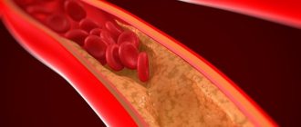
Failure of the proper functioning of the vascular system, caused by certain diseases or changes in the body (for example, pregnancy), prevents the normal supply of blood to the inner surface of the eyeball. Because of this, small vessels and capillaries narrow or expand, their walls become thin, brittle, lose elasticity, and there is a risk of rupture.
Impaired blood flow speed and improper nervous regulation cause spasm, followed by the systematic death of vascular tissue. The likelihood of retinal detachment increases many times over, after which it is no longer possible to restore adequate blood supply. The result is partial or complete loss of vision.
Important! Regular examination by an ophthalmologist will help recognize angiopathy at an early stage of development. If vascular defects are detected, it is worth undergoing a full medical diagnosis to establish the underlying disease. A consultation with a therapist, endocrinologist, neurologist, cardiologist, and rheumatologist is required.
Types of violation
Angiopathy is classified based on the type of disease or other factors that caused the development of the pathology. The violation can occur in the following ways:
- diabetic;
- hypertensive;
- hypotonic;
- traumatic;
- youthful.
The least common form of the anomaly is juvenile angiopathy, also known as Eales disease. This type of disorder occurs in the presence of an inflammatory process, often of unknown etiology.
Diabetic angiopathy (retinopathy)
Angiopathy that occurs when affected by diabetes mellitus is also known as retinopathy. This disorder is considered a severe complication, occurring in 90% of diabetics.
Diabetic angiopathy can occur in two forms. Experts distinguish:
- microangiopathy;
- macroangiopathy.
In the first case, the patient experiences damage to small capillaries. At the same time, thinning of their walls and subsequent hemorrhages occur.
Macroangiopathy occurs with impaired permeability of large blood vessels. If there is such a diagnosis, there is a high risk of damage to the veins or arteries that supply important organs, and partial or complete disability of the patient.
Retinopathy can be accompanied by a significant deterioration in visual perception and loss of ability to work. Patients with this pathology risk completely losing their vision ten times more than people without diabetes.
Hypertensive angiopathy
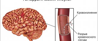
Retinal vascular angiopathy, which is a consequence of arterial hypertension, is often detected by a specialist examining the fundus of the eye. An increase in blood pressure leads to the destruction of vascular walls and damages the structure of the inner layer. Retinal vessels pressing on the veins disrupt microcirculation.
Under such conditions, hemorrhages occur and the process of thrombus formation begins. If the vessels rupture, the patient is diagnosed with retinopathy.
At the beginning of the development of hypertension, pathologies present in the fundus are detected in 60-70% of cases. With progressive hypertension, disturbances in the organs of vision are found in 90-97% of patients.
Hypotonic
The development of hypotensive angiopathy is associated with a decrease in systemic pressure. In the early stages of this type of disorder, there are often no obvious clinical signs. As the pathology progresses, a decrease in the transparency and tone of the vessels is observed, and they acquire branched shapes instead of straight ones. The fundus of the eye has an unhealthy pallor.
Constant hypotension causes the opposite effect - the formation of a hypertensive variant of angiopathy. This process occurs with the deposition of calcium salts and lipid compounds in the eye vessels, and the loss of the former elasticity of the vascular walls.
Traumatic
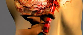
A traumatic type of pathological process often develops after intense physical impact on the sternum, skull bones, or when the cervical vertebrae are injured. Vascular damage in such cases is associated with a sharp rise in pressure.
In the case of the development of traumatic angiopathy, hemorrhage occurs in the retina, and the capillaries narrow. This type of pathology is dangerous due to a sharp weakening of vision without the possibility of its subsequent restoration.
Youth
This type of disorder gives rise to an inflammatory process, in most cases of unknown origin. With this type of angiopathy, hemorrhages occur, the sites of development of which are the retina or vitreous body. In patients with this pathology, connective tissue grows in the organs of vision, the retina detaches, and glaucoma or cataracts develop.
Of all types of angiopathy, the juvenile form of the pathology has the most unfavorable prognosis.
Symptoms
As a rule, at the beginning of the pathological process, retinal vascular angiopathy occurs without any symptoms and the person is not even aware of the presence and progression of the disease. Asymptomatic ophthalmological pathology poses a great danger to humans. Often, a person turns to a specialist for help when vision problems begin to appear. It is also not uncommon for patients to come in at the last stages of the disease, when nothing can be changed.
We suggest you familiarize yourself with the main symptoms that will help you suspect the presence of vascular pathology of the organs of vision:
- decreased visual acuity and rapidly progressive myopia ou;
- change in field of view;
- sharp and bursting pain appears in the eyes;
- feeling of pulsation;
- redness of the sclera due to vascular ruptures;
- loss of visual angles;
- fog before the eyes;
- flickering “flies” before the eyes.
Symptoms
The onset of angiopathy is often sudden, and occurs with a characteristic symptom - a sudden deterioration in visual perception. In addition to this negative phenomenon, the pathology is distinguished by the presence of other symptoms:
- progressive myopia;
- dizziness;
- headaches;
- cutting sensations in the eyes;
- flashes, “flies” in the field of view;
- bloody impurities in feces;
- intoxication syndrome.
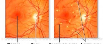
Additional symptoms are determined by the type of disorder. Diabetic angiopathy is characterized by the presence of destructive processes in the retinal tissue and yellow spots in the fundus. With the hypertensive type of pathology, the patient observes flickering before the eyes, sees surrounding objects vaguely, experiences internal heat, and suffers from coordination problems. The hypotonic variant of angiopathy occurs with pulsation and darkening in the eyes, dizziness, and loss of strength.
The traumatic form of the disorder is accompanied by pain and hemorrhages in the organs of vision. For the juvenile form of the pathological process, an unexplained impairment of visual function is typical (often with normal general health and the absence of any suspicious symptoms).
Diagnostics
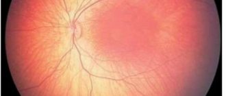
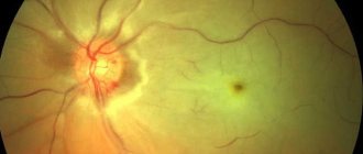
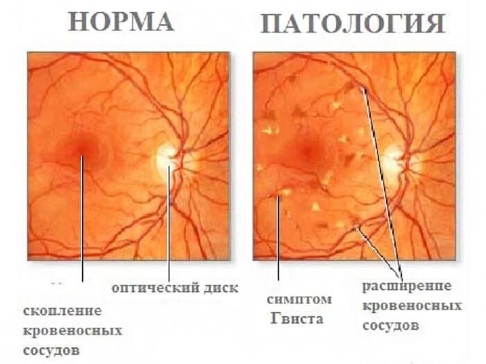
Retinal angiopathy can mainly be detected during an ophthalmological examination of the fundus. The retina of the eye is examined using a microscope, after inducing mydriasis (artificial dilatation of one or both pupils). During the examination, narrowed or dilated vessels, hemorrhages are revealed, and the position of the macula is determined.
Additional methods for diagnosing angiopathy include:
- Doppler ultrasound, which helps determine the speed of blood flow and the condition of the vascular walls;
- X-ray examination (this type of diagnosis is based on the introduction of contrast agents into the vessels);
- MRI, which evaluates the general condition of the tissue that forms the eyeballs.
The exact diagnosis is announced after studying the collected medical history and considering the results of the examination.
Retinal angiopathy in children
In children, angiopathy is also associated with serious illness. At an early age, pathology can manifest itself against the background of endocrine disorders (diabetic retinopathy), tuberculosis, kidney diseases, and inflammatory processes in the organs of vision. Spinal curvature and toxoplasmosis can lead to the development of the disorder.
In children, a traumatic form of angiopathy associated with damage to the eyeballs, or a juvenile version of the symptom, is predominantly detected. Diagnosis of this retinal disorder in younger patients is based on general principles used in adults.
Stages
Retinal angiopathy in children and adults is divided into stages. They have their own characteristics:
- First degree. Narrowing of the arteries and simultaneous expansion of the veins. The vessels become different sizes. The capillaries acquire a strong bend.
- Second degree. The bends and differences in the sizes of the vessels increase significantly. They also take on the appearance of copper wire. A defect in the capillaries causes them to appear as thin, whitish lines. Blood clots and bleeding appear. The bottom of the organ becomes waxy in color.
- Third degree. Excessive bleeding occurs. White spots form in the retina of the eye and swelling occurs. The boundaries of the optic nerve are blurred.
Treatment of this pathological process is mandatory. Both in children and adults. In the early stages of development, the disease is more amenable to therapeutic measures.
In newborns
An abnormality of the retina can be detected in the early postpartum period, but at this stage it is not considered a pathology of the visual system. Deviation is perceived as a violation at a later age.
It is quite difficult to detect pathology in a newborn without the participation of a doctor. Often the only obvious symptom is the presence in the eye of a network of red capillaries or subtle spots (characteristic signs of traumatic angiopathy).
Angiopathy and pregnancy
Many women expecting the birth of a baby are susceptible to the development of angiopathy. While carrying a child, the expectant mother’s body experiences an increase in the volume of circulating blood and dilation of blood vessels, leading to the appearance of pathology.
With a mild degree of angiopathy, there is no need for treatment - in the case of a favorable delivery, the anomaly disappears within 2-3 months and does not cause unwanted complications.
Angiopathy detected before pregnancy and associated with hypertension can become unsafe. This disorder can progress and provoke various complications at any stage of pregnancy. Patients with such pathology require constant monitoring of blood pressure and regular examination of the fundus. Prescribing medications to lower blood pressure becomes mandatory.
If angiopathy tends to progress and begins to pose a threat to life, termination of pregnancy is recommended. The patient may also be prescribed an operation to help safely deliver the baby (caesarean section).
Treatment methods
Treatment of angiopathy aims to eliminate the underlying disease that provoked the pathology. Drug therapy, physiotherapy, diet, and reduction of visual stress become relevant. For any type of disorder, medications must be used to ensure normal blood circulation in the organs of vision.
Treatment of angiopathy is carried out comprehensively, with the participation of several specialists (ophthalmologist, therapist, endocrinologist). The duration of the treatment course is individual and may require a considerable period of time.
Drug treatment
The main medications indicated for the development of angiopathy are:
- Means to improve blood circulation. Patients are prescribed oral or injection administration of Pentilin, Vasonit, Trental, Actovegin Pentoxifylline, Piracetam.
- Drugs that increase the permeability of vascular walls. It is possible to achieve a positive effect thanks to Parmidine, Ginkgo biloba, and calcium dobesylate.
- Medicines that prevent platelet aggregation. Prescribing Ticlodipine, Dipyridamole, and Acetylsalicylic acid helps to avoid such a violation.
- Eye drops. Additional nutrition and tone of the visual organs are provided by Lutein Complex and Anthocyanin Forte. To restore blood vessels, therapy with Emoxipin and Taufon is indicated.
During treatment, a course of vitamin therapy is required. The most beneficial for blood vessels and vision are vitamins C, P, E, and substances belonging to group B.
Some of the listed medications are not prescribed during pregnancy, lactation, or childhood. The treatment course lasts for 2-3 weeks and is most often repeated every 6 months.
For angiopathy caused by diabetes or hypertension, a balanced diet is important. It is recommended to refrain from consuming animal fats and to reduce the content of foods rich in carbohydrates in the menu. Controlling excess weight gain is of great importance.
Physiotherapy
Most specialists involved in the elimination of various types of angiopathy necessarily include physiotherapeutic procedures in the overall treatment regimen. If there is a disorder in the retinal area, sessions of acupuncture, magnetic therapy, and laser irradiation become effective. For pathology caused by hypotension, exercise therapy, hydrotherapy, and massage in the cervical-collar area are prescribed.
Physiotherapy not only enhances the effectiveness of the main treatment of angiopathy, but also helps improve the patient’s overall well-being.
Folk remedies
Alternative medicine suggests the use of infusions that accelerate the normalization of the condition in angiopathy. Such compositions are prepared from the following components:
- birch leaves and buds;
- horsetail;
- rosehip;
- ginseng;
- lingonberries.
Such infusions are especially effective for retinal disorders found in diabetic patients. For patients with hypertensive angiopathy, herbal mixtures from valerian roots and lemon balm, tinctures from the herb St. John's wort, and chamomile are recommended.
For all forms of pathology, the use of infusions of dill and caraway seeds, chokeberry tea, and currant leaves is beneficial.
Treatment
After we have learned about the symptoms of retinal angiopathy and what kind of disease it is, we move on to treatment. The doctor selects how to treat the pathology, thoroughly assessing the predisposing factors, concomitant pathology and stage of the disease. Treatment of retinal angiopathy begins with the cause of the disease. One way or another, the cause-and-effect factors that led to the formation of retinal angiopathy. Thus, in case of arterial hypertension, the first step is to prescribe drugs that lower the pressure to the optimal value. For diabetes mellitus, medications that regulate blood sugar levels.
If a patient has atherosclerosis of the vessels of the eye, then it is necessary to prescribe the use of statins.
Retinal treatment must be carried out comprehensively using conservative and surgical methods. To increase the effectiveness of the therapy, it is necessary to consult with related specialists.
Groups of drugs that are used for the conservative treatment of retinal angiopathy:
- Medicines that improve microcirculation in the bloodstream and have a general strengthening effect on the lining of veins and arteries.
- Medications to reduce the permeability of the ocular vessels.
- Vitamin therapy.
- Medicines that improve the rheological properties of blood and prevent thrombosis.
- Drugs to treat the underlying disease.
Taking medications begins only after consulting your doctor. Since self-medication can cause irreparable harm to the body.
An integral part of conservative treatment for angiopathy is the use of physiotherapeutic procedures. Such as: magnetic resonance therapy, as well as laser irradiation. If vasopathy continues to progress, then surgical intervention is resorted to.