Causes
Retinal vascular thrombosis is always a consequence of long-standing chronic eye pathology and/or systemic therapeutic diseases.
Risk factors and precursor diseases of retinal thrombosis:
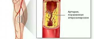
- Atherosclerosis . The deposition of “harmful” lipids in the inner lining (intima) of blood vessels leads to damage to their walls. In response to this, inflammation occurs, which provokes the migration of coagulation factors to the site of damage and increased thrombus formation.
- Diabetes. This disease not only aggravates the course of atherosclerosis, but also contributes to the fragility and pathological tortuosity of blood vessels. There is even the term “diabetic retinopathy” - pathological changes in the vessels of the retina as a result of damage by structurally altered glycosylated (saturated sugars) proteins.
- Arterial hypertension . People with high blood pressure should be especially wary of retinal vascular thrombosis. Due to hypertension, the smallest vessels are damaged, blood supply is disrupted and the formation of blood clots is accelerated.
- Vasculitis - from Latin the term literally translates as “inflammation of blood vessels.” It occurs as an allergic reaction or as a result of connective tissue and blood diseases (hemorrhagic vasculitis, systemic lupus erythematosus, scleroderma, etc.).
- Protruding eyes due to long-term and persistent thyrotoxicosis . Excess thyroid hormones affect the periorbital tissue - it begins to grow. The eyeball literally “sticks out” outward. The vessels cannot keep up with it - they burst and thrombose.
- Tumors . They can grow both from eye tissue and metastasize from other organs. Sometimes a piece of tumor that gets into the vessel blocks its lumen. Read more about neoplasms of the eyelids and eyeball →
Causes
As a rule, retinal thrombosis most often occurs in elderly people due to the following diseases:
- atherosclerosis;
- hypertension;
- diabetes mellitus;
- inflammatory and destructive processes of the walls of blood vessels (vasculitis);
- various disorders affecting blood clotting.
Possible causes of thrombosis of the central nervous system of the eye:
- complication after viral and infectious diseases;
- increased pressure inside the eye;
- swelling of the optic nerve;
- tumor inside the eye;
- autoimmune ophthalmopathy.
Patients at risk are:
- obese;
- with thyroid diseases;
- leading a sedentary lifestyle;
- alcohol abusers.
Stages and types of retinal thrombosis
Central retinal vein thrombosis (CRVT) can be of two types:
- thrombosis of the central vein, scientifically called central occlusion;
- thrombosis of one or more branches of the central vein - peripheral occlusion.
This division is necessary to evaluate the following parameters:
- Damage area . With thrombosis of the central vein, most of the retina is damaged, and if there is a thrombus in a small venule, only a small area may be affected.
- The severity of possible consequences and the urgency of hospitalization . Central vein thrombosis is dangerous due to significant loss of vision and requires immediate hospitalization. Thrombosis of the peripheral retinal veins, with early diagnosis and a small affected area, can be treated even on an outpatient basis.
- Scope of ophthalmological care . Treatment for central occlusion will be more prompt and extensive than for peripheral occlusion.
Occlusion (embolism, obstruction, thrombosis) of the central retinal artery
Before embarking on a review of the designated topic, it is necessary to clearly define the concepts included in the title and differentiate them - in order to avoid confusion that is so common in our time.
Any vessel or duct participating in the complex system of circulation of biological fluids in the body (be it blood, lymph, bile, urine, etc.) must perform its functions uninterrupted. To do this, the lumen of the vessel must remain exactly as nature created it - no more and no less - otherwise the laws of hydrodynamics will inevitably change the pressure of the transported liquid, which will lead to its excess or, conversely, its deficiency at the “destination”. For the body, this always means serious problems.
If we talk only about the circulatory system, then in a number of areas of medicine there is the term “vascular catastrophe,” meaning a sudden blockage of a blood vessel. More precisely, perhaps, you cannot say here: this is precisely a catastrophe in a local area, since a deficiency or cessation of blood supply (ischemia) quickly leads to necrosis, the death of working cells of a tissue or organ on a certain scale (depending on the caliber and significance of the blocked vessel and the organ being supplied ) – a heart attack occurs.
The most famous and clear examples of what such conditions mean for the body as a whole are coronary heart disease and myocardial infarction. But not even a complete, but only a partial reduction in the standard volume of blood that should flow through the arteries or drain through the veins ultimately leads to equally catastrophic consequences: dystrophy gradually develops (a state of constant deficiency of nutrients and oxygen bound by red blood cells), a decrease and “drying out” of the starving tissue/organ, then atrophic degeneration begins, i.e. disintegration of high-molecular proteins and degeneration, replacement of parenchymal (functional, specialized) cells with universal and useless, in this case, connective tissue, as well as its scarring (fibrosis) with growth in size and compaction. Various options for vascular obstruction are possible, and none of them are safe. Stenosis is a persistent narrowing of the lumen, for example, due to thickening of the vascular walls for any reason.
Thrombosis is a complete or partial blockage of a vessel, for example, by a detached and migrating atherosclerotic fatty plaque or a clot of coagulated blood. Occlusion is a complete and, in the literal sense, hopeless closure of a vessel, i.e. lack of even minimal lumen and blood flow. Thus, thrombosis in its extreme form means occlusion, but partial thrombosis does not. Embolism is a synonymous term used to designate blockage of a vessel by any body or particle in general; This is not necessarily a blood clot - an embolus can be, for example, a gas bubble that gets inside a vessel (the original meaning of the ancient Greek word “embolos” is “gag”, “plug”, “plug”, etc.).
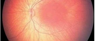
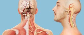
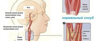
Returning to the topic, let’s imagine a very thin, very fragile, not very well protected and not equipped with uninterruptible power supply systems, but normally a perfectly tuned and balanced optical-biomechanical system called the eye. Along the inner surface of the back wall of the eyeball, - on the so-called. the fundus of the eye - its most important functional tissue, or retina, is located in a thin layer. The retina consists of receptor cells saturated with light-sensitive enzymes and is nourished by the underlying choroid. Approximately in the center of the most sensitive zone (“macula”, “macula”) the retina is fused with the receiving disc of the optic nerve (OND), where the optical image is “digitized” into electrochemical signals-impulses and in this form is transmitted further along the nerve to the visual cortex of the brain brain The largest vessels in the circulatory system of the eye are located along the optic nerve: this is the central retinal artery (CRA), from which blood saturated with oxygen and nutrition is supplied to the smaller branches and smallest capillaries of the retina, as well as a paired vessel for draining waste blood that has released nutrients, also carrying away metabolic products (metabolism) - the central retinal vein (CRV).
Since functional retinal (retinal) tissue is very thin and vulnerable, the slightest problems with its blood supply and nutrition lead to degenerative changes (the most common and well-known example is diabetic retinopathy), and with a stable and long-term worsening of such problems, the retina simply detaches from the choroid. Nutrition, oxygenation (oxygen saturation), as well as the processes of formation of visual images and transmission of signals to the brain stop at this point. Blindness sets in.
The attentive reader, now fully armed with an understanding of the terms, can then independently model the catastrophic picture of what happens to the retinal tissue during thrombosis, embolism and/or, in the worst case scenario, occlusion of the central vein or central retinal artery. The CAS is blocked approximately one and a half times less often than the central vascular system, but the consequences of thrombosis/occlusion of the artery develop more rapidly and turn out to be more (even more) severe than with blockage of the vein.
In addition to atherosclerosis and hypertension (arterial hypertension), as well as damage to the vascular walls in diabetes mellitus, arterial occlusion can be caused by acute and chronic vascular inflammation (vasculitis, giant cell arteritis), ocular ischemic syndrome due to stenosis, compression of the artery by an oncological neoplasm or fibrous tissue. Complete or partial blockage can occur both in the main artery itself and at any point in its basin, i.e. systems of branches and capillaries; in the first case, the ischemia is extensive and total, on a retinal scale, in nature, in the second - local, affecting a more or less limited area.
Stages of the thrombotic process in the retina
The development of the disease occurs in several stages:
- Prethrombosis . It is characterized by dilation and tortuosity of the veins, single point hemorrhages. At this stage there are no clinical manifestations yet, but periodic blurring before the eyes may appear.
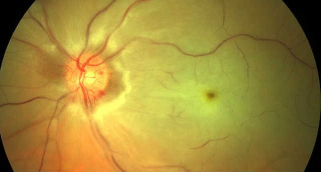
- Direct thrombosis . Numerous linear hemorrhages, swelling of the macula on the retina, which is responsible for color perception, and unclear boundaries of the optic nerve head are visible in the fundus. There is a sharp decrease in visual acuity and a persistent “veil” before the eyes.
- Postthrombotic changes . Traces of hemorrhages and newly formed vessels with thin walls are visible in the fundus. Visual acuity recovers slowly.
Causes of retinal vein thrombosis
It occurs as a result of blockage of a vessel, the causes of which may be atherosclerosis, hypertension, diabetes mellitus, especially often when there are jumps in blood pressure and blood sugar. In young people, thrombosis can be caused by infectious diseases, for example, influenza, sepsis, focal infections of the oral cavity and paranasal sinuses. Also important in the development of retinal vein thrombosis are ocular hypertension (increased intraocular pressure), papilledema, and external pressure on the eyeball (tumors).
Risk factors:
sedentary lifestyle, obesity, heart and vascular diseases, endocrine disorders, especially with inadequate treatment.
Clinical symptoms and diagnosis of retinal thrombosis
Symptoms largely depend on the location of the blood clot and the degree of narrowing of the vessel (occlusion).
If there is thrombosis of the central vein of the retina, at least 3/4 of the retina is damaged: there will be large multiple hemorrhages, rapid deterioration of vision and distortion of color perception.
If thrombosis of a branch of the central retinal vein (a small branching vessel) occurs, visual acuity decreases slowly and is often not regarded as an alarming symptom. Blurry black spots or a “fog” may appear in the field of vision.
Complete occlusion (occlusion of the vein lumen by 95% or more) has pronounced clinical symptoms. Fortunately, it is rare. Partial occlusion may not be clearly visible. The manifestation of signs of thrombosis begins when the lumen of the vessel narrows by 70 percent or more.
Thrombosis of the central retinal artery is always an urgent (emergency) condition that requires prompt qualified assistance! If with venous occlusion there is a chance to preserve vision, then with occlusion of the central nervous system there is a risk of complete blindness.
Diagnostics
To diagnose thrombosis of the vascular system of the eyes, you need to consult an ophthalmologist. Based on the results of a visual examination and instrumental diagnostic procedures, a specialist will be able to quickly make a diagnosis.
Thrombosis of the central retinal vein and peripheral vessels of the eye is diagnosed using the following methods:
- ophthalmoscopy - in the fundus, the doctor visualizes an increase in the venous pattern, pinpoint hemorrhages or the symptom of a “crushed tomato”;
- visometry - during the study a significant drop in central and peripheral vision will be established;
- tonometry - against the background of thrombosis of the vessels of the eye, a unilateral increase in intraocular pressure often occurs or, on the contrary, a strong drop;
- biomicroscopy - visual examination of intraocular structures and vessels under high magnification allows the doctor to visualize damaged vessels, determine the intensity of the ischemic process and detect a suspension of blood elements and exudate in the vitreous body;
- perimetry - measuring visual fields helps determine the approximate location of a thrombosed vessel;
- Angiography with contrast is an X-ray examination, based on the results of which the doctor receives clear images of the vessels of the eye, which show the localization of the blood clot, the outlines of the ischemic focus, the area with hemorrhage and other features;
- Electroretinography is a tool for determining the degree of retinal ischemia, which allows you to accurately predict the likelihood of complications and select adequate measures to prevent them.
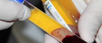
In addition to identifying the characteristics of the pathology, it is important for the ophthalmologist to know the causes of thrombosis. To do this, laboratory blood tests are prescribed: for glucose and cholesterol levels, the degree of coagulation (coagulogram and clotting factors). Additionally, consultation with an endocrinologist, therapist, cardiologist and other specialists may be required.
Treatment
Treatment is carried out in 4 stages:
- Restoring blood flow in a thrombosed vessel.
- Reducing retinal edema.
- Dissolution and elimination of formed hemorrhages (if they are small in area).
- Improving microcirculation in the retina.
Therapy methods
To treat retinal thrombosis, an integrated approach is used:
- The clot can be dissolved by Fibrinolysin or Plasminogen. They are administered using a syringe under the eye. The only caveat: no more than 2 hours should pass from the onset of clinical manifestations of thrombosis.
- Injectable heparin, warfarin or clopidogrel are used to prevent further thrombus formation and reduce blood clotting in small vessels.
- Trental can improve blood circulation and protect the walls of blood vessels from hypoxia. It is administered intravenously 2 times a day.
- Retinal edema is treated by injecting glucocorticosteroid solutions (Prednisolone, Hydrocortisone) into the tissue around the eye. For severe pain in the eye, anti-inflammatory drugs are prescribed intravenously.
Diagnosis and treatment of the disease
It is not difficult to establish the presence or absence of a disease such as eye thrombosis. First of all, the doctor conducts a survey of the patient, during which the general picture of his condition is clarified: what diseases and when the person was ill, what treatment methods were used, what changes in the condition have occurred recently, etc. Thus, the doctor assesses the possible impact of any causes and factors predisposing to the development of the disease.
Then the patient's intraocular pressure is measured, and the quality of visual function is determined, it is determined whether vision is narrowed, etc. Next, a special procedure is performed to examine the fundus of the eye - ophthalmoscopy - during which it is determined whether hemorrhages are occurring. In addition, diagnostic procedures such as fluorescein angiography and biomicroscopy are used. Blood is also taken for general and biochemical analysis. The doctor may prescribe other diagnostic methods at his discretion, for example, an ECG.
Despite the fact that vision loss due to eye thrombosis occurs gradually, treatment must begin immediately as soon as the first signs of the problem are discovered.
This rule applies equally to all diseases, and thrombosis is no exception. In this case, treatment is aimed at solving the following problems:
- blood clot removal;
- restoration of blood circulation;
- removal of swelling;
- resorption of hemorrhages;
- decrease in intraocular and blood pressure;
- stimulation of retinal nutrition.
This is followed by drug therapy aimed at preventing the recurrence of a blood clot in the vessels of the eye. Typically treatment includes the use of drugs such as:
fibrinolytics (Fibrinolysin);
- anticoagulants (Heparin, Clexane);
- antiplatelet agents (Verapamil, Pyrazoline);
- glucocorticoids (Dexamethasone);
- antispasmodics (No-shpa, Papaverine);
- vitamins (C, A, group B).
Blood pressure is reduced with the help of Phenigidine, Nifedipine, intraocular pressure - with the help of Cusimolol, Arutimol. However, the treatment of ocular thrombosis is not limited to the use of medications. After this, laser coagulation of the retina is performed. This procedure is a surgical intervention during which special equipment is used to influence the retina in order to treat various diseases and prevent complications.
In particular, laser coagulation significantly improves blood flow and restores visual function of the eye. The technique is recognized as very effective in the treatment of eye diseases.
Traditional therapy
In addition to traditional therapy, there are many traditional medicines. But they are used only for preventive purposes. To maintain the elasticity of the walls of blood vessels, nettle decoction, sage tincture, mint in all varieties (tincture, tea, juice) are suitable. Forest honey helps improve vision.
Drops made from freshly squeezed clover or cornflower juice are excellent in preventing eye diseases. Take 1 tablespoon of chopped herbs per glass of boiling water. The mixture is infused for 2 days, then filtered. You need to put 2 drops in each eye at least 4 times a day.
Natural remedies are, of course, good, but not for emergency help. They can slow down the rate of development of pathological changes. But in the presence of complications or severe neglect of the process, only traditional, scientifically proven methods can save.
Since complications of a blood clot in the retina include, at a minimum, decreased vision, and a maximum of optic nerve atrophy and complete blindness, it is important to recognize the symptoms in time and provide qualified assistance. But it’s easier to prevent negative consequences.
Methods of therapy
It is important that ocular thrombosis is treated as soon as possible after diagnosis. Within 6 hours the chances of recovery are above average. Blood thinners are mainly used. If the disease has already progressed, surgery may be a promising treatment as a treatment.
Small cuts made directly to the retina and optic nerve head stimulate circulation and can dissolve a blood clot in the eye. This procedure is also called "radial optic aneurotomy." It has been proven that significant improvement in vision can be achieved for many affected people.
Another surgical procedure is performed using a laser. Laser technologies help effectively prevent the proliferation of blood vessels. It also minimizes the risk of subsequent vitreous hemorrhage and cataracts.
Since 2011, there has been a method of inserting an implant directly into the eye. This therapy is also called "intravitreal injection" and is performed under local anesthesia. The implant delivers a precisely defined amount of cortisone over a long period of time. This treatment method is very effective and in most cases has mild side effects.
Prevention measures
Retinal thrombosis can actually be prevented. You just need to undergo annual examinations and follow your doctor’s prescriptions. Methods for preventing retinal vein thrombosis depend on the presence of a specific risk factor and concomitant pathology.
- In case of hypertension, medications are needed to normalize blood pressure. There are many of them; an individual combination is selected for each patient. You should consult a cardiologist regarding the effects of specific medications.
- For all types of diabetes, the main task is to achieve a constant normal blood glucose level. This can be achieved through diet, adequate physical activity and carefully selected medications. For type 1 diabetes, you need to establish the dosage of insulin, for type 2 diabetes, the type and frequency of use of glucose-lowering drugs.
- Any eye diseases require increased attention. Under no circumstances should glaucoma be advanced. Not only does it threaten thrombosis of the blood vessels of the eye, it also leads to a complete absence of lateral vision. People with various types of retinopathy (diabetic or hypertensive) need to be checked by an ophthalmologist once every six months.
- Correction of hormone levels. If the thyroid gland is overactive, medications that reduce thyroxine levels are needed. Women are not recommended to get carried away with oral contraceptives - they increase the risk of blood clots.
- Prevention of increased aggregation (“sticking together”) of platelets - take Aspirin (ThromboASS or Plavix) daily, 1 tablet per day. This is especially necessary for those who suffer from cardiovascular diseases.
Vision is a special sense organ, without which a person loses the ability for self-care and normal social life. Patients with eye diseases should understand that thrombosis of the blood vessels of the eyes leads to irreversible changes. No operation will return or “resurrect” the retinal neurons that died as a result of oxygen starvation. It is better to start preventing retinal thrombosis right now.
Author: Maria Vasilyeva, doctor, especially for Okulist.pro
Treatment options
Treatment of eye thrombosis can be carried out on an outpatient basis, but it is better to hospitalize the patient. The main goal in the treatment of thrombosis is to restore normal vision, blood supply and nutrition to the eyes. Treatment of pathologies that contribute to thrombus formation is also required.
Medication
The following drugs are used for treatment:
- lowering blood pressure;
- restoring normal blood circulation;
- to eliminate swelling;
- fibrinolytic - to thin the blood and eliminate fibrin from it.
After this, a course of anticoagulants is prescribed to prevent the formation of new blood clots. Most often, thrombosis is treated with the use of corticosteroids against inflammation and swelling. Local medications are also used: eye drops, ointments, creams.
The duration of the course and treatment regimen are prescribed by the attending physician according to individual indications.
Surgical intervention
If medications are not enough to cure or their use is impossible, laser coagulation of the eye may be prescribed. The laser breaks up the blood clot that is blocking the vessel. The operation is quick, painless and bloodless. You need to understand that a laser can eliminate a blood clot, but systematic conservative treatment of the underlying pathology and elimination of provoking factors of thrombosis must be carried out.
ethnoscience
It is not possible to cure eye thrombosis with folk remedies, but you can significantly speed up the healing process and also use them as preventive measures. Herbal decoctions, compresses and applications are used for treatment. For compresses, use black tea, decoctions of plantain, lemon balm, elecampane root, and sage.
The juice of fresh red clover grass helps a lot.
Home remedies can also include massage of the eye area. It prevents blood stagnation and improves its circulation. It is necessary to do eye exercises. They also promote normal blood circulation, train the eye muscles, and can have a significant positive effect on visual acuity.