© Author: Lyudmila Vasilievna Tomarova, edited by first category doctor Z. Nelly Vladimirovna, especially for SosudInfo.ru (about the authors)
As a rule, when talking about vascular diseases, they primarily mean damage to the walls of the arteries - atherosclerosis and changes in the diameter of the vessels of the venous bed - varicose veins. However, the entire human body is permeated with vessels, and not everyone realizes that the list of diseases related to the vascular area includes more than a dozen. This includes all kinds of phlebitis, thrombosis, tortuosity of arteries, lymphostasis of the extremities, called elephantiasis, pelvic varicose veins, hemorrhoids and much more. Therefore, checking vessels and their performance in a timely manner is everyone’s sacred task.
Who needs verification?
The passage of these vessels is a priority for elderly patients, in addition, for those, regardless of age, who are most susceptible to psycho-emotional influences and physical stress, and attacks of headaches. The presence of excess weight, poor nutrition, abuse of bad habits can serve as an indicator for periodic examination. The specialist chooses the diagnostic method depending on the symptoms and preliminary tests. As a rule, the following should be taken into account:
- patient reports for headaches, dizziness;
- previous injuries (head and cervical region);
- sudden manifestations of visual and auditory disturbances;
- causeless nosebleeds;
- the patient's tendency to faint with short-term loss of consciousness;
- the presence of a previous cerebral infarction, symptoms of chronic ischemia;
- head and hand tremors due to parkinsonism;
- there are suspicions of the development of a neoplasm;
- with developed encelopathy in order to clarify the role of blood supply in encephalopathies.
In addition, there are such indications that serve as a reason for mandatory vascular studies:
- Symptoms of problems with blood supply to the brain. They manifest themselves as sudden and severe headaches, nausea, impaired movement and other phenomena.
- Atherosclerosis. This disease is characterized by partial or complete blockage of blood vessels, which ultimately leads to serious complications.
- Diabetes. Patients suffering from this disease should have their blood vessels checked from time to time, since the disease contributes to many dysfunctions of a large number of organs and provokes the development of atherosclerosis.
- Brain tumors. Neoplasms, although not characteristic of the brain, increase in size and provoke intracranial pressure. They can be either benign or malignant. One of the main symptoms is a morning headache, often accompanied by vomiting. Then other signs begin to appear: memory impairment, mental disorders.
- Hypertension. Hypotension. High arterial and intracranial pressure increases the load on the vascular walls of blood vessels, and this in turn provokes disruptions in supplying the brain with nutrients and, as a result, pain. Low blood pressure also does not bode well for the functionality of blood vessels, since the brain, due to a lack of sufficient blood supply, begins to experience oxygen starvation.
- Sharp headaches. This should include pain, the nature of which is not sufficiently clarified. Because in some cases it is vascular disorders in the neck and head that provoke the development of headaches.
- Cervical osteochondrosis. With this disease, the tissue of the intervertebral discs is damaged and deformed. They begin to put pressure on the blood vessels, as a result, their normal functioning is disrupted due to a lack of blood supply to the brain cells, hence the pain.
- Vertebrobasilar insufficiency. Disturbances in brain function due to obstructed blood flow in these arteries are, as a rule, reversible; however, such phenomena should not be left to chance.
- Planned heart surgery. It is a necessary condition for preoperative examination to check the blood vessels of the neck and brain.
- Traumatic brain injury. As a result of dangerous head bruises or a resulting concussion, deformational displacement of blood vessels may occur due to mechanical damage to the tissues that frame the vessels. And as a result, they begin to put pressure on the vital vessels of the cervical region and brain.
- Stroke. A condition of the body characterized by acute circulatory disorders. This causes tissue damage and disruption of various functions. By type, stroke is divided into hemorrhagic and ischemic. Symptoms of impending pathology may be similar in both cases and are characterized by heaviness in the head, increasing tinnitus, weakness and dizziness. In such pre-stroke conditions, micro-strokes, immediate diagnosis and, if necessary, surgical intervention are required.
- Alzheimer's disease. As a result of the body's production of pathological proteins, nerve cells atrophy. The onset of the disease is almost impossible to recognize; the patient experiences absent-mindedness, memory impairment, and resorts to using notebooks.
- Epilepsy. Seizures often lead to loss of consciousness and convulsions appear. Seizures can be generalized and local. For the first case, damage to both hemispheres of the brain is noted, for the second - only one.
Currently, diagnostics can be used to detect both the disease itself and the causes of its occurrence. This gives a chance to start timely and adequate treatment. It is necessary to consider the main ways to study the structure of the brain.
Hardware examination methods
These research methods include all instrumental diagnostics. Indications for these studies depend on the patient’s age and assessment of his physical characteristics.
Absolutely every person over forty years of age must undergo these examinations annually, choosing the desired method, relying on medical recommendations and the capabilities of medical equipment.
Hardware research methods make it possible to find out:
- General condition of blood vessels;
- Narrowing of their lumens;
- Damage level;
- The presence of cholesterol plaques or blood clots in the vessels;
- Presence/absence of neoplasms.
Data obtained through timely diagnostics using hardware methods help prolong the patient’s life, and sometimes even save it.
How to check the vessels of the neck and brain
The following are some recommendations that will help you decide on the choice of examination. Any one of them should define:
- Does the patient have blockage or narrowing of blood vessels, what effect do they have on the supply and nutrition of brain cells with oxygen, as well as what pathologies may develop.
- What is the extent of the impact of the existing disease on the blood flow.
- In the absence of signs of the presence of dysfunction of the cervical and head vessels in the initial stages.
- The success of the treatment.
- The state of the tone of the vascular walls.
- Formation of aneurysm, spasms and various deformities.
- Congenital malformations of the structure of blood vessels.
- Disruptions in the blood flow of the veins.
Having established the necessary data, the specialist must identify the cause of vascular malfunctions, as well as the possible impact of the identified disease on the vessels now and in the future. If a patient is diagnosed, for example, with diseases such as atherosclerosis and aneurysm, as well as some other causes, then this in itself is a sufficient reason to perform vascular surgery.
Ultrasonic
Echoencephalography. This method is often called cerebral ultrasound. It is based on the use of an oscilloscope. Using ultrasound, the state of the brain is recorded and displayed in the form of a schematic image. A current standard has been developed, the requirements of which must be met by the echoencephalogram of a patient who does not have any disorders in the functioning of blood vessels and the brain. Otherwise, the drawing will disrupt the usual picture. Using this method, you can also study brain activity and the functionality of all parts of the brain. Consequently, echoencephalographic examination allows us to detect not only malfunctions in the cervical and cephalic vessels, but also the potential consequences of these malfunctions.
Doppler ultrasound. Combines classical ultrasound with Doppler ultrasound. This technique is considered the safest, at the same time very effective, however, not all medical institutions still have the appropriate equipment to conduct such studies. Therefore, you may have to make an effort to find a well-equipped clinic where it will be possible to undergo an ultrasound scan. It is considered the most popular method for studying the vessels of the head and neck, as it makes it possible to establish a large number of parameters of their activity:
- The intensity of movement of arterial and venous blood flow.
- The presence of atherosclerotic plaques and the presence of narrowing of the lumens.
- Detect arterial blockage.
- Identify the presence of changes in the direction of blood flow, which were provoked, for example, by damage to tissues adjacent to the vessels or due to osteochondrosis.
- Aneurysm formation.
Duplex scanning. Using this method, which is based on a Doppler coding system and spectral analysis, it is possible to conduct studies of vascular malfunctions. It makes it possible to color evaluate the lumens of blood vessels, measure the speed of blood flow in them, and in addition determine such conditions of the walls of blood vessels as:
- Strength and elasticity.
- Tone.
- Changes in thickness and structure.
- Mechanical damage. In addition, you can determine the formation of atherosclerotic plaques and blood clots, vessel branching and other important parameters.
On the screen, areas of the tissue being examined, differing in structural and echogenic characteristics, are displayed in different color shades.
Neurosonography. This diagnostic method is used to study infants and young children. As you know, in children from the moment of birth the large fontanelle remains open for some time; with its help, it is possible to better identify the shape of the bones and soft tissues of the head and neck, brain matter, nervous tissue and blood vessels. The method allows you to detect malfunctions of blood vessels, aneurysms, neoplasms, congenital pathologies and other disorders. To perform neurosonography using a fontanel, no additional equipment or special devices are needed: a typical ultrasound apparatus is used. For adults, this device is also applicable, although it is not used as often, and special equipment settings must be used. The examination is carried out through the temple bone. In addition, a comprehensive study is used that combines these two research methods. This approach makes it possible to avoid mistakes and achieve more accurate results.
Neurosonography is considered today the most effective and non-hazardous method; it is used for research in children. Transcranial Dopplerography. Allows you to achieve complete information and is used to obtain a complete picture of hemodynamics, the degree of fullness and functionality of blood vessels and other important information. This is the latest technology based on digital examination, which gives the chance to increase the depth of examination to nine centimeters and provide a more thorough picture of the arteries and veins. All scanning levels of this method are carried out layer by layer.
Which specialist should I contact?
Sometimes, feeling unwell, a person finds it difficult to choose the right specialist who can solve the problem. Although, in any settlement there will probably be a paramedic or therapist who accepts patients with any ailments. He will definitely tell you which doctor specializes in a particular disease.
In every regional center or more or less similar to an urban village, there is a neuropathologist or neurologist, which is essentially the same thing. Neurologist is a modernized name for the same specialist. This doctor will be able to help with injuries and hemorrhages in the brain, osteochondrosis, sleep disorders and coordination of movements. In a word, where vascular diseases of the nervous system occur.
In order to make an accurate diagnosis, the neurologist has the right to refer you for an examination of the cerebral vessels using the most modern methods. After all, a violation of the blood supply will immediately affect your overall well-being, which manifests itself in the form of frequent headaches, dizziness, and increasing irritation.
A specialist in vascular surgery, called an angiologist or angiosurgeon, will help you check the blood vessels in your legs. It treats blood and lymphatic vessels. He has the power to save humanity from diseases such as stroke, kidney failure, trophic ulcers, thrombophlebitis, thrombosis, and even impaired potency, if it is related to his specifics. It is important to know that such a diverse range of activities of a vascular surgeon is explained by the fact that his competence includes the treatment of lesions of veins, arteries and lymphatic vessels. You can also entrust the condition of the veins to a more specialized specialist - a phlebologist.
It seems that almost everyone is aware that in order to check the blood vessels of the heart, you should contact a cardiologist. Although, sometimes it is difficult to draw a clear line between true heart diseases and other vascular diseases that have an indirect effect on it. Thus, rheumatism and myocarditis are traditional heart diseases, and atherosclerosis and phlebitis cause more damage to arteries and veins. Finally, hypertension has a negative impact on the entire cardiovascular system.
Each of the above doctors can prescribe a comprehensive examination within the framework of their specialization.
Electrical measuring
Electroencephalography. This procedure is carried out using a device that records brain electrical impulses. And provides comprehensive diagnostics. The medulla, the network of nerve fibers, blood circulation, brain activity and some other tissues and vessels are checked. To prescribe the procedure, there are various indicators of disruption of the functionality of blood vessels, injury, night sleep disturbances and mental disorders. Large research parameters make it possible, using this device, not only to identify various disorders, but also their effect on brain tissue. It is possible to identify a region of the brain and the presence of oxygen deprivation volumes. Research using this method is considered absolutely harmless. Can be used by patients of all ages, even young children. To carry out the procedure, you need to prepare in advance. Patients taking antispasmodics and other similar drugs should avoid them for a while. The duration of the procedure depends on the symptoms and can last quite a long time, starting from a quarter of an hour. Diagnosis of a wide range of diseases and low cost of testing allow the device to be used among a wide range of patients.
Magnetic resonance
Magnetic resonance angiography. As with standard tomography, it involves the principle of using radio waves that affect an object placed in an artificially created magnetic field. Angiography reveals the state of the brain substance, nerve trunks and head vessels of the head and produces a detailed image at high power of the tomograph. To prescribe the procedure, serious vascular disorders are required, characterized by constant and severe headache. Other indications may include: New growths in the head. Presence of a microstroke. Suspicion of vascular thrombosis. Does not require special preparation. But the cost is relatively high. In this regard, MR angiography of the head and neck is prescribed only if there are sufficient indications. Before carrying out, any metal products must be removed. The patient must maintain a motionless body position.
X-ray
Contrast angiography. When examining this method, X-rays are used, and a contrast agent is also introduced into the patient’s body in order to obtain a better image of the vessels and the structure of their walls. For this purpose, an iodine-containing substance is used, which is administered by puncture or using a catheter. This research method is divided into:
- to general. This is when the entire vascular network is examined.
- selective. When one or more veins and arteries are examined. The last angiography is carried out for the purpose of a more thorough analysis.
The technique is considered the most popular when it is necessary to determine the magnitude of the threat when the patient has atherosclerotic blood clots in the vascular lumens. Depending on the indications, arterial, venous and capillary examination may be used. Indeed, with the help of this method, a unique opportunity is provided to display even the smallest vessels. It should not be forgotten that X-ray radiation has a negative effect on the patient’s body, so it may not be indicated for all patients.
Coronary angiography of the heart vessels: what is it, indications, performance
Cardiovascular diseases are a very common pathology for people over 40 years of age. And among these diseases, the most common are associated with imperfection of the vascular bed and limited nutrition of the heart muscle.
To clarify the causes of heart disease, there are many diagnostic methods. One of the most informative checks is coronary angiography of the heart vessels - what is it, is it dangerous to do it, and how is the examination performed?
general information
This is an invasive manipulation that serves to determine the condition of the vessels that carry blood and oxygen to the heart. They are called coronary. The left and right coronary arteries normally provide nutrition to the muscle and maintain the functionality of the entire organ.
In case of unfavorable development of events, these arteries, for various reasons, narrow (stenosis) or become blocked (occlusion) . The blood supply to the heart is significantly limited or stops altogether in a certain area, which causes coronary artery disease and heart attack.
To exclude such a defect or, if present, to determine its extent, coronary angiography is performed.
This is an X-ray examination of the lumen of the coronary vessels using an angiograph and a contrast agent introduced through a catheter precisely into the vestibule of the cardiac arteries. The survey is carried out from different angles, which allows you to create the most detailed picture of the condition of the object being examined.
Indications for the procedure
Routinely, coronary angiography is performed for:
- confirmation or refutation of the diagnosis of IHD;
- clarifying the diagnosis when other methods of determining the disease are ineffective;
- determining the nature and method of eliminating the defect during the upcoming operation;
- audit of the condition of the organ in preparation for open-heart surgery, for example, in case of a defect.
In emergency cases, the procedure is performed in the presence of the first signs and symptoms of a heart attack or in a pre-infarction condition that requires immediate intervention for life-saving reasons.
Let's look at how to prepare for coronary angiography of the heart, as well as how this procedure is done.
Preparation
Before prescribing coronary angiography, it is necessary to undergo a series of examinations in order to exclude or confirm the presence of factors that do not allow the use of this diagnostic method. Training program:
- blood tests (general, sugar, hepatitis B and C, bilirubin and other liver parameters, HIV, RW, group and Rh factor);
- urine test for the presence of renal pathology;
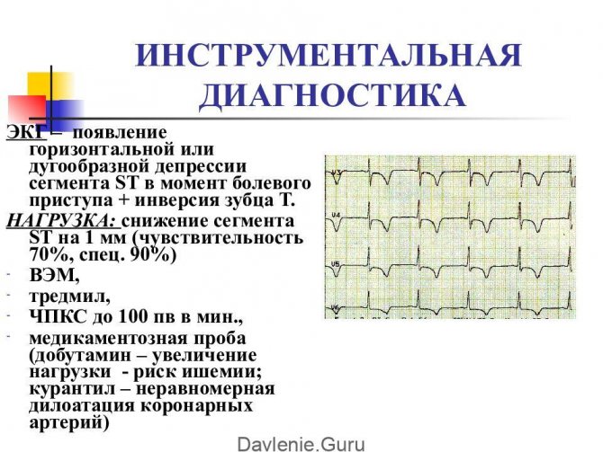
- ECG in 12 leads;
- examination and conclusion of specialists on existing chronic diseases.
Upon admission to manipulation, immediate preparation is carried out before the procedure :
- the doctor stops some medications in advance, for example, those that reduce blood clotting;
- exclude food intake on the day of diagnosis - to avoid complications such as vomiting, the study is carried out on an empty stomach;
- The doctor collects an allergic history and conducts a test with a contrast agent.
Immediately before coronary angiography, it is recommended to take a shower, shave the hair in the groin, remove jewelry from the body (earrings, rings, piercings), glasses, removable dentures, lenses, and use the toilet.
How it's made
The patient lies down on a special table. Heart monitors are attached to his chest. In the area where the catheter is inserted, local anesthesia and skin disinfection . A micro-incision is made in the vein through which a catheter is inserted.
The catheter is passed through the vessels under the control of an angiograph to the mouth of the coronary arteries. A contrast agent is injected into each of them one by one, which outlines the internal space of these vessels. are carried out from different positions . The location of stenosis or occlusion is determined.
During the operation, the patient is conscious and his condition is monitored by observing his appearance, heart rate and verbal behavior.
Once monitoring is complete, the catheter is carefully removed from the vein. The wound is carefully sutured. The patient remains lying down for some time, and the doctor writes a conclusion .
It indicates the size of the smallest lumens in the vessels, the degree of narrowing and the recommended method of correcting the situation - stenting or bypass surgery of the heart vessels.
If there are no problem areas, a general description of the coronary arteries is given.
about how outpatient coronary angiography of the heart vessels is performed:
Conditions
Most often, coronary angiography is performed in a hospital setting as part of a routine examination for coronary artery disease. In this case, all tests are taken here, a few days before the intervention.
Diagnostics can also be carried out on an outpatient basis. But the patient must first independently undergo all the examinations on the list, obtain a cardiologist’s opinion on the possibility of coronary angiography and a referral for it, indicating the purpose of the study.
In an outpatient setting, the insertion of a catheter for coronary angiography is most often carried out through the wrist vein and in the arm - in the postoperative period, the load on it, in contrast to invasion through the femoral vessel, can be minimized in order to avoid dangerous bleeding.
Contraindications
A number of conditions do not allow the use of this diagnostic method, so they resort to alternative ones. Preliminary examination may reveal these conditions:
- uncontrolled arterial hypertension - intervention can provoke stress, resulting in a hypertensive crisis;
- post-stroke condition - anxiety can cause a re-attack of the disease;
- internal bleeding in any organ - with invasion, blood loss may increase;
- infectious diseases - the virus can contribute to the formation of blood clots at the incision site, as well as peeling of areas on the walls of blood vessels;
- diabetes mellitus in the stage of decompensation is a state of significant kidney damage, high blood sugar levels, and the possibility of a heart attack;
- elevated temperature of any origin - accompanying high blood pressure and rapid heartbeat can lead to heart problems during and after the procedure;
- severe kidney disease - the contrast agent can cause organ damage or worsen the disease;
- intolerance to contrast agent - a test is carried out on the eve of diagnosis;
- increased or decreased blood clotting - can cause thrombosis or blood loss.
With preliminary preparation, all these conditions are identified and treatment is prescribed to compensate for them. There are no absolute contraindications to the procedure. Once stabilization has been achieved, the procedure can be performed in a hospital setting.
Risks, complications and consequences
Coronary angiography, like any invasion, can have side effects caused by the body’s improper response to the intervention and the patient’s stress. Rarely, but the following events occur:
Pre-procedural testing is intended to prevent these conditions, but sometimes they happen. The doctors involved in the examination cope with the situation, the procedure is stopped at the first unfavorable signs , the patient is removed from the dangerous state and transferred to a hospital for observation.
Recommendations after completion
Based on the conclusion of the doctor who conducted the study, the cardiologist determines the path of treatment for the patient . If there are indications, the time for stent installation is scheduled (in the same way as coronary angiography - using a catheter).
Sometimes this procedure is performed directly during diagnosis, if there is the prior consent of the patient. The cardiologist may also prescribe outpatient treatment or coronary artery bypass surgery.
Diagnostic cost
If you have a compulsory medical insurance policy, coronary angiography according to indications is performed free of charge. But the equipment of most hospitals does not allow covering everyone with this diagnostic method in a short time. Usually the queue lasts for months, because... Limited quotas for examination are provided. It is possible to undergo this study on a commercial basis.
The cost in Russia is in a wide range - from 10 to 45 thousand rubles. Abroad, this intervention is also not always covered by insurance and is also not cheap - from 300 dollars to 2500 euros.
Coronary angiography is included in the mandatory list of diagnostic procedures to determine the degree of damage to the heart vessels. The procedure has been worked out and standardized for a long time - this serves as a guarantee of patient safety. The level of cardiology in the country makes it possible to identify pathology at an early stage and take measures to eliminate it or prevent its development.
Source: https://oserdce.com/diagnostika/sosudov/koronarografiya.html