Causes of uveitis
Factors of inflammation of the choroid can be various reasons: infections, metabolic disorders, autoimmune processes, suppressed immunity, etc. Sometimes uveitis is a secondary disease, and the main focus of inflammation is located far from the eyes, and the infection enters the eyeball through the bloodstream (with tonsillitis and sinusitis, septic endocarditis, meningitis).
It is not uncommon for uveitis to develop after a penetrating injury to the eye. However, pathogens and viruses are the most common cause of uveitis. As a rule, inflammation is caused by chlamydia, herpes virus, Koch's bacillus, toxoplasma, brucella and cytomegalovirus. Uveitis often occurs against the background of systemic connective tissue pathologies (rheumatoid arthritis, SLE, rheumatism, Reiter's syndrome, etc.
https://youtube.com/watch?v=RjmKtqXHGPU
To determine what treatment to prescribe to a patient, the doctor must first determine the cause of autoimmune uveitis. In most cases, uveitis begins after hypothermia, and is also a consequence of metabolic disorders in the human body. Impaired immunity is the main cause of the development of the disease.
Pathological autoimmune processes occurring in the human body lead to damage to the choroid. When autoimmune uveitis occurs, inflammation can penetrate the anterior part of the eye membrane. In this case, the patient develops anterior uveitis, causing damage to the ciliary body and iris.
Due to the occurrence of autoimmune uveitis, in most cases only one eye is affected. But there are cases of damage to both eyes, then doctors diagnose peripheral uveitis.
Often, the occurrence of autoimmune uveitis occurs as a result of damage to the body by various infectious diseases. Diseases that may contribute to the development of uveitis include the following:
- tuberculosis;
- toxoplasmosis;
- syphilis;
- chlamydia;
- brucellosis and some other diseases.
According to modern medicine research, viral uveitis can develop when a person develops viral diseases caused by various bacteria or fungi.
In an infant under one year of age, uveitis can develop when infected with the ECHO-19 virus. As a result of damage to the child’s body by this virus, enteroviral uveitis can develop. It was discovered by doctors recently. This disease is very severe and can lead to the formation of cataracts or other pathologies.
Scientists have now proven that autoimmune uveitis can be caused by diseases such as:
- rheumatism;
- Reiter's syndrome;
- Bekhterev's disease;
- rheumatoid arthritis.
There are cases when even qualified ophthalmologists cannot determine the cause of uveitis when diagnosing a patient.
Types of uveitis
According to the location of the inflammatory phenomena, uveitis is:
- Anterior (among them - iritis, anterior cyclitis, iridocyclitis).
- Posterior (this group includes choroiditis, retinitis, neurouveitis, chorioretinitis).
- Median (including pars planitis, posterior cyclitis, peripheral uveitis).
- Generalized.
If a patient develops anterior uveitis, then the ciliary body and iris are involved in the pathological process. This type of disease is the most common.
Median uveitis leads to damage to the choroid and ciliary body, as well as the retina and vitreous body. If posterior uveitis is diagnosed, then in addition to the retina and choroid, the optic nerve is affected.
If inflammatory processes cover all parts of the uveal tract, then panuveitis, or generalized uveitis, develops.
According to the type of inflammation, uveitis can be purulent, serous, fibrinous, hemorrhagic, mixed. Due to its occurrence, the pathology can be primary (associated with systemic diseases), secondary (develops against the background of other eye diseases), as well as endogenous and exogenous.
According to the nature of the course, uveitis is differentiated into acute, chronic, and recurrent. Based on the type of changes in the choroid, diseases are classified into non-granulomatous, or toxic-allergic widespread, and granulomatous, or local metastatic.
Diagnostics
Before treatment, it is necessary to conduct a diagnosis that will help determine the area of the lesion, stage and the presence of other complications and pathologies. For this purpose:
- Optical coherence tomography;
- Visometry and perimetry;
- Ophthalmoscopy;
- Biomicroscopy;
- Gonioscopy;
- Measuring intraocular pressure;
- Ultrasound of the eye;
- Retinography;
- Electroretinography.
First of all, the doctor conducts an external examination to determine the condition of the conjunctiva and eyelids. The pupillary reaction is examined. Other methods for examining the eye and its parts depend on a number of factors. For example, if it is not possible to perform ophthalmoscopy due to clouding of the optical media, then ultrasound is performed.
It is necessary to understand that the cost of full diagnostics in private clinics can reach serious amounts. But if government institutions do not have the ability to carry out the necessary procedures and research, then you should turn to private institutions. In such cases, it is possible to conduct a partial diagnosis by paying only for those diagnostic methods that are not available in a free queue at a government agency.
It is also worth noting that not all treatment can be carried out in a regular hospital. For example, administering parabulbar injections involves the use of special drugs. They are introduced under the supervision of specialists, and therefore require certain experience, skills, and appropriate conditions, which not every institution can provide. Therefore, you should not save on your health, since uveitis often ends in blindness or decreased visual function.
To differentiate the pathology from other inflammatory diseases with similar symptoms, a decision may be made to perform an optical CT scan, as well as angiography of vessels in the retinal area. Laser scanning tomography is often used. If necessary, the doctor may decide to take a biopsy sample for laboratory testing of the material. If there are suspicions of concomitant pathologies, the patient may be referred to other specialists, for example, for tuberculosis - to a phthisiatrician, for allergies - to an allergist, and so on. In addition, Mantoux reaction tests, fluorography, X-rays of the spine, MRI of the brain, and so on can be performed. It all depends on the expected pathology.
Classification
The division of the pathological process is possible according to different criteria; all typing methods are used by practitioners to describe the disorder, its precise coding according to the classifier, specifying the diagnosis and identifying a clear vector for medical correction of the condition.
Uveitis can be divided according to its origin:
- Primary. As an independent disorder. For example, after an infectious disease, due to a decrease in general and local immunity. It is relatively rare. Such cases account for up to 25% of the total clinical situations.
- Secondary. Particularly common. It is defined as uveitis formed as a result of other eye diseases. Even banal conjunctivitis, as well as minimal trauma, can play a role here. For example, a cut or scratch to the organ of vision while performing duties, interacting with animals, etc.
Both options are equally dangerous.
Depending on the location, a large group of pathologies is distinguished. It must be borne in mind that the uveal tract “stretches” over the entire eye and is represented by a group of structures: the ciliary (ciliary) body, the iris, and the choroid (choroid).
Based on anatomical features, three regions are distinguished: anterior, median and posterior, which is closest to the retina and optic nerve.
Treatment
All therapeutic procedures must be carried out by an ophthalmologist; sometimes the involvement of other specialists is required. The time factor plays a very important role in the treatment of this disease - it is important to establish a diagnosis as early as possible and begin etiotropic and pathogenetic treatment. All these measures are aimed at reducing the likelihood of complications that can lead to significant vision loss or blindness. At the same time, treatment is prescribed aimed at eliminating the cause of the disease.
As for treatment with folk remedies, this approach is extremely undesirable - this method of treatment almost always leads to a deterioration in the patient’s condition. Treatment of inflammatory processes should include the prescription of medications - mydriatics, steroid hormones and drugs that suppress inflammatory processes at the systemic level.
Mydriatics are prescribed to reduce tension in the ciliary muscle, which makes it possible to reduce the risk of developing fusion between the lens capsule and the iris (synechia). Drugs in this group include the following:
- tropicamide;
- phenylephrine;
- cyclopentolate;
- atropine.
When the infectious nature of uveitis is established, antibiotics or antiviral agents are prescribed (depending on the type of infectious agent). If the cause of the disease is systemic inflammatory diseases, then NSAIDs and cytostatics are used. For allergies, antihistamines are prescribed.
An important role in the treatment of uveitis is played by the local use of steroid hormones (prednisolone, dexamethasone, betamethasone). For this purpose, drugs are used in the form of ointments, subconjunctival, sub-Tenon, parabulbar injections are made, and instillations are carried out into the conjunctival sac. If steroid therapy fails, systemic immunosuppressive agents are prescribed.
Surgical treatment is required if complications develop. In this case, the synechiae of the iris are dissected, and cataracts, glaucoma, and retinal detachment are treated. If iridocyclochoroiditis develops, a vitrectomy (removal of the vitreous) or removal of the entire eyeball may be necessary.
Anterior uveitis
This includes two types of inflammation:
Iritis. Inflammatory lesion of the iris itself. In isolated form, it is extremely rare due to the general innervation and local circulatory system with surrounding structures. Identification is difficult and requires a comprehensive assessment.
Iridocyclitis. A much more common problem. In addition to the iris, part of the ciliary body is involved. Without quality medical care, it quickly leads to the formation of adhesions, areas connected to each other by special fibrin strands that prevent the eye from functioning normally.
These two forms are variants of damage to the anterior region of the uveal tract.
Uveitis therapy
The treatment strategy for uveitis should be determined by the cause of the disease. However, it is often not possible to establish the cause of uveitis, so treatment regimens include symptomatic medications or those prescribed empirically until the nature of the inflammation is established. Specific therapy is applied after determining the cause of the disease.
Throughout the world, the “gold standard” in the treatment of uveitis is the use of corticosteroids. The main goals of prescribing hormonal therapy are to reduce exudation, suppress the production of inflammatory mediators, as well as the lymphocytic reaction, and stabilize cell membranes. The choice of a drug of this group and the determination of the method of administration take into account certain factors: the activity of the inflammatory process, the possibility of raising IOP, etc. Today, both local and systemic administration of corticosteroids is practiced; in addition, it is possible to install an implant that releases small doses of the drug throughout long period into the eye cavity or under the eye membranes.
For uveitis, drugs with cycloplegic and mydriatic effects are also very often prescribed. Their use is necessary to prevent the occurrence of synechiae, which are pathological fusions of the iris with surrounding tissues. Also, these drugs relieve spasm of the pupillary and ciliary muscles, which prevents the occurrence of pain, stabilizes the blood-ophthalmic barrier, and prevents further effusion of protein into the intraocular fluid.
NSAIDs are second-line drugs for the treatment of uveitis. Compared to steroids, they have less anti-inflammatory activity, but are very useful in stopping the inflammatory response and pain. In addition, NSAIDs prevent relapses of the disease and macular edema accompanying uveitis in some cases. The combined administration of NSAIDs and corticosteroids makes it possible to reduce the doses of the latter during long-term therapy of some chronic forms of uveitis. NSAIDs can be prescribed in different forms - tablets, powders, eye drops.
Special mention should be made of drugs from a relatively new group - immunomodulators, which are successfully used today in the case of certain forms of uveitis (for example, caused by Behçet's disease, when the process involves the posterior segment of the eye; necrotizing scleritis or Wegener's granulomatosis). This group of drugs includes antimetabolites (Azathioprine, Methotrexate, Mycophenolate mofetil), T-lymphocyte inhibitors (Tacrolimus, Cyclosporine), alkylating agents (Chlorambucil, Cyclophosphamide). Such therapy is aimed at targeted inhibition of certain mechanisms of the inflammatory response of the immune system (immunosuppression), which caused damage to the organ of vision. These medications can be used with or without corticosteroids, which will reduce the negative effects of the latter on the body.
For specific forms of uveitis such as shotgun chorioretinitis, serpinginous choroiditis, sympathetic ophthalmia; uveitis caused by Behcet's disease, Vogt-Koyanagi-Harada disease, seronegative spondyloarthropathy or juvenile idiopathic arthritis, the so-called biological therapy - the use of drugs that suppress tumor growth factor-α. The most popular of them include: Adalimumab and Infliximab. Biological therapy drugs in the treatment of these diseases have become “second-line” drugs, i.e. are used only in cases where previous therapy did not have a positive effect.
Traditional medicine methods
During the treatment of uveitis, you can use some folk recipes. However, before any manipulation it is necessary to consult a doctor .
The following recipes will help effectively eliminate inflammation:
- washing the eyes with a medicinal decoction. It is necessary to take equal quantities of chamomile, calendula, and sage flowers. Grind the raw materials. Take 3 tablespoons of the mixture and pour a glass of boiling water. The composition is infused for 1 hour. Strain the resulting product and rinse the eyes with the decoction;
- aloe juice is diluted with cold boiled water in a ratio of 1:10. The resulting solution is dripped 1 drop no more than 3 times a day into the affected eye;
- marshmallow root lotions. The raw materials should be crushed, pour 3-4 tablespoons of 200 ml of cold water. The product is infused for 8 hours, then used for lotions.
Diagnostics
Experts consider a complete and thorough history taking to be very important in diagnosing uveitis. Often, an available medical history helps to save the patient from unnecessary examination methods. Many ophthalmologists even use various questionnaires that include key queries. This makes it possible to standardize the survey and provide sufficient coverage of the medical history.
There are no mandatory specific methods for ophthalmological diagnosis of uveitis. A general examination makes it possible to identify certain signs characteristic of the disease.
Be sure to pay attention to the level of IOP, which, according to clinical observations, tends to increase in almost half of the patients. An examination with a slit lamp of the anterior segment of the eye remains indispensable, which helps to detect precipitates, hypopyon or pseudohypopyon, changes in the iris and other characteristic signs on the posterior surface of the cornea
To differentiate existing changes in the posterior segment of the eye, along with a standard examination of the fundus, FA and OCT are recommended.
Laboratory tests (HLA typing, PCR, etc.), X-ray examination, MRI, as well as cytology, are prescribed according to indications to identify the cause of uveitis.
In 2005, the International Group for Standardizing Uveitis Nomenclature developed recommendations on the scope of diagnostic tests used for different forms of the disease. They include a list of necessary studies in each clinical case and allow you to avoid performing unnecessary ones. The scope of diagnostic appointments always depends on the potential cause of the disease.
Recognition of the “masquerade” syndrome, imitating the symptoms of uveitis, has a special place in the diagnosis of the disease. It should be suspected if there is a response to minimally aggressive drug therapy.
It is important to understand that in case of uveitis, the purpose of the examination is not only to identify the cause of the disease, it is also to exclude pathology for the treatment of which it is inappropriate to use certain drugs (for example, infectious, not identified by specific tests, “masquerade” syndrome). In addition, diagnostics involves identifying systemic diseases that can worsen the patient’s condition, determines the prognosis for recovery, and, if necessary, helps adjust the prescribed treatment regimen.
Etiology
Many factors can provoke inflammation of the uveal tract - allergic reactions, syndromic and autoimmune diseases, injuries to the visual apparatus of varying severity, metabolic and hormonal disorders, pathological activity of infectious agents.
In 43% of clinical cases, the “trigger mechanism” for the development of this disease was the pathogenic activity of microorganisms. Most often, uveitis develops due to penetration of the following infectious agents into the uveal tract:
- streptococci;
- Koch's wand;
- toxoplasma;
- cytomegalovirus;
- fungi;
- herpesvirus;
- Treponema pallidum.
Allergic uveitis progresses due to the body's increased sensitivity to certain substances. It develops against the background of hay fever, food or drug allergies, and it is also possible that it occurs after the introduction of various serums or vaccines into the body. Only in this case we are talking about serum uveitis.
Also, inflammation of the uveal tract can progress against the background of autoimmune, syndromic or systemic pathologies (the etiological connection between the pathologies has already been proven). Most often the disease is diagnosed when:
- ulcerative colitis;
- spondyloarthrosis;
- glomerulonephritis;
- rheumatoid arthritis (in this case, rheumatoid uveitis develops);
- multiple sclerosis;
- autoimmune thyroiditis;
- Reiter's syndrome;
- psoriasis;
- sarcoidosis.
The development of inflammation of the uveal tract can be facilitated by various hormonal imbalances, metabolic disorders, pathologies of the hematopoietic system, as well as ailments of the visual apparatus (scleritis, blepharitis, conjunctivitis).
Symptoms of the disease
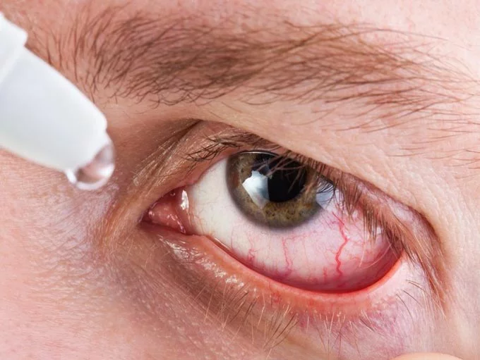
Clinical manifestations depend on the localization of the inflammatory process and the specific pathogen. The acute form of the disease occurs with the following symptoms:
- pain;
- severe redness and irritation of the eyes;
- lacrimation;
- constriction of the pupil;
- decreased quality of vision;
- photophobia;
- increased intraocular pressure.
In chronic anterior recurrent uveitis, there are no characteristic signs. Slight redness of the eyes may occur under the influence of unfavorable exogenous or endogenous factors.
Peripheral uveitis is accompanied by a bilateral inflammatory process. Patients complain of single or multiple blurred vision and deterioration of central vision. The posterior form of the disease can lead to retinal detachment, macular edema and severe ischemic process.
It is extremely important to begin treatment for acute or indolent uveitis in a timely manner to avoid complications.
Iridocyclochoroiditis is the most severe. It develops against the background of sepsis and is accompanied by an abscess of all internal structures of the eye and accumulation of pus in the vitreous body. Inflammation of the choroid against the background of Vogt-Koyanagi-Harada syndrome leads to the development of sensorineural hearing loss, alopecia, vitiligo, and the appearance of severe psychoses.
Complications
In the absence of adequate treatment, uveitis can become more complicated:
- Cataract: clouding of the lens of the eye. Causes vision impairment up to its loss.
- Secondary glaucoma: increased intraocular pressure, which can lead to blindness.
- The appearance of adhesions (synechias) with impaired circulation of intraocular fluid
- Overgrowth of the pupil with subsequent changes in the structure of the lens
- Persistent vitreous opacification
- Neovascularization of the retinal vessels followed by retinal detachment
- Damage to the optic nerve
Treatment of uveitis
Uveitis should be treated only by an ophthalmologist; due to the possibility of severe complications (including loss of vision), self-medication is not recommended. Treatment is prescribed in accordance with the main cause that caused the appearance of uveitis.
In case of infection, antibacterial and anti-inflammatory drugs are used: locally - in the form of drops, ointments, parabulbar injections (for example, Floxal), as well as in the form of systemic therapy - in tablets or injections
It is imperative to prescribe medications that dilate the pupil; their use is especially important for anterior uveitis in the first day from the onset of the disease. Treatment for uveitis is most often performed with corticosteroids
Anterior uveitis is treated with hormonal corticosteroid therapy. Posterior uveitis is treated with corticosteroid injections into the skin of the lower eyelid. If corticosteroid therapy does not produce results, then the drug cyclosporine is added to treatment.
Treatment for uveitis is most often done with corticosteroids. Anterior uveitis is treated with hormonal corticosteroid therapy. Posterior uveitis is treated with corticosteroid injections into the skin of the lower eyelid. If corticosteroid therapy does not produce results, then the drug cyclosporine is added to treatment.
The prescription of drugs depends on the causative agent of uveitis:
- Syphilitic uveitis: doxycycline, tetracycline, erythromycin, benzylpenicillin compounds.
- Leptospirosis uveitis: gamma globulins, doxycillin, amoxicillin, sulfone.
- Uveitis caused by parasites: treatment will consist of thiabenzene and mebentazole.
- Brucellosis uveitis: drugs of the sulfonamide, tetracycline, aminoglycoside group.
- Tuberculous uveitis: isoniazid, rifampicin.
- Uveitis caused by toxoplasmosis: drugs pyrimethamine, sulfadimezine, folic acid.
- Uveitis caused by herpes: acyclovir, valacyclovir.
The use of vasodilators and immunostimulants is indicated; physiotherapeutic treatment gives good results. If uveitis is accompanied by an increase in intraocular pressure, antiglaucoma agents are additionally used.
When choosing a clinic for the treatment of uveitis, you should pay attention to the ability of a particular clinic to provide timely and complete diagnosis and the most modern and effective methods of therapy. Pay attention to the level of equipment of the clinic and the qualifications of the specialists working in it, because it is the attention and experience of the clinic’s doctors that allows you to achieve the best results in the treatment of eye diseases.
Treatment of uveitis with medications
Most often, ophthalmologists have to treat viral uveitis (tuberculosis, toxoplasmosis, syphilitic) using modern drugs. An integrated approach to the selection of medications is encouraged. An ophthalmologist selects medications for the treatment of uveitis. Their action should be aimed at preventing complications, including loss of vision.
It is important to not only treat viral uveitis and combat acute manifestations of pathology, but also eliminate the influence of concomitant diseases.
Common medications used to combat inflammation of the choroid:
- mydriatics (atropine);
- antibiotics (fluoroquinolones, cephalosporins);
- steroid hormones (prednisolone);
- antiviral agents (polyoxidonium, arbidol);
- NSAIDs (for combined joint damage);
- antihistamines;
- antimicrobial agents.
Read in a separate article: Anisometropia: what it is, rules and methods for its correction in children and adults
The listed groups of medications are used both in the form of tablets and injections, and in the form of local agents - ointments and drops. Drugs for the treatment of uveitis are prescribed for 5-10 days. The doctor selects the specific dosage. Local agents in the form of drops are instilled 4-5 times a day into both eyes. It is better to apply the ointment at night.
Some drugs to treat uveitis
It is important to start treatment in a timely manner, especially for sluggish endogenous uveitis in children and adults. In childhood, the disease often causes complications, accompanied by fever and decreased vision. If a child develops symptoms of the disease, it is urgent to determine the causative agent of the infection and prescribe appropriate treatment.
When ocular inflammation is combined with rheumatoid lesions, immunocorrective drugs (Humira) are prescribed. Such drugs reduce the risk of recurrence of both the underlying pathology and concomitant ophthalmological disorders. Humira is a fairly serious immunosuppressive drug available in a solution for subcutaneous administration. For rheumatoid arthritis it is combined with methotrexate. The drug Humira is administered subcutaneously into the abdomen or thigh, 0.8 ml (1 ampoule).
Causes of uveitis
Often it is not possible to determine the cause of uveitis (idiopathic uveitis). Provoking factors can be genetic, immune or infectious diseases, injuries.
It is believed that the cause of uveitis after injury is the development of an immune reaction that damages the cells of the uveal tract in response to microbial contamination and the accumulation of decay products of damaged tissue. When the disease is infectious, the immune system begins to destroy not only foreign molecules and antigens, but also its own cells. In cases where uveitis occurs against the background of an autoimmune disease, the cause may be damage to the choroid's own cells by immune complexes, as a result of a hypersensitivity reaction.
Diseases that most often contribute to the occurrence of uveitis include: seronegative arthropathy (ankylosing spondylitis, Reiter's syndrome, psoriatic arthropathy, inflammatory bowel disease (Crohn's disease, ulcerative colitis)), rheumatoid arthritis, systemic lupus erythematosus, Behçet's disease, sarcoidosis, tuberculosis, syphilis, herpes virus, toxoplasmosis, cytomegalovirus, AIDS.
According to Rodrigues A. et al. (1994), idiopathic uveitis predominates among other forms and accounts for about 34%. Seronegative spondyloarthropathy causes the disease in 10.4% of cases, sarcoidosis - in 9.6%, juvenile rheumatoid arthritis - in 5.6%, systemic lupus erythematosus - in 4.8%, Behçet's disease - in 2.5%, AIDS - at 2.4%. According to the same author, anterior uveitis is the most common (51.6%), posterior - in 19.4% of cases.
When identifying symptoms of uveitis in a patient, it is necessary to remember about the “masquerade” syndrome, which imitates the disease. It can be of either a non-tumor nature (with intraocular foreign bodies, retinal detachments, myopic dystrophies, pigment dispersion syndrome, retinal dystrophies, circulatory disorders in the eye, reactions to the administration of medications), or tumor (with such oncological diseases as intraocular lymphomas, leukemia , uveal melanoma, metastases of tumors of other localizations, paraneoplastic syndrome, cancer-associated retinopathy, retinoblastoma).
Main reasons
All causes leading to the development of the disease are usually divided into two groups: exogenous, when the infection enters the body from the outside, and endogenous, if the source of the pathology is located inside the body. Among them, the most common violations are the following:
- Infections of various etiologies. The causative agents of the disease can be streptococci, Treponema pallidum, tuberculosis microbacteria, cytomegalovirus, etc.
- Allergic reactions caused by taking medications or certain foods.
- Damage to the visual apparatus associated with burns or cuts.
- Systemic diseases (sarcoidosis, psoriasis, multiple sclerosis).
- Hormonal disorders.
- Eye pathologies (retinal detachment, conjunctivitis, keratitis).
In children and elderly patients, the causes of uveitis in most cases are hidden in infectious processes. However, the trigger for their development can be stress and allergies.
A common cause is an infectious disease in which the pathogen reaches the eyes through the vessels, provoking a local inflammatory process. In this case, the main factor is a current illness that has weakened the immune system. For example, this happens with tuberculosis, sinusitis, sepsis, caries, syphilis, viral infections and any others that occur at a designated point in time.
The causative agents are:
- cytomegalovirus;
- fungus;
- Herpes virus;
- toxoplasma;
- treponema pallidum;
- Mycobacterium tuberculosis.
Allergic uveitis of the eye can be caused by a specific allergic reaction to drugs, food, the external environment or serum (drugs, vaccines).
Systemic or syndromic diseases are a favorable factor for infection and can also provoke the disease. Among such diagnoses: psoriasis, rheumatism, multiple sclerosis, rheumatoid arthritis and others.
There are also post-traumatic forms of the disease, which are characterized by the damage experienced: burns, introduction of foreign bodies, mechanical damage.
Other pathologies that cause a chronic decrease in immune defense can also contribute to the occurrence of inflammatory processes in the eyes.
Prevention measures
This disease can be prevented.
To do this, you must follow the following recommendations:
- promptly treat infectious diseases;
- Wear safety glasses when performing work that is hazardous to the eyes;
- exclude injuries;
- prevent eye burns;
- visit an ophthalmologist periodically;
- monitor hormonal levels;
- do not contact with allergens;
- lead a healthy lifestyle.
The most common causes of uveitis are infection, trauma, and systemic disease. They need to be prevented or treated in the early stages. Most often, uveitis is a complication of another pathology. Prevention should be carried out from a young age. To protect children from this pathology, it is necessary to prevent bacterial and viral infections.
Symptoms of uveitis of the eye
The clinical picture of uveitis directly depends on the anatomical location of the inflammatory process.
Anterior uveitis
Anterior uveitis is accompanied by a feeling as if a person is looking through a thick fog.
Hyperemia (redness) of the mucous membrane appears, and pain increases. Over time, fear of light and profuse lacrimation develop. Visual acuity gradually decreases. Anterior uveitis can cause increased intraocular pressure.
Posterior uveitis
Posterior uveitis is accompanied by less striking manifestations. This is largely due to the fact that the choroid does not contain nerve endings.
This form is characterized by a progressive decrease in vision and distortion of the contours of objects. Some patients may complain of floating spots or spots appearing in their field of vision.
Posterior uveitis can affect the retina and even the optic nerve. It is manifested by symptoms of a sharp decrease in vision, loss of visual fields, photopsia (luminous spots in front of the eyes) and even a violation of color perception - the patient ceases to distinguish colors or their shades. This is due to hypoxia of the retina and nerve due to vascular damage.
Generalized uveitis
The most severe course is generalized uveitis. As a rule, it occurs against the background of severe sepsis (blood poisoning) and poses a serious threat to the patient’s life.
With generalized uveitis, the inflammatory process involves all structures of the eye containing blood vessels: the iris, choroid and even the retina.
Therefore, the symptoms will be pronounced: pain in the eyes, decreased vision, lacrimation, photophobia. On examination, injections (dilations) of blood vessels are visible, redness of the eyes is pronounced.
Treatment Options
The main goal of traditional treatment of ocular uveitis is the elimination of inflammatory infiltrates. Therefore, the course of therapy may include the following drugs:
- Broad-spectrum antibiotics. The choice of drug depends on the causative agent of the disease. To do this, a microbiological examination of the discharged eye is carried out for microflora, and then the sensitivity of the isolated microbe to antibiotics is determined.
- For viral uveitis, it is considered advisable to prescribe antiviral drugs (Acyclovir, Zovirax in combination with Viferon, Cycloferon). They are used in the form of intravitreal injections and orally.
- Non-steroidal anti-inflammatory drugs, glucocorticoids (Prednisolone, Ibuprofen).
- If anti-inflammatory therapy is ineffective, immunosuppressants (Methotrexate, Cyclosporine) are used.
- Fibrinolytic drugs (Lidaza, Wobenzym). They have a resolving effect.
- Antihistamines (Claritin, Suprastin).
In particularly serious cases or when complications develop, treatment of uveitis with drugs is ineffective. The patient is scheduled for surgery. Depending on the clinical picture, the adhesions between the lens and the iris are dissected, glaucoma and cataracts are removed. The outcome of such interventions is not always positive. In some cases, an exacerbation of the inflammatory process is observed.
In the remission stage, physiotherapy is indicated. The following procedures are characterized by the greatest effectiveness: phonophoresis, electrophoresis, ultraviolet irradiation, cryotherapy, laser coagulation.
Diagnosis of uveitis
The most important thing in diagnosing uveitis is a correct and complete history taking. This saves the patient from undergoing unnecessary types of examination. Many experts have even proposed various questionnaires containing key questions for implementation. They help to standardize the survey and avoid incomplete clarification of the medical history.
There are no mandatory specific ophthalmological methods for diagnosing uveitis. A general complete examination will reveal certain characteristic signs of the disease.
It is important to pay attention to the level of intraocular pressure, which, according to Herbert, tends to increase in approximately 42% of patients. Inspection of the anterior segment is indispensable, which will help identify precipitates on the posterior surface of the cornea, hypopyon or pseudohypopyon, changes in the iris and other characteristic changes. To differentiate changes in the posterior segment of the eye, in addition to the standard examination of the fundus, FA and OCT can be used
To differentiate changes in the posterior segment of the eye, in addition to the standard examination of the fundus, FA and OCT can be used.
Laboratory diagnostics (PCR, HLA typing and others), X-ray, MRI and cytological methods of examination are carried out according to indications depending on the suspected cause of uveitis.
In 2005, a working group to standardize the nomenclature of uveitis developed recommendations on the scope of diagnostic measures for various forms of uveitis (see Appendix). They contain a list of the main examinations necessary in each specific clinical case and help to avoid prescribing unfounded ones.
A special place is occupied by the diagnosis of “masquerade” syndrome, which imitates the symptoms of uveitis. It should be suspected in cases of minimal response to aggressive drug therapy. The scope of diagnostic procedures depends on the suspected cause.
It is important to understand that the purpose of examination for uveitis can be not only to establish the cause of the disease, but also to exclude pathology, the treatment of which is excluded by certain drugs (for example, infectious, in particular those that cannot be identified by specific tests, “masquerade” syndrome ); systemic diseases that can worsen the patient’s general condition, prognosis of recovery, require correction of the treatment regimen
Symptoms
Symptoms of uveitis may not be obvious. Sometimes children complain of increased sensitivity to light or blurred vision. The child's vision appears red or cloudy. These symptoms usually develop slowly and are characteristic of anterior uveitis.
Iridocyclitis is characterized by the formation of exudative and cellular deposits, which contain epithelial cells known as Koeppe's nodes. Intraocular pressure at an early stage of development is normal or slightly higher.
Acute iridocyclitis is manifested by sudden pain in the head and eye on the affected side. Palpation causes severe pain. The following signs appear:
- photophobia;
- blepharospasm;
- lacrimation;
- hyperemia;
- swelling of the eyelids;
- constriction and sluggish reaction of the pupil to light.
In acute iridocyclitis, the acuity of visual perception does not change or decrease. The injection of the eyeball is deep. Precipitates and exudation in the humor of the anterior chamber are detected in the media. The iris is swollen and changes color. The child’s general condition does not change, but if such symptoms are present, he becomes irritable, moody and sleeps poorly.
With the viral form of the disease, rashes appear and body temperature rises to 39–40 degrees. The child experiences severe redness and swelling of the iris.
The tuberculous form of the pathological condition is characterized by lethargy and frequent relapses. The disease provokes the appearance of large sebaceous precipitates on the posterior surface of the cornea, and new vessels appear on the iris. This form is characterized by clouding of the vitreous body and the formation of tubercles of a yellowish-gray hue.