Hemorrhagic stroke is a serious disease that, if untimely or inadequate care is provided, can quickly lead to death. Therefore, everyone should be familiar with the symptoms of a stroke in order to call for help in time and help save a human life.
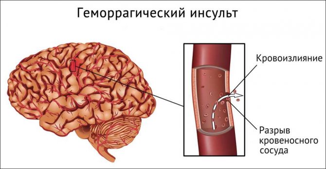
Hemorrhagic stroke is a type of acute cerebrovascular accident, which is based on bleeding from the vessels supplying the brain, the outcome of which is hemorrhage into the brain itself or into the spaces between its membranes.
Among all strokes, hemorrhagic ones account for approximately 20.2%.
What is hemorrhagic stroke
A hemorrhagic stroke is a sudden disruption of blood circulation in the brain, in which a hematoma forms or the nerve tissue becomes saturated with blood.
As a result of a number of reasons, the walls of the vessels that provide nutrition to the brain are disrupted, and either a rupture occurs or the permeability of the vessel increases. Thus, blood freely “spills” into the area of the human brain, compresses it, and depending on the location of the hematoma, many vital functions of the body are disrupted in the patient. This pathology in its development goes through the following stages:
- The appearance of pathological permeability of the vessel or complete rupture of its wall.
- Blood entering directly into the brain cavity.
- Formation of a blood clot (hematoma) or soaking of part of the brain matter with spilled blood.
- Partial or complete destruction of adjacent brain cells.
- Compression of nearby nerve centers in the brain.
- Development of edema of the affected brain.
The patient's entire body is plunged into a terrible catastrophe. Mortality in the first month, with this development of the situation, reaches 80%. Over the next year, another 60 to 80% die, and of those who survive, more than half become disabled. The severity of the disease will directly depend on the size of the hemorrhage and the speed of emergency medical care.
Clinical manifestations
Hemorrhagic stroke develops acutely, usually during the day with maximum human activity. Clinical signs are divided into several groups, depending on the stage of development of the disease.
Harbingers of defeat
In 30-40% of patients, symptoms preceding a stroke appear within minutes, hours and days:
- constant or paroxysmal headache;
- causeless weakness and fatigue during mental stress;
- nausea turning into vomiting;
- the appearance of areas of skin with increased or decreased sensitivity, the development of paresthesia is possible - unpleasant sensations in the form of tingling, “crawling”, etc.;
- increased sensitivity to loud sounds and bright lights;
- unilateral muscle weakness;
- changes in facial features associated with changes in the tone of facial and chewing muscles;
- loss of certain areas of the visual fields of a transient nature.
If these signs are detected, you should immediately seek medical help. Comprehensive diagnostics allows you to timely identify the source of impaired blood circulation and prescribe treatment.

Signs of a stroke
The height of the disease
The development of hemorrhage in the cranial cavity during a hemorrhagic stroke is accompanied by a detailed clinical picture:
- disturbances of the patient's consciousness - stupor, accompanied by general lethargy, or coma;
- convulsive attacks affecting a separate muscle group or having a generalized unilateral nature;
- the symmetry of the face is disturbed, it is distorted, and if the stroke affected the right side of the brain, then muscle tone disorders will be on the left side, and vice versa; the patient has a drooping corner of the mouth and upper eyelid on the affected side; when trying to inflate the cheeks, air escapes due to incomplete closure of the lips;
- the eyeballs are turned towards the lesion or their chaotic movement is observed;
- the pupil on the affected side is dilated;
- paresis or paralysis are observed, in the first case, muscle strength in certain muscle groups is reduced, paralysis is characterized by its complete absence, and an increase or decrease in reflexes in the arms or legs is also observed.
Reasons why a cerebral hemorrhage may develop
Considering that hemorrhagic stroke is a long-known and very common disease, it has been very well studied. All the mechanisms of development of this disease are clear, but it is almost impossible to prevent and carry out preventive diagnostics.
This disease usually affects people after they reach 40–50 years of age. Often we simply do not pay attention to the first alarm bells that arise. People are not even aware of the presence of problems that can lead to such dire consequences as a stroke.
Neurologists identify the following reasons why cerebral hemorrhage may occur:
- persistent very high blood pressure (arterial hypertension),
- the presence of arteriovenous malformations and vascular aneurysms.
So, the first and most common cause of brain damage by hemorrhagic stroke is high blood pressure. It is under the influence of high pressure that the walls of blood vessels undergo flattening and impregnation with proteins; in some cases, even the appearance of necrotic areas of the inner wall of blood vessels can be observed. Over time, the affected vessels become very brittle and highly permeable. It is in these areas that blood can penetrate into the human brain, and a hematoma occurs at the site of this penetration.
Another option may be the rupture of a fragile and thinned vessel under the influence of a sharp increase in pressure, which is commonly called a hypertensive crisis. Such ruptures of arteries or arterioles most often occur in older people.
In young adults, hemorrhagic stroke is usually associated with the presence of aneurysms or vascular malformations.
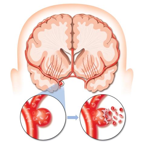
An aneurysm is a congenital or acquired pathology of a vessel, in which there is an increase in its lumen by more than 2 times. This occurs as a result of its stretching or thinning.
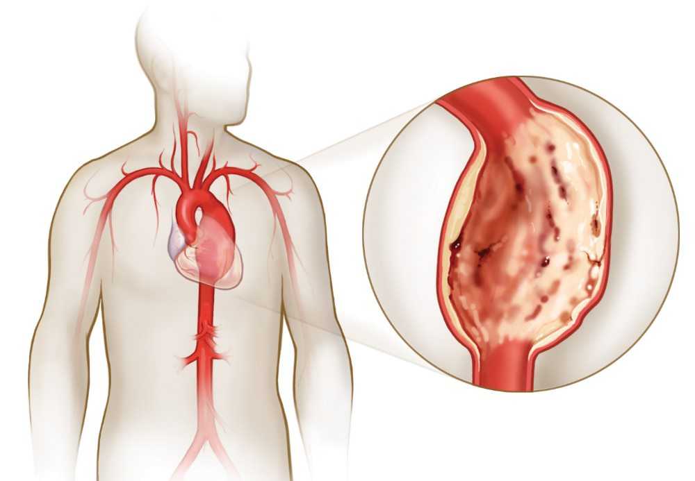
Abdominal aortic aneurysm
Vascular malformations - this congenital pathology includes the formation of vascular tangles and various weaves, up to direct connections of arteries and veins, where blood is discharged directly.
If high pressure always makes itself felt, and it is simply impossible not to notice it, then the presence of aneurysms or malformations can only be detected by examining the vessels, which most often happens by chance. It is this factor that is absolutely asymptomatic, and patients learn about the presence of pathology only after a stroke. This problem most acutely concerns children and adolescents; this factor can leave a young person disabled for life.
Other rarer causes leading to hemorrhages sometimes include diseases such as:
- changes in vascular walls as a result of diseases such as vasculitis and amyloid angiopathy,
- diseases that affect blood clotting, such as thrombocytopenia or hemophilia,
- cirrhosis of the liver,
- long-term use of certain medications that reduce blood clotting and viscosity.
It is worth noting that the second group of factors refers to the causes of secondary hemorrhage, and this will not apply to a classic stroke.
In addition to the reasons that relate to disorders in the vascular and circulatory system, one can also note factors that predispose to the occurrence of the above reasons. These factors include:
- bad habits – excessive alcohol consumption, drug addiction, smoking,
- violation of nutritional standards, leading to obesity and abnormal lipid metabolism,
- age – after 50 years,
- hereditary factor
- diabetes.
Quite a lot of attention in this aspect should be paid to the presence of atherosclerotic changes in the vessels. A more common result of vasoconstriction due to the growth of lipid plaques is ischemic stroke or cerebral infarction. However, with such forms of damage to the walls of blood vessels, they become thinner and ulcerated, which, in turn, can also result in rupture and hemorrhage.
Reasons for development
The causes of hemorrhagic stroke are in one way or another related to disturbances in the condition or functioning of blood vessels. First of all, the following causes of cerebral hemorrhage should be noted:
- High blood pressure, especially its long-term chronic form. Constant impact on the vessels has a traumatic effect, thins them, makes the walls fragile and leads to rupture.
- Atherosclerotic disorders. A lipid plaque forms inside the vessel, partially blocking the blood flow. The pressure on the area of the vessel before it is constantly growing, its walls are stretched and thinned. With significant overload, for example, stress, heavy lifting, physical strain, drinking alcohol or smoking, the vessel may burst.
- Pathological processes in blood vessels due to chronic inflammatory processes caused by vasculitis or a systemic disease such as lupus erythematosus, constant intoxication due to work in hazardous industries, lack of vitamin C or other vitamins in the body.
- Aneurysms – congenital or acquired. These are expansions in vessels with very thin walls that can rupture at any time.
- Arteriovenous malformations are a congenital disorder of the connection of arteries with veins bypassing the capillary network. The pressure in the veins constantly increases, causing their walls to stretch. When overloaded, a rupture occurs.
- Amyloid angiopathy. This is the deposition on the walls of a vessel of a special type of protein, which causes hardening of the vascular walls and provokes their fragility.
- Blood diseases.
- Taking too many anticoagulants leads to excessive blood thinning.
- Drugs that increase blood thickness - narcotic drugs, contraceptives.
- Damage to the vessels that fed the tumor. They are particularly prone to minor damage.
Additional factors can trigger a stroke: severe stress, physical overload, overheating, taking large doses of strong alcohol, smoking too many cigarettes in a short period of time, hypertensive crisis.
Another important reason for hemorrhagic stroke is a tendency to such a disease. It can be hereditary if the patient has relatives with a similar pathology; age over 60 years and being male are also risk factors.
Classification of the main types of hemorrhagic strokes
In medical practice, all strokes of the hemorrhagic type are usually divided according to the location of the hemorrhage and structural changes in the brain. The following classification applies to them:
- Parenchymal hemorrhage is the most common type of hemorrhoidal stroke. With it, blood flows directly into the brain. With this option, two different types of disease development are distinguished: hematoma formation or hemorrhagic impregnation.
Hematoma – Formation of a blood clot in the space between the membrane and the brain itself. Complete cell death occurs in this zone. With this option, you can clearly observe a characteristic clinical picture with all the symptoms, and, unfortunately, a fatal outcome is very likely.
Hemorrhagic impregnation - characterized by blood saturation of the nervous tissue of the brain. With this option, neurons do not die en masse and do not entail such consequences as the formation of a hematoma. Most often, such hemorrhage occurs as a result of improper treatment with anticoagulants.
- Subarachnoid hemorrhage occurs due to the effusion of blood under the soft membrane, which consists of a huge number of vessels covering the brain. In this case, they most often speak of a ruptured aneurysm or malformation.
- A ventricular stroke is usually the result of rupture of the choroid plexuses entering their cavity. This usually happens as a consequence of already formed hematomas of the cerebral hemispheres, which block the outflow of intracerebral fluid. In this case, cerebral edema increases sharply, and the patient has practically no chance of survival. Death occurs within the first two days.
- Sub- and epidural (non-traumatic).
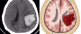
Brain tomography for stroke
Based on the location of the hemorrhagic stroke that occurs, the following strokes can be distinguished:
- lobar - affects only one lobe of the brain,
- cerebellar stroke,
- deep stroke - the deep parts of the brain, subcortical nuclei or internal capsule are affected,
- hemorrhagic stroke in the brain stem, this is the central part of the brain responsible for the processes of breathing and heartbeat.
Therapeutic measures
A patient delivered to a medical facility undergoes an urgent medical examination to clarify the location of the outbreak and the possible cause of the development of the pathology, after which the patient is placed in the intensive care unit or intensive care unit.
Depending on the type of blood accumulation, there are 2 ways to eliminate the focus:
- Operational. Necessary for large (more than 30 ml) hematomas to prevent compression and tissue necrosis.
- Conservative. Used when the brain structure is soaked in blood, small hematomas, or after surgery.
For cerebral hemorrhage, conservative treatment consists of several stages:
- Elimination of cerebral edema. To reduce intracranial pressure, diuretics (Furosemide) and corticosteroids (Dexamethasone, Prednisolone) are used.
- Stopping bleeding. Hemostatic medications are used to prevent possible intracerebral bleeding, using Dicinon, Rutin or Troxevasin. At the same time, blood clotting tests are regularly taken. This is necessary to reduce the risk of recurrent stroke due to increased blood clots.
- Pressure stabilization. One of the reasons why hemorrhagic stroke occurs is frequent increases in blood pressure. Individual selection of antihypertensive drugs will help reduce the risk of complications.
- Providing peace. In the first days, extraneous irritants aggravate brain damage and, by placing the patient in the intensive care unit or intensive care unit, the person is provided with maximum peace. To relieve agitation, mild tranquilizers (Elenium, Phenazepam) are prescribed.
The conservative treatment algorithm is the same for small hemorrhages and for restorative therapy after neurosurgical removal of the hematoma.
Stages of development of cerebral hemorrhage
Hemorrhagic stroke of any origin and severity always goes through the following stages:
- acute period,
- recovery period,
- period of residual effects.
In the first period, cerebral symptoms caused by increased intracranial pressure and emerging and developing bleeding are pronounced. Blood accumulates, resulting in damage to nerve brain tissue. Quite often necrotic areas appear in these places. This period lasts approximately 2 – 4 weeks. With the most negative course of the disease, cerebral edema and ultimately death can develop. It is during this period that most people who are diagnosed with a major stroke die.
In the second period, blood is gradually removed from the brain matter, restoration processes and the proliferation of neurological cells begin. The length of this period will depend on the severity of the lesion and may last several months.
The third period is the period of so-called residual manifestations of the disease. It lasts for the rest of your life. With high-quality treatment and good rehabilitation, it is possible to restore all body functions and sometimes even performance.
Diagnostic methods
If a stroke is suspected, a doctor is immediately called to the patient. For a professional, diagnosing this pathology is usually not difficult, since all the symptoms have a clearly defined picture.
After the patient is admitted to the hospital, he is examined by a neuropathologist. To confirm the diagnosis and to determine the extent of brain damage, the size and location of damaged areas, the following diagnostic methods are usually used - computed tomography (CT), MRI - magnetic resonance imaging and examination of cerebrospinal fluid through lumbar puncture. Sometimes angiography is necessary. The introduction of a contrast agent into the vessels helps determine the presence of an anomaly and the nature of the blood flow.

At the stage of clinical examination, the question often arises as to whether a hemorrhagic or ischemic stroke occurred to the patient, especially if he is in serious condition and cannot answer a number of clarifying questions. It is in such cases that accurate diagnostic methods are especially important.
Symptoms
Clinical symptoms of the disease, their severity and duration depend on the location of the lesion and its volume. The main signs of cerebral hemorrhage are:
- severe headaches;
- vomit;
- impaired coordination of movements (walking, standing, sitting);
- facial redness;
- disturbance of consciousness (stupor, stupor, coma);
- paresis and paralysis - impaired movement of the limbs on one half of the body, since they are constantly in a half-bent position and it is impossible to straighten them;
- speech disorder;
- convulsive syndrome;
- decreased muscle tone;
- mental disorders and irritability;
- visual impairment up to complete blindness;
- facial distortion;
- weakened breathing;
- hemiplegia, hemiparesis, hemihypesthesia;
- numbness of the skin of the limbs, face;
- vegetative state (lack of response to external stimuli and signs of brain activity in the presence of pulse and breathing).
Treatment of hemorrhage
Before the ambulance team arrives, the patient must be provided with emergency first aid. It will include several mandatory steps:
- lay the patient down and place a pillow under his head and shoulders,
- open the window and provide fresh air,
- loosen all tight clothing,
- measure blood pressure,
- the patient’s head must be turned to the side in order to avoid vomit from entering the respiratory tract,
- in case of loss of consciousness, try to prevent the tongue from retracting.

First aid for stroke
After the ambulance arrives, the patient is provided with prehospital care:
- medications are administered to normalize blood pressure,
- medications are used to prevent cerebral edema,
- if necessary, assistance is provided in maintaining breathing and heart function,
- agents are administered to stop bleeding.
Treatment of stroke patients can only occur in a hospital setting in a specialized neurological department. At the first stage of the disease, a person is placed in intensive care, where intensive therapy is started and additional examination is carried out. After passing the acute stage of the disease, the patient is transferred to the general ward of a specialized department.
Treatment of hemorrhagic stroke after admission to hospital can be either surgical or conservative.
Conservative treatment will include the use of various drugs from different pharmacological groups. This will include drugs such as blood pressure reducers, antipyretics, to prevent cerebral edema and a number of other medications.
Surgical intervention is also not uncommon in the treatment of hemorrhagic stroke. It is usually used in the first three days. Indications for urgent surgery would be the formation of large hemispheric hematomas, effusion of blood into the ventricles of the brain and rupture of an aneurysm. The purpose of such a surgical intervention will be to remove blood and reduce pressure on the brain substance itself, which leads to the death of nerve cells. This manipulation can significantly improve the prognosis for saving the patient’s life.
First aid
The main goal of first aid for a stroke is to maintain breathing, heartbeat and urgent hospitalization. When providing first aid for a stroke, you must do the following:
- If there is no pulse in the peripheral arteries, heartbeat and breathing, chest compressions and artificial respiration should be performed.
- If there are convulsions, it is necessary to lay the victim on his side and place something soft under his head.
- Measure and adjust blood pressure.
- Apply an ice pack to your head: this will help constrict blood vessels and stop bleeding.
What is the prognosis for hemorrhagic stroke?
The consequences of a hemorrhagic stroke are much more severe than the consequences of an ischemic stroke. Unfortunately, the prognosis of a hemorrhagic stroke is not very comforting, especially if there was a coma at the beginning of the attack. With this disease, it is very rare for anyone to recover completely. Both the patient himself and his relatives face a very long road of rehabilitation, the goal of which will be to restore the ability to care for oneself and, with a good outcome, to partially restore working capacity.
Rehabilitation measures in this situation will be as follows:
- massage,
- physiotherapy,
- physiotherapy.
To achieve a good result, the positive attitude of the patient himself and his desire to restore his physical capabilities are very important. Sometimes, if there are psychological problems, psychotherapists are involved in working with the patient.
The first signs and symptoms of a recurrent stroke
Expert opinion Alena Ariko Expert in the field of cardiology
The peculiarity of repeated brain damage is that most often the focus is located near the primary one. However, this pattern is not observed in all patients, and there is no guarantee that the relapse will be identical in form - after the ischemic one there may be a hemorrhagic variant and vice versa.
Initial manifestations may be similar to the first attack or be supplemented by new symptoms. Acute cerebral ischemia is characterized by:
- severe weakness in the arm and leg, muscles of half the face;
- visual acuity impairment;
- difficulty speaking and understanding words;
- dizziness;
- loss of skin sensitivity;
- loss of the ability to maintain body balance.

Hemorrhage in the brain usually appears against the background of a hypertensive crisis. Accompanied by severe headache with nausea and vomiting, convulsions. The same signs may also occur with extensive cerebral infarction, damage to the brainstem or cerebellum.
With a repeated hemorrhagic stroke, the patient often falls into a coma from the first minutes of its development. Rapidly progressing cerebral edema leads to wedging of the brain table into the foramen magnum, which threatens the patient’s life if emergency measures are not taken.