A frequent type of cardiac muscle contraction rhythm disorder, in which the localization of the arrhythmogenic focus is in the atrial myocardium, is called atrial tachycardia. It is also called supraventricular tachycardia. Such arrhythmia can occur in people with and without pathologies of the cardiovascular system. If an attack of rapid heartbeat does not last long, then it can be left untreated, but if it lasts for a long time, you need to go to the hospital for help, since this condition leads to exhaustion of the heart muscle.
Types of atrial tachycardia
The following types are distinguished:
Enter your pressure
Move the sliders
120
on
80
- Monofocal. It is characterized by an accelerated heartbeat (from 100 to 250 beats per minute), but the rhythm is regular.
- Multifocal. The rhythm is irregular, the heartbeat is slower, P waves in 3 forms.
Classification according to the location of the pulse generation area
| View | Localization |
| Sinoatrial reciprocal | Impulses are created in the sinoatrial region |
| Reciprocal | In the myocardium in the atria |
| Ectopic | One or more sections of automatic pulse generation |
Classification according to the mechanism of occurrence of the pathological impulse
| View | Mechanism of occurrence |
| Reciprocal | Occurs as a result of heart disease. Also with the wrong choice of drugs and procedures for treatment. Heart rate from 90 to 120 beats per minute. |
| Automatic | Occurs in young people. Occurs after physical stress. |
| Trigger | Occurs in older people. Occurs as a result of taking cardiac glycosides and after physical overexertion. |
| Polytopic | Appears after serious pulmonary disease and heart failure. |
Classification according to the nature of the flow
| View | Peculiarities |
| Paroxysmal atrial tachycardia | Characterized by paroxysmal course. It appears suddenly and ends just as suddenly. Attacks can vary in duration. In this case, a regular rhythm is noted. |
| Non-paroxysmal tachycardia | Rarely seen. It appears in two forms. The first form is characterized by a long course, and the second - by a continuously relapsing course. |
Diagnostics
To diagnose atrial tachycardia, both laboratory and instrumental methods are used. They do a laboratory blood test; it is important to know the concentration of red blood cells and hemoglobin. This is necessary in order to exclude diseases such as leukemia, anemia, etc. It is also important to do a thyroid hormone test and a urine test. A urine test determines the breakdown products of adrenaline.
Instrumental methods include an electrocardiogram.
With its help, the work of the heart and the type of tachycardia are determined. They can also use a Holter ECG - this is a study in which the work of the heart is recorded over a long period of time (24, 48 days, more if necessary).
ECHO-CG is important and more informative. This study evaluates the work of the myocardium as a whole and the functionality of the heart valves.
Also, using ECHO-CG and ultrasound of the heart, it is possible to diagnose heart defects and other chronic diseases that can provoke atrial tachycardia.
Causes of pathology
Excess weight is the cause of many diseases.
myocardial inflammation;- excess weight;
- high blood pressure;
- circulatory disorders;
- development of heart failure;
- poor metabolism;
- presence of heart disease;
- endocrine diseases;
- operations;
- chronic lung diseases;
- intoxication syndrome;
- physical stress;
- side effects when taking medications;
- drinking large amounts of alcoholic beverages;
- drug use.
Causes of the disease
The most common causes of atrial tachycardia include:
- heart failure;
- myocarditis;
- persistent increase in blood pressure;
- congenital and acquired heart defects.
In addition to the listed reasons that provoke the development of atrial tachycardia, you should also know the risk factors that can lead to disturbances in the functioning of the atria. Risk factors include metabolic disorders, damage to the thyroid gland, the presence of extra pounds, damage to the lungs and bronchi (especially with a chronic course), and excessively increased activity of the adrenal glands. Atrial tachycardia can also be provoked by the presence of surgical intervention on the heart, since it significantly weakens the heart muscle.
In some cases, attacks of right-sided tachycardia can be triggered by taking certain medications. For example, glycoside drugs and antiarrhythmic drugs. This is also most common in older people who are taking medications to eliminate manifestations of arrhythmia (for example, Novocainamide).
Diagnostic features
- When the first signs of an attack appear, be sure to contact a therapist and cardiologist. They will conduct an examination, differential diagnosis and make a diagnosis.
- General blood analysis.
- General urine analysis.
- Blood chemistry.
- Electrocardiography using the Holter method.
- EchoCG.
- Ultrasound of the heart.
- Hormone analysis.
Differential diagnosis
Atrial tachycardia on the ECG is characterized by the following signs:
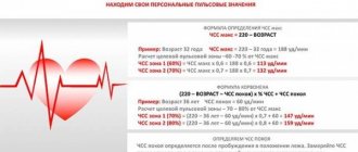
- The correct rhythm is noted and the heart rate ranges from 150 to 250 beats per minute.
- The appearance of a gradual increase in rhythm frequency and the absence of PQ.
- Uneven duration of PP intervals.
- The P wave is negative or level with the T wave.
Before prescribing treatment, a diagnosis is made between atrial flutter and atrial tachycardia. If the heart rate in adults is more than 220 beats per minute and in children more than 250 beats per minute, this confirms the diagnosis of Atrial Flutter. When an isoelectric line is recorded between the P waves in II, III and aVF, atrial tachycardia is diagnosed.
It is imperative to carry out a differential diagnosis of atrial tachycardia from sinus tachycardia and sinus-atrial paroxysmal tachycardia. In the sinus form, the heart rate reaches 160 beats per minute. It also has a gradual development and passes in the same way. With sinus-atrial paroxysmal tachycardia, the ECG shows a normal P wave configuration, a milder course, which can be stopped with the help of vagal tests and the use of antiarrhythmic drugs.
Symptoms
The symptoms of atrial tachycardia completely coincide with arrhythmia. Namely this:
- weakness, malaise;
- dizziness, darkening of the eyes;
- shortness of breath, lack of air;
- pain in the heart area;
- increased heart rate (in attacks) from 140 to 250 beats per minute;
- Often with atrial tachycardia, people feel a feeling of anxiety and fear.
Often such tachycardia does not manifest itself at all.
Treatment of pathology
Treatment must be prescribed by a doctor.
If the patient has manifestations of atrial tachycardia, you need to contact a specialist. They will collect all complaints upon admission, conduct diagnostics and prescribe treatment. If an attack of atrial tachycardia occurs, doctors prescribe the use of vagal tests, drug treatment, and also give dietary recommendations. In rare cases, to stop an attack, if the use of vagal tests and drug therapy does not help, electrical stimulation is used.
Drug treatment
If a patient experiences an attack of rapid heartbeat, the following medications are prescribed:
- Beta blockers (“Propranolol”, “Metoprolol”).
- Calcium channel blockers (Verapamil).
- Endogenous antiari).
- Cardiac glycosides (Digoxin).
Clinical picture of the disease
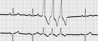
The formation of an ectoscopic focus in the supraventricular zone provokes an increase in the number of nerve impulses sent to the heart, which cause its additional contraction. An increase in the frequency of heart flutter significantly increases the wear and tear of the myocardium, causing its weakness. At the same time, the foci of the formation of such impulses additionally stimulate the heart muscle due to their formation in the atria.
In older age, the more acquired diseases a person has, the greater the likelihood of such pathological sources of impulses appearing. Multiple areas of excitation force large areas of the atria to react, which becomes noticeable when performing an electrocardiogram of the heart: on the resulting cardiogram they are shown in the form of P waves, which are separated by an isoline. Atrial tachycardia on an ECG is most easily diagnosed, which makes this method of identifying diseases of the cardiac system the most convenient and informative.
The development of the considered pathology of the heart most often occurs with damage to the right atrium, less often - to the left. The risk zone should be considered elderly people with existing abnormalities in the functioning of the heart, its severe weakness. Therefore, after reaching the age of 55, you should be more attentive to the state of your own cardiac system.
Atrial tachycardia: mechanism and causes, diagnosis, treatment, prognosis
© Sazykina Oksana Yurievna, cardiologist, especially for SosudInfo.ru (about the authors)
Anyone can experience a fast heartbeat called tachycardia.
But not every person knows when tachycardia does not pose a threat to life and health, and when one should immediately seek medical help if tachycardia is one of the dangerous types of heart rhythm disorders.
This is especially true for atrial tachycardia, since in the absence of timely examination of the patient, time may be lost in treating the underlying disease that led to the occurrence of this type of tachycardia.
However, atrial tachycardia in itself is not a severe disorder causing hemodynamic disorders and is not a life-threatening condition.
What happens with atrial tachycardia?
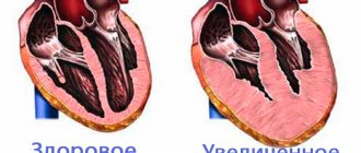
normal heart contraction
So, atrial tachycardia appears when an additional focus appears in the atrial tissue (right or left), in which conditions are created for the circulation of the impulse, or for the formation of a mechanism for the re-entry of the excitation wave.
In another way, this mechanism is called re-entry. The following happens.
If there is any block to conduct electrical excitation along the fibers of the atria, the electrical signals are forced to return back and then move again towards the existing block.
Also, such a mechanism can form if an extrasystole occurs in the atrial tissue, which has a critical coupling interval, due to which conduction along the atrium fibers is prolonged. In this case, the impulse is again forced to return back, and then again towards the atrioventricular node - a re-entry of the excitation wave is formed.
With each such repeated impulse, the atrium tissue contracts synchronously in the correct rhythm, but much more often than normal. In addition to the atrial one, such a mechanism can also be provoked by a ventricular extrasystole if it “managed” to return from the ventricles to the atria through the atrioventricular node.
It is precisely because the impulse moves in the opposite direction that atrial tachycardias are called reciprocal.
contraction of the heart along the normal conduction path (left) and the development of tachycardia due to the occurrence of a re-entry loop (right)
Classification of atrial tachycardias
Atrial tachycardia refers to supraventricular tachycardia, which also includes tachycardia from the atrioventricular junction. The only difference is in the location of the re-entry loop (atrium or AV node, respectively), as well as in the ECG signs. In turn, atrial tachycardia is divided into the following options:
- By localization - from the right and left atria, as well as from their upper or lower parts - ectopic atrial tachycardia,
- According to the form – mono- and polyfocal (from one or several parts of the atria at the same time),
- According to the nature of the course - paroxysmal (paroxysmal) and non-paroxysmal (with a long or continuously recurrent course).
Causes of atrial tachycardia
Such rhythm disturbances can occur in young people without any serious pathology after physical overload.
More often, people with vegetative-vascular dystonia, especially the hypertensive type, are susceptible to such paroxysms that occur spontaneously and disappear without treatment.
They experience unstable atrial tachycardia (in 3-6% of healthy individuals during 24-hour blood pressure and ECG monitoring).
In older people, especially in elderly patients, atrial tachycardia can be caused by organic damage to the heart muscle, lung disease, as well as general disorders in the body.
Of the heart diseases that can provoke this disturbance of heart rhythm, inflammatory diseases (myocarditis), ischemic and post-infarction changes (ischemic heart disease, previous infarction), changes in the architectonics of the heart due to defects (usually stenosis or insufficiency of the mitral valve), with cardiomyopathies and with hypertrophy of the left ventricle due to hypertension. These diseases lead to the replacement of normal atrial tissue with scar or hypertrophied tissue, resulting in electrically inert foci that are incapable of conducting impulses along the path of signals.
In addition to cardiac pathology, long-term chronic diseases of the bronchopulmonary system - obstructive bronchitis, bronchial asthma, prolonged recurrent pneumonia, bronchiectasis and emphysema can lead to the occurrence of atrial tachycardia. In these diseases, cor pulmonale is formed, characterized by hypertrophy of the right atrium, in which an ectopic rhythm often occurs.
Separately, it should be noted general disorders in the body, such as fever, intoxication (alcohol and its surrogates, drugs), traumatic brain injuries, pathology of the thyroid gland (hyperthyroidism and thyrotoxicosis with the formation of dyshormonal cardiomyopathy and thyrotoxic heart), as well as intoxication in malignant tumors with polychemotherapy (PCT).
How does atrial tachycardia manifest?
The clinical manifestations of atrial tachycardia are determined by the nature of the course of this rhythm disorder, as well as the type of the underlying causative disease.
Paroxysms of atrial tachycardia are characterized by a sudden, sharp onset of rapid heartbeat, which is accompanied by a pulse rate of 140 to 250 beats per minute.
As a rule, paroxysm of atrial tachycardia does not cause a significant aggravation of the patient’s general well-being and condition, as happens with atrial fibrillation or flutter.
However, the patient has a certain discomfort in the chest, accompanied by a feeling of lack of air, sticky cold sweat, and general weakness.
Atrial tachycardia with a long or continuously recurrent course is accompanied by a not so high heart rate, and episodes of rapid heartbeat alternate with a normal pulse rate. In general, these types of tachycardia are well tolerated by the patient, since he is already adapted to this heart rate.
During the interictal period, the patient may be bothered by complaints caused by the underlying disease, which is characterized by chest pain, shortness of breath caused by physical exertion, as well as swelling and other signs.
Diagnosis and differential diagnosis
The diagnosis of atrial tachycardia is established on the basis of an electrocardiogram and its modifications (24-hour ECG monitoring, exercise ECG, TEE - transesophageal electrophysiological study).
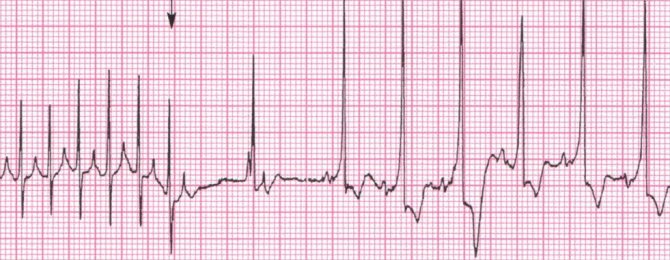
The main sign of atrial tachycardia on the ECG is a high heart rate of more than 140 beats per minute, as well as the presence of biphasic or negative P waves before or after each ventricular complex.
Often on the ECG you can see how the normal sinus rhythm is interrupted by runs of atrial tachycardia, again returning to normal heart rate - this is the so-called recurrent, or salvo, atrial tachycardia.
In addition to an ECG, it is necessary to conduct an ultrasound of the heart (echocardioscopy), and, if necessary, coronary angiography, fluoroscopy of the chest cavity, pulmonary function testing and other research methods to clarify the nature of the causative disease.
In terms of differential diagnosis, the physician describing the ECG should be aware of the following cardiac arrhythmias that may be similar to atrial tachycardia:
- Sinus tachycardia - characterized by a high, but lower than with atrial tachycardia, heart rate (100-120 per minute).
- Reciprocal tachycardia from the AV junction, characterized by biphasic P waves following only the QRST complex.
- Atrial fibrillation - with atrial fibrillation, the contraction frequency can be less than normal, normal and above normal (brady-, normo- and tachysystolic variants, respectively), but the ECG shows different RR intervals (between adjacent ventricular complexes, as well as the absence of P waves before each complex is a pathognomonic sign of non-sinus rhythm). Atrial flutter, in turn, is distinguished by a higher heart rate (more than 250 per minute, the CC intervals can be almost the same, as with the rhythmic form of atrial flutter, for example).
Tachycardia
Tachycardia
– one of the most common heart rhythm disorders, manifested by an increase in heart rate (HR) of more than 90 beats per minute.
When tachycardia appears, a person experiences a feeling of palpitations; in some cases, pulsation of the vessels of the neck, anxiety, dizziness, and rarely fainting are possible.
In patients with cardiovascular pathology, this arrhythmia can worsen the prognosis of life and provoke the development of complications such as heart failure.
The main mechanism for the development of tachycardia is to increase the automatism of the sinus node, which normally sets the correct rhythm of the heart.
If a person feels his heartbeat becoming faster and stronger, this is not always evidence of the existence of problems.
In practically healthy people, tachycardia can be caused by the action of physiological compensatory mechanisms in response to the release of adrenaline into the blood and activation of the sympathetic nervous system, which cause an increase in heart rate, which is a response to one or another external factor. The cessation of the action of the latter leads to a gradual return of the heart rate to normal.
In healthy people, tachycardia occurs
:
- as a result of stressful situations, physical activity and emotional arousal;
- when the air temperature rises;
- when consuming certain medications, strong tea, coffee or alcohol;
- from a sudden change in body position, etc.
In preschool children, tachycardia is considered a physiological norm.
At the same time, the course of certain pathological conditions is often accompanied by tachycardia.
Causes
Cases of sinus tachycardia occur in all age groups, both healthy people and patients with certain diseases. Its occurrence is facilitated by intracardial or extracardiac etiological factors (cardiac or extracardiac, respectively).
In patients with cardiovascular diseases, sinus tachycardia can be a manifestation of any heart pathology: coronary heart disease, arterial hypertension, myocardial infarction, acute and chronic heart failure, rheumatic and congenital heart defects, myocarditis, cardiomyopathies, cardiosclerosis, infective endocarditis, exudative and adhesive pericarditis.
Physiological extracardiac factors contributing to the development of tachycardia include emotional stress and physical activity.
The majority of extracardiac arrhythmias are neurogenic tachycardias, which are associated with primary dysfunction of the cortex and subcortical nodes of the brain, and disorders of the autonomic nervous system: affective psychoses, neuroses, neurocirculatory dystonia. Young people with lability of the nervous system are characterized by the greatest susceptibility to them.
Other factors of extracardial tachycardia are represented by endocrine disorders (thyrotoxicosis, increased production of adrenaline in pheochromocytoma), anemia, acute vascular insufficiency (shock, collapse, acute blood loss, fainting), hypoxemia, acute painful attacks.
Tachycardia may appear as a result of fever that develops in the context of various infectious and inflammatory diseases (pneumonia, tonsillitis, tuberculosis, sepsis, focal infection). For every 1 °C increase in body temperature, there is an increase in heart rate by 10-15 beats/min. in children and 8-9 beats/min. in adults (compared to normal).
The occurrence of pharmacological (drug) and toxic sinus tachycardia is caused by the influence of various medicinal and other chemical substances on the function of the sinus node.
These include sympathomimetics (adrenaline and norepinephrine), vagolytics (atropine), aminophylline, corticosteroids, thyroid-stimulating hormones, diuretics, antihypertensive drugs, caffeine, alcohol, nicotine, poisons, etc.
Certain substances that do not have a direct effect on the function of the sinus node increase the tone of the sympathetic nervous system and cause the so-called reflex tachycardia.
There are adequate and inadequate sinus tachycardia. The latter is characterized by the ability to remain at rest, lack of dependence on exercise and medication.
Such tachycardia may be accompanied by sensations of shortness of breath and palpitations.
Experts suggest that this rare and little-studied disease of unknown origin is related to primary damage to the sinus node.
Symptoms
Clinical symptoms of sinus tachycardia appear depending on how severe and prolonged it is, as well as on the nature of the underlying disease.
Subjective signs of sinus tachycardia may be completely absent; sometimes there may be a feeling of palpitations, a feeling of heaviness or pain in the heart area.
With inadequate sinus tachycardia, persistent palpitations, a feeling of lack of air, shortness of breath, weakness and frequent dizziness are observed. Fatigue, insomnia, worsening mood, decreased appetite and decreased performance may occur.
The severity of subjective symptoms depends on the sensitivity threshold of the nervous system and the underlying disease. In patients with diseases of the cardiovascular system (coronary atherosclerosis, etc.), an increase in heart rate can provoke the occurrence of angina attacks and aggravate decompensation of heart failure.
Forecast
Physiological sinus tachycardia in healthy individuals (including those with pronounced subjective manifestations) has a good prognosis and is not life-threatening.
In patients with heart disease, the prognosis can be very serious, since sinus tachycardia can worsen the course of chronic heart failure.
Prevention
Prevention of sinus tachycardia means early diagnosis and timely treatment of pathology, as well as the elimination of extracardiac factors in the development of arrhythmias.
The serious consequences of tachycardia can be avoided by strictly following the recommendations for maintaining a healthy lifestyle.
Read more about: prevention of cardiac tachycardia.
Where to get advice
Moscow, St. Petersburg, Barnaul, Vladivostok, Volgograd, Voronezh, Izhevsk, Kazan, Nizhny Novgorod, Rostov on Don, Stavropol, Yaroslavl, Blagoveshchensk, Irkutsk, Krasnoyarsk, Novosibirsk, Essentuki, Krasnodar, Tomsk, Ekaterinburg, Omsk, Perm, Ryazan , Samara, Ufa, Khabarovsk, Chelyabinsk, Caucasian mineral waters
Ask a Question
Classification of supraventricular tachycardias
- sinus reciprocal paroxysmal tachycardia
- atrial tachycardia
- reciprocal
- paroxysmal
- chronic: constantly recurrent, continuously relapsing, atrial tachycardia
- focal paroxysmal or chronic atrial tachycardia
- focal, less often reciprocal paroxysmal or chronic atrial tachycardia with anterograde AV block of the second degree, including the digitalis toxic form of Laun-Lvain.
- multifocal or multifocal/chaotic atrial tachycardia, including its pulmonary hypoxic Lipson-Name form:
- prefibrillator form
- post-defibrillation form