Symptoms
As a rule, any disorder associated with a lack of oxygen or nutrients to the brain leads to various pathologies or neurological abnormalities, expressed by corresponding symptoms:
- Headache, dizziness, fainting.
- Disease in the area of the divergence of the artery leads to visual impairment.
- Memory disturbances, sleep disturbances, absent-mindedness, irritation and some other psychological disorders.
- If the area of expansion touches the nerves, then this place begins to hurt.
- Bleeding from the nose and mouth, difficulty inhaling and exhaling, difficulty speaking, all this may indicate that the artery has increased in diameter so that it affects nearby organs.

Headache and fainting can be symptoms of a carotid artery aneurysm.
In addition, symptoms of a carotid artery aneurysm may occur, such as pain along the trigeminal nerve, impaired sensitivity and oculomotor function.
The main symptoms of a carotid aneurysm in the neck are associated with its complications. An aneurysm may not cause any sensation and is discovered by chance during a medical examination or ultrasound of the neck.
Due to repeated detachments of small blood clots, ocular symptoms of a carotid artery aneurysm may develop: blurred vision, double vision, dilated pupils, loss of visual fields.
A sudden and severe headache may be a sign of a ruptured carotid aneurysm, as well as other arteries in the brain. This pain is so severe that most patients describe it as “unbearable and most excruciating pain.” Headache is usually accompanied by nausea and vomiting, tension in the neck muscles, and loss of consciousness and coma often occur.
Vertebral artery diseases
Vertebral artery syndrome worsens a person's well-being
There is the following classification of pathologies of the vertebral artery:
- Occlusive conditions: atherosclerosis, thrombosis, embolism, arteritis, aneurysm.
- Extravasal compression: bone vessel abnormalities, including osteophytes and tumors.
- Congenital or acquired abnormal deformation: pathological tortuosity of the arteries, high entry of the vertebral artery into the bone canal, non-fusion of the arteries into the ostial basilar branch, duplication, hypoplasia, etc.
Each disease has its own causes of development and characteristic symptoms.
Occlusive conditions
Cholesterol deposits in the vertebral artery interfere with blood supply to the brain
Occlusion of the right vertebral artery (or left) is a condition characterized by partial or complete occlusion of the lumen. This is possible against the background of the formation of an atherosclerotic plaque or thrombus in the cavity, as well as against the background of embolism and arteritis.
Atherosclerosis of the vertebral artery is a disease that occurs due to the deposition of cholesterol on the wall. Common causes include metabolic disorders, chronic hypertension, smoking, hormonal imbalance, overweight and obesity, and poor nutrition.
Signs of atherosclerosis of the vertebral arterial branch:
- headache;
- impairment of visual and auditory function;
- noise in ears;
- short-term loss of consciousness;
- at a late stage in elderly people - dementia, memory impairment (due to damage to intracranial branches), impaired coordination of movements.
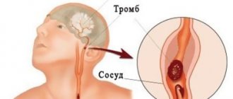
Embolism of the vertebral artery occurs against the background of blockage of the lumen by an embolus - a formation that should not be present in the bloodstream, but after its occurrence spreads with the bloodstream into the corresponding artery. It could be a piece of fat, tissue, an air bubble, or a blood clot that is causing hypoperfusion (reduced blood flow).
Thrombosis is characterized by blockage of the lumen of an artery with a blood clot, which is formed as a result of increased coagulability of the blood fluid. Both thrombosis and embolism are not independent diseases, but symptoms of another primary process:
- atherosclerosis;
- congenital heart disease, infective endocarditis, atrial fibrillation;
- infectious arteritis, hypertension;
- pneumonia;
- blood diseases, etc.
A vertebral artery aneurysm can lead to spinal cord infarction
A blocked artery causes a decrease or cessation of blood flow to the brain. This contributes to the development of acute ischemia (oxygen starvation) of the cerebral region, which causes headaches, death of organ cells, and gangrene.
Arteritis is an inflammatory process in a vessel, which is accompanied by a narrowing of the lumen and impaired blood supply to the brain. Among the main causes of development are smoking and leading a sedentary lifestyle. Characteristic symptoms are pain in the neck and head, impaired visual function, and malaise.
A vertebral artery aneurysm is an expansion of the lumen with dysplasia and thinning of the wall, which increases the risk of vessel rupture and ischemic and hemorrhagic stroke. The causes of the pathology are a congenital defect in the structure of the artery, a history of trauma, hypertension, atherosclerosis. Characteristic symptoms of fusiform (fusiform) and other types of aneurysm are pain in the neck and head, dizziness, and impaired visual function.
Extravasal compressions
With pathologies of the spine, dysfunction of the vertebral artery is possible
Compression of the vertebral artery most often occurs through compression by osteophytes or a tumor.
Osteophytes are bone growths that are localized on the vertebrae in the cervical region, causing compression of the vertebral artery. New growths on the bones are formed due to:
- development of an inflammatory process of infectious or traumatic etiology in the spine;
- osteochondrosis, arthrosis, malignant tumor in bone tissue;
- excessive physical activity;
- hereditary predisposition;
- disruption at the hormonal level;
- compression fracture.
Symptoms of compression of the vertebral artery occur at a late stage of the primary pathological process, when compression of the nerve roots and vessels has occurred:
- headache and dizziness, ringing and noise in the ears;
- limited range of motion in the cervical region and numbness;
- impairment of visual and auditory function;
- nausea;
- impaired coordination of movements;
- increased sweat production;
- hypertension;
- aching discomfort in the neck, muscle spasms, degenerative processes in muscle structures.
Compression of the vertebral artery on the left or right is a symptom of a tumor process in the spine or soft tissues of the cervical spine. Among the provoking factors:
- hereditary predisposition;
- radioactive and chemical effects on the body (both options provoke an acceleration of the pathological division of normal cells);
- smoking and alcohol abuse;
- failure to maintain proper nutrition.
In most cases, compression of two or one artery is caused by a malignant tumor of the exophytic type. In the cervical region, the development of the oncological process is accompanied by neurological symptoms: headache, dizziness, impaired consciousness, pain, numbness and muscle weakness.
Artery deformities
Arterial deformations impair blood patency
Kinking of the vertebral artery is a pathological process in which the branch lengthens and sharply twists, kinks form in it and patency is impaired. There are several types of twisting and bending of the branch (kinking syndrome), taking into account the shape of the artery:
- C-shaped;
- S-shaped (wavy);
- loop-forming.
In most cases, pathological tortuosity occurs as a complication of combined atherosclerosis and arterial hypertension. Characteristic symptoms of tortuosity of the arterial canal are temporary paralysis of the arms, impaired speech and visual function, headache and dizziness, and short-term fainting.
1Aneurysm of the vessels of the neck and lungs
Schematic representation of the carotid artery
When talking about aneurysm of the neck vessels, we are talking about the common carotid artery. This is a large vessel of the neck, which is divided into 2 branches - the external and internal carotid arteries. The disease is quite rare and accounts for only 13% of all aneurysms of the branches of the thoracic aorta.
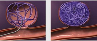
Pulmonary aneurysm is a congenital or acquired disease. It would be more correct to call it an arteriovenous aneurysm, since in this disease a communication is formed between the wall of the artery and vein. This formation takes on the appearance of a chaotically woven vascular glomerulus. Both men and women get sick equally. The disease can be detected in childhood or adulthood.
This disease has a number of synonyms, so it can be found under the following names:
- Arteriovenous fistulas (fistulas).
- Arteriovenous malformations.
- Lung hemangiomas.
- Cavernous angiomas of the lungs.
- Pulmonary telangiectasias.
What is the disease
The carotid arteries are large vascular trunks that supply blood to the neck and all parts of the brain. Some internal and external factors can negatively affect the walls of the artery, damaging them and leading to expansion. The result is a small bulge that resembles a sac, called an aneurysm. The sac contains blood or thrombotic masses. Over time, the vascular wall becomes thinner, which can burst due to changes in blood pressure.
Carotid artery aneurysm is a disease that has pronounced typical symptoms. Often the disorder is organic in nature and reappears even with timely treatment. Against the background of pathology, cerebral circulation may be impaired due to a lack of oxygen and cell nutrition.
Diagnostics
If you have this disease, you should contact a neurologist who will offer to undergo a course of examination to accurately determine the type of disease.
A carotid aneurysm can be determined using the following diagnostics:
- To detect pathologies of the skull, X-rays are taken. It is important to do several from different sides for a complete examination of the head.
- To confirm the disease of the blood vessels, in connection with which the main problem has arisen, you should undergo electroencephalography.
- Magnetic resonance imaging will confirm the violation and indicate the location.
- In addition to MRI, angiography is used to determine the location of the expansion.
- Thanks to these diagnostic methods, an accurate diagnosis is made with further treatment of the patient.
How is an aneurysm diagnosed?
When patients go to a medical facility, the doctor conducts an initial examination and palpation. When pressing on the vessels, pain may be felt, which signals the development of pathology. However, no one will dare to make a diagnosis immediately after the examination; a comprehensive examination is carried out, since the symptoms of an aneurysm are similar to many other diseases.
The patient is prescribed tests and diagnostics using several methods:
- MRI is performed both during the initial examination to make an accurate diagnosis, and before the surgical procedure. Allows you to identify the size and location of the aneurysm, and make a prognosis of development;
- Computed tomography – is highly accurate in examining arteries and vessels;
- X-ray – pictures of the skull are taken from all sides, helping to identify changes in the skull.
Based on the research results, the doctor must make an accurate diagnosis, determining what pathology is developing. This could be an aneurysm of the carotid artery, vessels of the neck, brain, or a completely different disease.
5How to diagnose the disease?
Pulmonary aneurysm
The reasons leading to the development of aneurysm of the neck vessels are divided into 2 main groups:
- Congenital malformations of the vessel wall.
- Purchased:
- Atherosclerosis.
- Injury.
- Previous operations on the carotid artery.
- Infectious lesion of the artery.
Arteriovenous pulmonary aneurysm may have the following causes:
- Congenital - 70% of cases. The disease is based on a defect in the genes responsible for the normal growth and development of the vessel. Such an aneurysm may be a manifestation of Randu-Osler disease.
- Acquired - 30% of cases.
The causes of their occurrence can be the following diseases and conditions:
- Cirrhosis of the liver.
- Metastatic carcinoma.
- Congenital heart defects.
- Injuries.
- Actinomycosis is an infectious disease caused by fungal microorganisms.
Intraoperative angiography
Diagnosis of aneurysm of the neck vessels, in particular the common carotid artery, is carried out using instrumental methods. Although already at the stage of examining the patient, a pulsating formation can be noticed. But at this stage, the doctor can only suspect an aneurysm. It must be confirmed by other instrumental methods, which include:
- Ultrasound duplex scanning. The method combines two research modes, in which it is possible to obtain information about the structure of the vessel wall, the state of its lumen, and also evaluate the impact of these changes on blood flow. Using this method, arteriovenous aneurysms of the common carotid artery are very well diagnosed.
- Angiography. It is an x-ray examination method in which a contrast agent is injected into the vessel. Angiography can be used to assess the condition of the vessel wall in the area of the aneurysm.
CT image of the lungs
Diagnosis of pulmonary arteriovenous aneurysm is based on the following instrumental methods:
- Chest X-ray. The most accessible method to identify the disease. In this case, a shadow of varying sizes appears in the image. Its important feature is its connection with the root of the lung.
- Perfusion scintigraphy. A method to determine the presence of a connection between an artery and a vein. Its essence lies in the fact that a substance connected to a radioactive element is introduced into the vessel. When there is communication between an artery and a vein, the substance moves beyond the lung tissue.
- CT scan. A method that allows you to accurately diagnose an arteriovenous aneurysm.
- Pulmonary angiography is an X-ray method for studying the blood vessels of the lungs using a contrast agent.
- Ultrasound of the heart with contrast. The method is based on the introduction of microparticles (gas bubbles) into a vessel.
Is it possible to prevent aneurysm?
It is impossible to accurately prevent the development of a carotid artery aneurysm, but you can reduce the chances of pathology occurring. The patient should adhere to the following rules:
- Lead a healthy lifestyle;
- To refuse from bad habits;
- Control your diet by eating healthy foods and vitamins;
- Eliminate a sedentary lifestyle;
- Treat infectious and other diseases in a timely manner.
The main preventive measure is to immediately consult a doctor when the first symptoms appear.
With a timely visit to the medical center, the prognosis is favorable, and the chances of recovery increase. Although in any case the result depends on the patient’s age, his state of health and related factors.
Carotid artery aneurysm is a serious pathology that requires immediate treatment. If headaches occur, you should not take painkillers, since they will not solve the original source and the problem. The only way out is to visit a doctor and get proper treatment.
Source: moisosudy.ru
Treatment
In order to cure an arterial aneurysm, operations aimed at restoring blood flow are currently used.
Treatment options for carotid artery aneurysm include three types of surgery:
- Endovascular. There are many complications after this type, so it is used extremely rarely.
- Removal of the pathological part of the dilated vessel and its replacement with a prosthetic one.
- If removal is not possible, then vascular bypass is performed, inserting an additional path for blood flow to the head of the human body.
- High-tech endovascular techniques. Here the same thing happens as in the second case, only with the help of angiosurgery.
Vascular bypass
All methods completely relieve a person from the disease and its characteristic symptoms, freeing up the lumen for blood flow.
One of the most risky operations for human life is surgery on the cardiovascular system.
There are several complications that may occur after or during carotid artery surgery:
- Severe bleeding, in which the patient may die on the spot;
- When sewing on a prosthetic area, the “native” vessel may rupture;
- Formation of a blood clot in the vessel where the foreign fragment was installed;
- A new increase in diameter in the artery.
Disconnection of arteriovenous communication and suturing of the arterial wall and veins
Treatment of aneurysm of the common carotid artery is only surgical. If the disease does not have any symptoms, a wait-and-see approach is chosen. The patient is subject to constant monitoring. If the condition worsens, the issue of performing an operation is decided. Surgical treatment methods include the following:
- Marginal resection of the aneurysm with installation of a synthetic patch. This type of operation is used for arterial aneurysm. During the operation, the dilated section of the artery is isolated and part of the aneurysmal sac is removed. The wall defect is closed with a synthetic patch.
- Disconnection of arteriovenous communication and suturing of the artery wall and veins. This operation is performed in the presence of an arteriovenous aneurysm.
The following types of surgical interventions are used in the treatment of pulmonary aneurysm:
- Removal of a lobe of the lung along with the aneurysm. The operation is performed if there is one isolated formation.
- Endoscopic surgery. A probe is inserted through a large vessel and passed into the cavity of the aneurysm. Then the cavity is filled with synthetic material. Thus, the aneurysm is switched off from the bloodstream.
In the presence of multiple aneurysms, unfortunately, surgical intervention is often impossible. Therefore, in this case, only supportive drug therapy is carried out.
Aneurysm of the carotid artery of the neck and brain (common, ICA, ECA): symptoms and treatment
Carotid artery aneurysms account for 12.5-18% of all arterial aneurysms.
The disease is typical for people over 45 years of age who suffer from cardiovascular pathology and metabolic disorders. The course is long-lasting and slowly progressing. If left untreated, the disease causes acute cerebral ischemia in 34% of patients.
Carotid artery aneurysm - characteristics depending on location
Carotid artery aneurysm (ICD-10 code - I72.0) is a local increase in its diameter by more than 2 times. It is a limited protrusion formed by a stretched vascular wall.
The average age of patients is 47-53 years. Women suffer 2-2.5 times more often than men.
An aneurysm forms secondary to chronic diseases affecting the arterial bed. In the first stage, under the influence of a causative factor, nonspecific inflammation develops in the vessel wall: edema, destruction of endothelial cells and the muscle layer.
In the second stage, enzymes in the lesion lead to the destruction of collagen fibers. The artery wall becomes thinner and stretches. In the third stage, under the influence of a pulse wave, the thinned area is pressed and forms a protrusion.
Common artery
The vessel extends from the aortic arch to the angle of the maxilla and is affected in 27-30% of cases . Causes: atherosclerosis, Takayasu syndrome.
Symptoms are associated with compression of the tissues and organs of the neck, and cerebral ischemia on the affected side. Formation within 2-4 weeks. The prognosis is relatively unfavorable, with frequent relapses.
Surgical treatment is indicated for rapid progression and increasing clinical symptoms.
In the area of the angle of the mandible, the common carotid artery is divided into external and internal branches.
External SA
The ESA does not penetrate the cranial cavity, but supplies blood to the soft tissues of the face, the auricle, and the tissues of the nose. Affected in 12-14% of cases . Causes: Takayasu disease, congenital anomalies.
Symptoms are associated with ischemia of the facial muscles, orbit, nasal cartilage, and external ear. Formation is slow, over 2-4 months. The prognosis is relatively favorable.
Treatment is symptomatic and surgical.
Internal carotid artery (ICA)
An aneurysm of the internal carotid artery of the brain can be in one of the sections of the ICA - the cavernous sinus, supraclinoid section and bifurcation.
- Cavernous sinus . Aneurysms of this location are located below the sella turcica of the skull. The frequency of occurrence is 15-17%. Etiology: hypertension, Takayasu's disease. Formation within 3-4 weeks. Symptoms are associated with ischemia of the facial muscles and trigeminal nerve. The prognosis is relatively favorable, relapses are rare. Treatment is conservative and surgical.
- Supraclinoid region . Located behind the eyeballs. Affected in 17-20% of cases. The causes of aneurysm of the supraclinoid part of the ICA are vascular anomalies, hypertension, and trauma. Formation within 1 month. Manifestations are associated with ischemia of the 2nd, 3rd, 4th and 6th pairs of cranial nerves innervating the eyeballs. The prognosis is unfavorable; irreversible vision loss occurs in 12-15% of patients. Treatment is surgical.
- At the bifurcation site . Damage to this area occurs in 14-15% of cases and is localized in the center of the base of the skull. Causes: vascular abnormalities, genetic syndromes. Formation within 4-6 months. Symptoms: intense headache, decreased or loss of vision. The prognosis is unfavorable. Treatment is surgical.
Causes and risk groups
Causes:
- Atherosclerosis;
- Takayasu's disease;
- Hypertonic disease;
- Congenital vascular anomalies;
- Marfan syndrome;
- Syphilis;
- Tuberculosis;
- Connective tissue diseases;
- Periarteritis nodosa;
- Vasculitis.
Persons at risk are:
- With a family history;
- Over 50 years old;
- Suffering from cardiac ischemia and valve defects;
- Having had a heart attack or stroke;
- People with increased thrombosis.
Classification
Types of current:
- Acute - characterized by a sudden bright clinic, general cerebral symptoms, loss of consciousness. Observed during complications.
- Chronic - perennial, erased, wavy or asymptomatic.
Carotid aneurysms can be:
- Saccular - consist of a neck (through which they are connected to the vessel), a spherical body and a dome. This variety is more susceptible to rupture. The selection operation is clipping. The prognosis is relatively unfavorable.
- Fusiform - smoothly transition into a healthy vascular wall. Delamination is more typical for this variety. The operation of choice is resection. The prognosis is relatively favorable.
- Mixed - show signs of both forms. They are characterized by high vulnerability and rapid delamination. The operation of choice is resection. The prognosis is unfavorable.
Forms of the disease:
- Tumor-like – clinically resembles tumor growth. The course is chronic, the symptoms are associated with compression of anatomical structures and increasing compression syndrome.
- Apoplexy – clinically resembles apoplexy (convulsive syndrome). The course is acute, convulsions and loss of consciousness may be the first and only manifestation.
We wrote about the classification of cerebral aneurysm here.
Possible complications
Complications may include dissection and rupture, thrombosis, stroke, compression of the esophagus, loss of vision or hearing, memory loss, thyroiditis, paralysis of half the body.
Rupture is a common complication of the natural course of the disease . It occurs due to overstretching of the carotid artery, which has undergone irreversible dystrophic changes.
Causes:
- Hypertensive crisis;
- Injuries;
- Detachment of an atherosclerotic plaque.
Risk factors are hypertension, smoking, diabetes, stress, physical overexertion.
Symptoms of a rupture:
- Anxiety;
- Dyspnea;
- Tachycardia;
- Increased pressure;
- Violation of visual, auditory, cognitive functions;
- Paralysis of half the body;
- Lack of response to external stimuli;
- Loss of consciousness.
Treatment in 100% of cases is surgical. The prognosis is unfavorable. Stroke occurs in a third of patients.
Symptoms of the disease
First symptoms:
- Bursting headaches and neck pains.
- Fatigue, tiredness.
- Increased pressure.
Symptoms depending on the location of the lesion:
- Common carotid artery - pain, feeling of a foreign body in the neck, hoarseness, pain when swallowing.
- NSA – loss of facial sensitivity, nosebleeds, pain in facial muscles.
- Cavernous sinus – decreased sensitivity of the tongue, facial skin, pain in the orbit.
- Supraclinoid region – paralysis of the eyeball, myopia.
- Bifurcation - flickering before the eyes, narrowing of the visual field.
Left and right side affected
Right-sided lesion appears:
- Loss of speech functions.
- Epilepsy.
- Impaired sensitivity (numbness, “crawling goosebumps”).
- Visual disorders.
Left-sided lesion is characterized:
- Migraine;
- Cramps;
- Fainting;
- Disorders of consciousness;
- Hypertension;
- Dizziness.
Signs of deterioration
As it progresses, symptoms of cerebral ischemia appear:
- Fainting;
- Decreased hearing, memory, attention;
- Frontal and occipital pain;
- Changes in personality traits;
- When the CCA and ICA are affected - paresis of half the body, hypertension;
- When the neck veins are compressed, they become swollen, swollen, and blue in the upper half of the body;
- When the ECA is affected, there is paralysis of the facial muscles, disturbances in swallowing and chewing, and jaw pain.
The threat of complications is indicated by increased headache, persistent hypertension, loss of consciousness, migraine with aura, convulsions, stupor, stupor, decreased reflexes.
Diagnosis of neck and brain disease
Examination algorithm:
- Survey and inspection . Complaints of bursting pain, fainting, decreased vision and hearing, sensory disturbances; history of vascular pathology. On examination, there is pastiness, facial hyperemia, and swelling of the veins of the neck.
- Objective research . Swelling and enlargement of the neck and thyroid gland, vascular murmur, hypertension.
- Laboratory data . Increased levels of platelets, fibrinogen, cholesterol.
- Angiography of neck vessels . Defect in the contour of the vascular wall, contrast leakage beyond the vessel.
- Ultrasound of neck vessels with duplex scanning . Limited vascular expansion, tortuosity of the branches of the carotid artery, atherosclerotic plaques, blood flow turbulence.
- CT (MRI) . Local edema, atrophy of cranial nerves 2, 3, 4, 6, hemorrhage, cidrocephaly, compression of the esophagus and trachea, foci of calcification.
- EEG . Disappearance of alpha waves, registration of theta and delta ranges.
You can read how to diagnose a cerebral aneurysm in a separate material.
Therapy tactics
Treatment of carotid artery aneurysm is divided into conservative and surgical. Indications for conservative therapy:
- Preparation for surgery;
- No complaints;
- Size up to 1 cm;
- No progression.
Conservative therapy includes:
- Statins, antihypertensive, diuretic drugs;
- Symptomatic drugs (analgesics, NSAIDs);
- Vasodilators and nootropics.
Surgery
Indications for surgery:
- Growth more than 4 mm in 6 months;
- The appearance of the clinic;
- Hypertension;
- Increased thrombus formation;
- History of heart attack or stroke;
- Risk of complications.
Contraindications for surgery:
- Exacerbation of infectious diseases;
- Multiple organ failure;
- Sepsis;
- High risk of surgical complications.
Types of operations:
- Clipping is the removal of an aneurysm from the bloodstream by placing a clip on it. Selection operation for pouch shape;
- Resection – removal of the enlarged area and replacing it with a prosthesis. The prosthesis can be a segment of a peripheral artery or a synthetic tube;
- Endovascular embolization is the filling of an aneurysm with surgical glue through a balloon inserted from the femoral artery. The operation of choice in uncomplicated patients.
Conservative treatment is indicated for patients for whom surgery is contraindicated. In case of increased intracranial pressure, a palliative operation is performed - installation of drainage for the outflow of cerebrospinal fluid.
Emergency assistance in case of rupture
If signs of rupture appear, you must call an ambulance.
Algorithm of actions:
- Provide the patient with rest and fresh air;
- Offer an analgesic or sedative;
- Help the patient take a horizontal position with the head down.
When an ambulance appears, the following is carried out:
- Transportation;
- Stabilization of hemodynamics;
- Introduction of plasma substitutes;
- If clinical death develops, resuscitation measures are taken.
Emergency diagnostics (CT, ultrasound) and surgery are performed in the hospital. The intervention is carried out by a surgical team under angiography control.
Life with a SA aneurysm and after its removal
Recommendations for patients when making a diagnosis:
- Normalization of blood pressure and weight;
- Control of blood sugar and lipid levels;
- Treatment of concomitant diseases;
- Vascular ultrasound and surgeon consultations 2 times a year.
After surgery, patients must register for lifelong dispensary registration. Ultrasound and CT scans are performed at least once a year to monitor cure. Physical overexertion and stress should be avoided.
Preventive actions
Primary prevention:
- To give up smoking.
- Normalization of blood pressure.
- Low-salt diet.
- For persons over 50 years old - annual medical examinations with ultrasound.
- Laboratory screening for the detection of atherosclerosis, diabetes mellitus, hypertension.
Secondary prevention:
- Clinical examination.
- Dynamic monitoring of the condition of the aneurysm (ultrasound, CT).
- Treatment of concomitant diseases.
Aneurysm of the carotid artery and its branches is a common complication of chronic vascular diseases. The pathology is distinguished by a progressive course and an increasing clinical picture. Treatment is surgical. Prevention is aimed at early detection and treatment of cardiovascular pathology.
Source: https://oserdce.com/sosudy/anevrizmy/sosudov-golovy-i-shei/sonnoj-arterii-i-ee-vetvej.html
3Why is an aneurysm dangerous?
Aneurysm rupture
The most dangerous complication of the disease is the rupture of an aneurysm. It occurs due to weakness of the vessel wall at the site of protrusion or expansion. Therefore, when the pressure in the vessel increases, the thinned wall may rupture and bleeding may occur.
The most serious complications of arteriovenous pulmonary aneurysm are:
- Abscess (purulent focus) of the brain.
- Cerebral infarction.
- Rupture of an aneurysm with the development of pulmonary hemorrhage.
- Hemothorax. In this complication, blood from a ruptured aneurysm accumulates in the pleural cavity and, in large quantities, compresses the lung. As a result, signs of blood loss and symptoms of difficulty breathing develop.
Types of pathology
Pathology is classified based on:
- The shape of the aneurysmal sac, which can be saccular, fusiform, or fusiform. Saccular aneurysm is called the most common form of the disease. It appears in the form of a hollow formation connected by a narrow or wide base to the vascular lumen. With a fusiform aneurysm, the vascular wall protrudes from all sides; with a fusiform aneurysm, the formations have a vague shape.
- Size. Tumors are miliary, ordinary, large and giant.
- Structures - single-chamber and multi-chamber formations.
- Places. They are divided into aneurysms of the external and internal carotid arteries.
- Prevalence. They can be diffuse and migratory.
The disease can occur in acute and chronic forms. In the acute form, the clinical picture worsens very quickly and often the patient cannot be saved. The chronic course is characteristic of hereditary forms of pathology. It is less dangerous, characterized by a neoplasm that rarely changes throughout life.
It is important not to hesitate when symptoms of the disease appear, but to contact a specialist who will prescribe a series of diagnostic measures and select the correct treatment.
Prevention
To prevent the appearance of a vascular aneurysm, you need to undergo an examination every year, avoid injuries to the cervical spine and get rid of cholesterol plaques in the vessels.
Thus, carotid artery aneurysm is a serious disease that cannot be neglected. There are three varieties of it, distinguished in accordance with the localization of the site of vessel dilation.
Pathology is identified using four methods that help accurately determine the location of the pathology and help prescribe one of four methods of operating on the patient. The basis for prevention is regular visits to medical institutions for a general examination of the body.
Causes
Factors contributing to the development of aneurysm are identified: high blood pressure, atherosclerotic and thrombotic processes, genetic predisposition, neck injuries, abnormal physical activity, and surgery on the carotid artery.
Diseases that contribute to the formation of arterial aneurysm:
- Atherosclerosis,
- Hypertension,
- Stroke,
- Embolism,
- Nodular periarteritis,
- Tuberculous lesion,
- Syphilis,
- Systemic autoimmune diseases,
- Coronary heart disease, myocardial infarction, heart defects,
- Parasitic infestation
- Infectious pathologies of the ear, nose and throat.
In the absence of adequate and timely treatment, thrombophlebitis develops, which complicates the course of the underlying pathology and lengthens the rehabilitation process. The disease, which is asymptomatic for a long time, leads to the death of the patient from internal bleeding.
Prevention recommendations
To maintain blood vessels in normal tone and try to reduce the risk of an aneurysm, you should adhere to the following recommendations:
- Monitor your body weight.
- Follow your diet.
- To refuse from bad habits.
- Try to eat as little smoked meats and fatty foods as possible.
- Avoid stress, conflicts, worries, and excessive physical strain.
- Get proper rest and sleep.
- Regularly undergo preventive examinations and, if necessary, periodically take a course of blood thinning medications.
If a person belongs to the risk group of patients who may develop an aneurysm, he should be especially careful about following the recommendations. This will help preserve not only your health, but possibly your life.
Vessels
Arteries of the Head and Neck: Anatomy, Diagram, Atherosclerosis
Feb 06, 2020 Kokh V. A.
15927
Vessels
Anterior Cerebral Artery: Segments, Resistance Index, Normal, Aneurysm, Branches
Feb 03, 2020 Kokh V. A.
11008
Vessels
Symptoms of a saccular aneurysm
The disease does not manifest itself in any way by external signs, even considering that the vessels themselves on the neck are clearly visible due to the thin skin. In order to confirm the presence of the disease, you need to undergo a certain diagnosis using the necessary equipment. In rare cases, the skin around the area with a saccular aneurysm may take on a different color.
One of the body’s likely warnings about the progression of pathology may be general malaise and weakness.
The clinical picture of an aneurysm is as follows:
- migraines that occur suddenly and quite often;
- sleep disorders, insomnia;
- tinnitus and dizziness;
- when pressing on nearby nerves, pain is possible in the area of the shoulder and neck, as well as the back of the head;
- pulsation of blood vessels, clearly felt in the head and ears.
Even the use of acoustic spectral analysis to identify an aneurysm is not always appropriate. Even though it is recognized by its specific noise, other background noise may interfere with hearing sounds.
The disease is also accompanied by heart pain and difficulty breathing. There are also such signs of an aneurysm:
- various visual impairments and oculomotor reactions;
- pain syndrome in the trigeminal nerve.
Symptoms include paresis of the oculomotor nerve or its branches in the left eye.
All manifestations can be determined only after diagnosis, namely, palpating the affected area by a specialist, contrast X-ray examination of blood vessels (angiography), ultrasound examinations and X-ray tomography.
There are no symptoms of an aneurysm until the formed aneurysmal sac has grown to its maximum size and caused a rupture.
When the saccular area enlarges, the following occurs:
- paresthesia or facial paralysis;
- a veil before the eyes;
- pain in the visual area;
- mydriasis (dilated pupils).
Rupture of the walls of the aneurysmal sac may be accompanied by:
- severe, sudden headache;
- diplopia (double vision of one object in the eyes);
- nausea, vomiting;
- increased tone of the neck muscles;
- drooping upper eyelid, painful sensitivity of the eyes to light;
- convulsions;
- unreasonable feelings of anxiety;
- fainting;
- in rare cases - coma.
The rupture can occur instantly or be warned about it for some time through migraines.
Symptoms of the disease
Clinical manifestations of a carotid artery aneurysm may not appear for a long time. If the size of the aneurysmal sac is small, there are no signs, and in this case the disease can be identified only after a series of diagnostic examinations.
With large aneurysms (pulsating tumors) in the neck, you can hear a noise above them that is constantly interrupted and appears again.
If there is blood in the aneurysmal sac, it will be elastic, if there are blood clots in it, it will be hard.
Symptoms of the disease may appear as:
- chronic fatigue;
- causeless headaches;
- insomnia;
- dizziness;
- tinnitus.
The larger the internal carotid artery aneurysm becomes, the more pronounced the symptoms of the pathology are. There is an increase in frequency and intensification of headaches, painful and unpleasant sensations in the chest, shortness of breath, and decreased vision. Other symptoms include numbness, hoarseness, loss of coordination, and a feeling of pulsation of blood vessels radiating to the head area.
As the tumor grows, the nerve fibers in the neck are compressed and painful symptoms occur, which can spread to the shoulders and back of the head. With a large aneurysm, symptoms occur in the form of functional disorders of the esophagus, dysphonia, and nosebleeds. With an aneurysm of the internal carotid artery, the jugular vein is compressed, which is manifested by blue discoloration of the skin of the face. If adjacent nerve trunks are pinched, acute pain appears, and paralysis and paresis may develop.
With an aneurysm of the left carotid artery, motor aphasia, paresthesia, and epileptiform seizures occur. An aneurysm of the right carotid artery is characterized by general cerebral disorders, manifested by headache, dyspepsia, fainting, psychomotor agitation, dizziness, and leg cramps.
Tissue rupture is accompanied by acute headache, nausea, vomiting, convulsions, partial or complete paralysis, confusion, and bluish skin in the neck area. A change in mental state occurs - patients develop severe anxiety and may lose consciousness.