- general characteristics
- Classification of the disease
- Causes
- Symptoms of ischemic cardiomyopathy
- Diagnostic methods
- Approaches to the treatment of ischemic cardiomyopathy
- Forecast
- Prevention measures
Ischemic cardiomyopathy is a myocardial disease in which the cavities of the heart enlarge and the heart rhythm is disturbed. The cause of this pathology is chronic circulatory failure. The danger of the disease lies in the absence of specific signs.
general characteristics
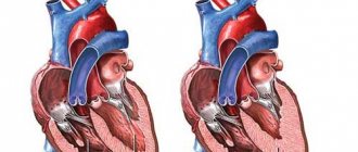
Ischemic cardiomyopathy is a pathological change in the myocardium, the most common form of such a deviation. As the disease develops, the cavities of the heart muscle become enlarged and its walls thicken.
Most often, damage to the left side of the organ is observed. In some cases, with ischemic cardiomyopathy, the interventricular septum thickens.
This disorder is diagnosed in men much more often than in women. Pathology occurs at the age of 45-55 years.
Pathology manifests itself in the form of attacks, sometimes quite rare. Since the patient does not seek medical help, this causes the progression of the disease, aggravates the course of the pathology and can cause death.
Ischemic dilated cardiomyopathy
What is ischemic dilated cardiomyopathy -
Ischemic cardiomyopathy is a myocardial disease characterized by an increase in the size of the heart cavities and clinical symptoms of CHF, caused by atherosclerotic lesions of the coronary arteries. In foreign medical literature, ischemic dilated cardiomyopathy is understood as a myocardial disease, characterized by an enlargement of all chambers of the heart to the degree of cardiomegaly, with uneven thickening of its walls and phenomena of diffuse or focal fibrosis, developing against the background of atherosclerotic lesions of the coronary arteries.
In ICD-10, ischemic cardiomyopathy is presented in class IX “Diseases of the circulatory system” in heading I 25.5 as a form of chronic ischemic heart disease. In the classification of cardiomyopathy (WHO/MOFC, 1995), ischemic cardiomyopathy is classified as a group of specific cardiomyopathies. Ischemic dilated cardiomyopathy is a myocardial lesion caused by diffuse, significant atherosclerosis of the coronary arteries, manifested by cardiomegaly and symptoms of congestive heart failure. Patients with ischemic dilated cardiomyopathy make up about 5-8% of the total number of patients suffering from clinically significant forms of coronary artery disease. Among all cases of cardiomyopathies, ischemic ones account for about 11-13%. Ischemic cardiomyopathy occurs mainly at the age of 45-55 years; among all patients, men make up 90%.
What provokes / Causes of Ischemic Dilated Cardiomyopathy:
The cause of the disease is multiple atherosclerotic lesions of the epicardial or intramural branches of the coronary arteries. Ischemic cardiomyopathy is characterized by cardiomegaly (due to dilation of the chambers of the heart and left ventricle primarily) and congestive heart failure.
Pathogenesis (what happens?) during Ischemic Dilated Cardiomyopathy:
The pathogenesis of the disease includes several important mechanisms: hypoxia of the heart muscle due to a decrease in coronary blood flow due to the atherosclerotic process in the coronary arteries and a decrease in the volume of blood flow per unit of myocardial mass as a result of its hypertrophy and a decrease in coronary perfusion in the subendocardial layers; myocardial hibernation - a local decrease in the contractility of the left ventricular myocardium caused by its prolonged hypoperfusion; ischemic contracture of myocardial myofibrils, which develops due to insufficient blood supply, contributes to impaired myocardial contractile function and the development of heart failure; ischemic areas of the myocardium during systole are stretched with subsequent development of dilatation of the heart cavities; ventricular remodeling (dilatation, myocardial hypertrophy, development of fibrosis); hypertrophy of cardiomyocytes develops, fibroblasts and fibrogenesis processes in the myocardium are activated; diffuse myocardial fibrosis is involved in the development of heart failure; Myocardial apoptosis is activated due to ischemia and contributes to the onset of heart failure and the development of cavity dilatation.
The development of the disease involves factors that play an important role in the pathogenesis of CHF: an imbalance in the production of vasoconstrictors and vasodilators by the endothelium with insufficient synthesis of the latter, activation of neurohormonal factors, hyperproduction of cytokines, tumor necrosis factor.
Symptoms of Ischemic Dilated Cardiomyopathy:
It most often develops in men over 45-55 years of age. Usually we are talking about patients who have previously suffered a myocardial infarction or suffer from angina pectoris. However, in some cases, ischemic cardiomyopathy develops in patients who have not suffered a myocardial infarction and do not suffer from angina. It is possible that such patients have silent myocardial ischemia that has not been previously diagnosed. In typical cases, the clinical picture is characterized by a triad of symptoms: angina pectoris, cardiomegaly, CHF. Many patients have no clinical or ECG signs of angina.
The clinical symptoms of CHF do not have any specific features and are basically identical to the manifestations of CHF in patients with idiopathic dilated cardiomyopathy. Heart failure progresses more rapidly in ischemic cardiomyopathy compared to dilated cardiomyopathy. Usually we are talking about the systolic form of HF, but it is possible to develop diastolic HF or a combination of both forms.
Cardiomegaly on physical examination is characterized by expansion of all borders of the heart and predominantly the left one. During auscultation, attention is drawn to tachycardia, often various arrhythmias, dullness of heart sounds, and protodiastolic gallop rhythm. Arrhythmia is detected in ischemic cardiomyopathy much less frequently (17%) than in idiopathic dilated cardiomyopathy. Signs of thromboembolic complications in the clinical picture of ischemic cardiomyopathy are observed somewhat less frequently than in idiopathic dilated cardiomyopathy.
Diagnosis of ischemic dilated cardiomyopathy:
LABORATORY AND INSTRUMENTAL DIAGNOSTICS
Blood chemistry
Characterized by an increase in the blood levels of total cholesterol, low-density lipoprotein cholesterol, and triglycerides, which is characteristic of atherosclerosis.
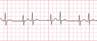
Electrocardiography
Cicatricial changes may be detected after previous myocardial infarctions or signs of ischemia in the form of horizontal displacement downwards from the ST interval isoline in various parts of the myocardium. Many patients exhibit nonspecific diffuse changes in the myocardium in the form of a decrease or smoothing of the T wave. Sometimes the T wave is negative, asymmetrical or symmetrical. Signs of myocardial hypertrophy of the left ventricle or other parts of the heart are also characteristic. Various arrhythmias (usually extrasystole, atrial fibrillation) or conduction disturbances are recorded. Daily Holter ECG monitoring often reveals silent, painless myocardial ischemia.
EchoCG reveals dilatation of the cardiac cavities, slight myocardial hypertrophy, an increase in end-diastolic volume, diffuse hypokinesia of the walls of the left ventricle, and a decrease in ejection fraction. The ejection fraction of the right ventricle in patients with ischemic cardiopathy, compared with the ejection fraction of the left ventricle, is reduced to a lesser extent than in idiopathic dilated cardiomyopathy.
In the presence of chronic myocardial ischemia, the rigidity and rigidity of the walls of the left ventricle significantly increases, and their elasticity decreases. This is due to a deficiency of high-energy compounds due to insufficient oxygen supply to the myocardium. Which leads to a slowdown in the process of early diastolic relaxation of the left ventricular myocardium. These circumstances lead to the development of the diastolic form of heart failure. Left ventricular diastolic dysfunction in coronary artery disease can occur without impairment of systolic function.
According to Doppler echocardiography, there are two main types of left ventricular diastolic dysfunction - early and restrictive. The early type is characterized by a violation of the early phase of diastolic filling of the left ventricle. During this phase, the speed and volume of blood flow through the mitral orifice decrease (peak E) and the volume and speed of blood flow increase during atrial systole (peak A). The time of isometric relaxation of the left ventricular myocardium increases and the time of deceleration of the E flow increases, the E/A ratio < 1. With the restrictive type of diastolic dysfunction of the left ventricle, the diastolic pressure in it significantly increases, the pressure in the left atrium increases, the E peak increases, the A peak decreases, and shortens left ventricular isometric relaxation time and flow deceleration time E, E/A ratio > 2.
With ischemic cardiomyopathy, the development of diastolic dysfunction is possible; the restrictive type is observed much less frequently. With the development of isolated diastolic HF, left ventricular systolic function is preserved and the ejection fraction is normal. With ischemic cardiomyopathy, isolated diastolic failure is rare; more often, with severe congestive heart failure, we are talking about combined systolic and diastolic dysfunction of the left ventricle.
X-ray examination
Determines a significant increase in the size of all chambers of the heart.
Radioisotope scintigraphy
Detects small foci of impaired accumulation of thallium-201 in the myocardium, which reflects myocardial ischemia and fibrosis.
Coronary angiography
Detects significantly pronounced atherosclerotic lesions of the coronary arteries. In this case, one of the arteries can be narrowed by more than 50%.
The diagnosis of the disease is made on the basis of the above clinical picture and data from instrumental studies. First of all, the presence of angina pectoris, anamnestic data on myocardial infarction, cardiomegaly, and congestive heart failure are taken into account. Diagnosis of ischemic dilated cardiomyopathy is based on the diagnostic criteria outlined in Table 9.
Table 9. Diagnostic criteria for ischemic dilated cardiomyopathy
Classification of the disease
There are the following forms of ischemic cardiomyopathy:
- symmetrical: both chambers of the heart muscle increase evenly;
- asymmetric: as the pathology develops, the left chamber of the heart predominantly enlarges.
Pathology can develop as an independent deviation or against the background of existing disturbances in the functioning of the cardiovascular system.

Clinical picture
Quite often, patients with ICMP have a history of angina pectoris or a previous myocardial infarction, but it can manifest itself for the first time without previous pathology. Ischemic cardiomyopathy is characterized by a triad of symptoms: cardiomegaly, exertional angina, CHF.
Cardiomegaly during physical examination can be suspected by changes in the apical impulse during examination and palpation: it is diffuse (large in area), high (amplitude), resistant (dense), and may be shifted to the left. Percussion determines the expansion of the boundaries of cardiac dullness.
Read also: Coronary heart disease arrhythmic variant
Confirmation of cardiomegaly requires imaging. The simplest thing to do in this situation is a chest x-ray. Angina pectoris has classic symptoms here: chest pain with characteristic features:
- localization - behind the sternum;
- character - burning, oppressive;
- duration - up to 15 minutes (if longer, a heart attack should be suspected);
- provoking factors: physical tension, stress;
- How it is treated: rest, nitrates (Nitroglycerin, Isoket)
Symptoms of heart failure are varied. Symptoms are identified for stagnation in the pulmonary and systemic circulation. In the first case (congestion in the lungs), patients are bothered by shortness of breath, suffocation, and often have a cough with sputum (characteristic “rusty” color - hemosiderin). With stagnation in the large circle, swelling of the legs, enlargement of the liver and nagging pain in the area where it is located, digestive disorders, etc. are noted.
Causes
Ischemic cardiomyopathy develops with damage to the coronary arteries, which most often occurs against the background of atherosclerotic changes. The latter cause vasoconstriction, which results in persistent tissue hypoxia. Against this background, thickening of the wall of the heart muscle occurs.
Risk factors include the following:
- history of myocardial infarction;
- high blood pressure;
- bad habits;
- burdened heredity;
- increased blood cholesterol levels;
- obesity;
- diabetes;
- left ventricular aneurysm;
- sedentary lifestyle;
- increased physical activity;
- drug use;
- psycho-emotional stress;
- pathologies of the adrenal cortex;
- thyroid disease.
The development of the disease is also facilitated by pathological phenomena such as myocardial hypertrophy, cardiac hypoxia, and damage to myocardial myofibrils.
Causes of the disease
The cause of heart damage in this case is widespread atherosclerosis of the coronary arteries, which leads to a hemodynamically significant narrowing of their lumen and the development of ischemic damage to cardiomyocytes. It is not so much the main trunk of the arteries that changes pathologically, but rather the small intramural and subepicardial branches. There is a chronic progressive course of the disease, the outcome of which is congestive CHF.
Risk factors
They are similar for all cardiac diseases, since the cause of cardiac pathology in the vast majority of cases is hypertension and atherosclerosis. To justify preventive measures, they are usually divided into changeable (modifiable) and unchangeable (non-modifiable).
- Factors that can be influenced (modifiable):
- high body weight,
- bad habits,
- blood pressure level (can be corrected with medication),
- poor nutrition
- stress and others.
- Natural, hereditary factors: risk age (45-55), gender, genetic characteristics cannot be changed. These are non-modifiable factors.
Pathogenesis
Several main pathogenetic links in the development of the process can be identified.
- Hypoxia of cardiomyocytes. It develops, firstly, as a result of insufficient blood supply to muscle cells through pathological vessels. Secondly, it is worth noting that healthy areas of the heart muscle are forced to endure increased loads, as a result of which their hypertrophy occurs, and the myocardium, increased in volume and mass, is again more difficult to “feed”.
- Myocardial hibernation is a protective mechanism of the body, which consists in reducing the contractility of muscle cells due to its hypoperfusion. In this way, the organ tries to “survive” an unfavorable period of low blood supply, but blood flow through the altered vessels will not be restored on its own, so this compensatory mechanism in this case becomes a link in pathogenesis.
- Ischemic contracture of myofibrils caused by hypoxia contributes to an even more pronounced decrease in contractile function and the development of organ failure.
- Ischemic cardiac fibers begin to stretch during systole, as myocardial tone is reduced. Dilatation of the cavities begins to form (morphologically, ICMP is a dilated cardiomyopathy)
- With excessive accumulation of carbon dioxide and metabolic products in cells, fibroblasts are always activated, which cause the synthesis of connective tissue in place of muscle - diffuse fibrosis develops.
All these changes are united by one term - cardiac remodeling. It should be noted that the remodeling process is determined by an imbalance of cellular regulatory factors: vasoactive substances, neurohormonal factors, cytokines, etc.
Classification
There are 2 forms based on the structural (morphological) features of the process:
- Symmetrical ischemic cardiomyopathy - with it there is a uniform increase in both the left and right halves of the organ. This form is rare.
- Asymmetrical - a form characterized by a predominant enlargement of the left chambers of the heart. This is what is observed in most cases.
Symptoms of ischemic cardiomyopathy
Pathology can be expressed by such signs
- shortness of breath, which is especially pronounced during physical exertion and emotional experiences;
- heart rhythm disturbance;
- headache;
- increased fatigue;
- attacks of suffocation at night;
- causeless weight gain;
- rare urination;
- swelling of the lower extremities;
- dizziness;
- increased heart rate;
- productive cough, streaks of blood may be observed in the sputum;
- sleep disturbance;
- fainting;
- feeling of stabbing pain and squeezing in the chest;
- loss of appetite;
- dull pain in the right hypochondrium;
- different forms of arrhythmia.
In the early stages of pathology development, signs of cardiac dysfunction may be absent.
The clinical picture of ischemic cardiomyopathy is nonspecific, which makes diagnosing the disease difficult.
Treatment of ischemic cardiomyopathies
Attention. All treatment should be prescribed exclusively by a cardiologist. Self-medication is unacceptable.
Therapy of the disease includes treatment of chronic heart failure, arrhythmic disorders, prevention of the development of blood clots and thromboembolism, as well as reducing the rate of disease progression.
Patients are prescribed a diet, a protective regimen, and limited fluid and salt intake.
Cardiac glycosides , loop diuretics, aldosterone antagonists, and ACE inhibitors are also prescribed
Diagnostic methods
To make a diagnosis, a number of studies are carried out, including:
- collection of family history;
- physical examination;
- listening to the heart;
- general and biochemical blood tests;
- general urine analysis;
- ECG;
- coronary angiography;
- chest x-ray;
- daily ECG monitoring;
- EchoECG;
- MRI of the heart muscle;
- if necessary, additional scintigraphic examination and biopsy are prescribed.
Also, during the research, differential diagnosis is required. Ischemic cardiomyopathy must be distinguished from other types of cardiomyopathies.
Diagnosis of ischemic cardiomyopathy
First, the cardiologist performs a physical examination. At the same time, it determines the presence of heart murmurs. Further diagnosis of the patient is carried out using laboratory and instrumental methods.
Laboratory diagnostic methods mean a blood test that will help identify the level of triglycerides, lipoproteins, and cholesterol in the blood.
As for instrumental examination methods, these include:
- Electrocardiography is a method that allows you to diagnose coronary heart disease and its numerous other lesions.
- Daily monitoring is a very important diagnostic method, since it allows you to identify pathologies at the very beginning of development, before any symptoms appear.
- Echocardiography allows visualization of the heart. This method determines hypertrophy, enlargement of the heart cavities and many other pathologies.
- Coronary angiography, which allows you to determine the condition of the coronary vessels and identify atherosclerotic plaques in them.
- PET (positron emission tomography) is performed to evaluate metabolic changes in the myocardium.
- MRI (magnetic resonance imaging) is used when another diagnostic method is not sufficiently informative.
Instrumental diagnostics reveals any morphological changes in the heart.
Approaches to the treatment of ischemic cardiomyopathy
The basis of therapy is taking medications. Surgical intervention is performed only in exceptional cases, as it poses a threat to the patient's life.
Drug treatment
Patients will be prescribed medications such as:
- antiarrhythmic drugs (Digoxin);
- beta-blockers (Metaprolol, Carvedilol);
- ACE inhibitors (Capoten);
- diuretics (Lasix);
- anticoagulants (acetylsalicylic acid).
Recommendations for correcting the regime
In parallel with taking special medications, the patient must comply with the following rules:
- limit to a minimum the amount of fat and salt consumed;
- refuse increased physical activity;
- avoid stress and emotional tension;
- leave enough time for proper rest;
- to refuse from bad habits.
Methods of surgical treatment
Surgery for ischemic cardiomyopathy is indicated only if conservative methods of therapy have failed.
The following methods of surgical treatment of pathology are distinguished:
- Pacemaker implantation. An implantable device is necessary when detecting heart failure. Thanks to the pacemaker, the heart rhythm is normalized. The device also reduces the risk of sudden death caused by ischemic cardiomyopathy.
- Implantation of a cardioverter-defibrillator. This measure is required for ventricular arrhythmia, a condition that threatens the patient’s life. The device monitors the rhythm of the heart muscle.
- Coronary bypass surgery. During the operation, another, previously removed, vessel is inserted next to the blocked artery. It is through it that the blood flow is directed. This event stimulates blood flow to the heart muscle.
- Angioplasty is an operation to widen the arteries to improve their patency.
- Installation of stents to expand the lumen of blood vessels.
- Atherectomy is a manipulation aimed at cleansing the walls of the arteries from plaques and plaque that impede the flow of blood.
- In the most severe cases, the patient requires a heart transplant.
Nutritional Features
People suffering from ischemic cardiomyopathy are recommended to include the following foods and drinks in their diet:
- baked goods without salt;
- low-fat dairy products;
- vegetables processed in various ways;
- seafood;
- lean fish;
- cereals;
- pasta from durum wheat;
- vegetable soups;
- berries and fruits, fresh or dry;
- honey;
- vegetable oils;
- green leaf tea;
- fresh juices.
You should not consume the following foods and drinks that negatively affect the condition of the heart muscle:
- meat and fish of fatty varieties;
- confectionery;
- salty and spicy dishes;
- fatty broths;
- canned food;
- smoked meats;
- onion garlic;
- semi-finished products;
- strong coffee and tea;
- alcohol.
Folk remedies
The point of using folk recipes for ischemic cardiomyopathy is to alleviate the patient’s condition. Such therapies cannot cure the disease and should under no circumstances be the basis of therapy.
The following recipes are useful:
- Viburnum berry tincture. To prepare, you need to take 40 g of raw materials (the berries must be ripe), pour a glass of hot water. Let the liquid brew for 2 hours, then strain. The resulting volume is the daily dose of the medicine. You need to drink it 2 times during the day.
- A decoction based on motherwort. You should take 15 g of raw material and fill it with 0.5 liters of hot water. The product must be infused for 7 hours. After this, the liquid must be filtered. Take the prepared infusion throughout the day. The norm is a glass of decoction, the volume of which should be divided into 4 doses.
- Herbal collection. It is necessary to prepare 2 tablespoons of nettle leaves and a tablespoon of motherwort, mix the ingredients, pour 250 liters of boiling water. Infuse the product for an hour, then strain. Take half a glass twice a day.
- Infusion of flax seeds. To prepare the product, you need to pour 4 teaspoons of flax seed into a liter of water. Place the mixture on the stove and let it boil. The product should be infused in a water bath for an hour. After this, the broth must be filtered. Take the finished product warm, half a glass, up to 5 times a day.
During the treatment process, the doctor regularly monitors the most important indicators: heart rate, pulse rhythm, and the patient’s blood pressure level.
Treatment
Treatment may vary dramatically depending on the diagnostic results, the form and degree of development of the disease, the presence of complications and other factors.
Therapeutic
The main goal of therapeutic treatment is to influence cardiac ischemia, which is the main factor in the development of cardiomyopathy. Therapeutic treatment is prescribed to all patients with this diagnosis. It may be combined with other treatment methods. The essence of therapy is as follows:
- changing your lifestyle (giving up bad habits, normalizing your daily routine and normalizing your workload);
- diet (low cholesterol, animal fats, salt);
- active pastime (swimming pool, long walks, yoga, physical therapy).
Medication
Such treatment can be aimed at suppressing unpleasant symptoms, strengthening the cardiovascular system and preventing the development of complications. Typically, cardiologists prescribe the following medications:
- beta blockers help lower blood pressure and heart rate;
- calcium antagonists also reduce blood pressure, but also dilate the coronary arteries;
- Aldosterone inhibitors allow you to remove excess fluid from the body and can be replaced with diuretics;
- anticoagulants reduce the risk of blood clots;
- drugs that normalize heart rhythm;
- Plasmapheresis is prescribed in rare cases.
Operation
Such treatment methods are rarely resorted to when other options are not effective and the disease poses a serious threat to the patient’s life. The main surgical treatment methods include the following:
- installation of a stimulator or defibrillator, which allows normalization of heart function;
- angioplasty, which counteracts the development of vascular stenosis;
- Stenting of coronary arteries restores the functioning of vessels blocked by plaques;
- atherectomy allows you to restore lumen in the arteries;
- A heart transplant is a last resort method that is used for severe muscle damage.
Folk remedies
No traditional medicine recipe can replace drug treatment. Such methods can be used to maintain the body, strengthen the heart and vascular system. Patients with ischemic cardiomyopathy benefit from decoctions and infusions of the following natural ingredients:
- viburnum;
- flax seeds;
- hare cabbage;
- oats;
- jaundice.
Hare cabbage
The result of such treatment can be a decrease in blood pressure, relief from the symptoms of heart failure and normalization of the heart rhythm. Any method of traditional medicine is chosen together with the attending physician. Self-medication is unacceptable.
Forecast
If left untreated, as ischemic cardiomyopathy progresses, the risk of complications increases. These include the following:
- blood clot formation;
- myocardial infarction;
- acute heart failure;
- pulmonary edema;
- arrhythmia.
The listed conditions can cause the death of the patient, as they develop rapidly.
The ischemic form of cardiomyopathy is characterized by a much more unfavorable prognosis compared to other forms of this pathology. The incidence of deaths during conservative therapy provided myocardial viability is 16%. With successful surgery, this figure drops to 3.2%.
The five-year survival rate of patients is more than 30%.
With timely treatment of the pathology, the chance of preventing dangerous complications increases.
Classification
All cardiomyopathies are divided into primary and secondary.
Primary diseases are divided into groups:
- Congenital, which appear during the period of intrauterine development, mainly due to maternal smoking, consumption of alcohol and medications, stress and anxiety, improper or insufficient nutrition.
- Acquired, arising after exposure to toxins, metabolic disorders or viruses.
- Mixed, combining both types of negative impact.
Secondary pathologies are caused by the following diseases or conditions:
- the effects of certain medications, including chemotherapy;
- alcoholism;
- endocrine diseases that provoke myocardial dysfunction;
- obesity;
- diseases of the gastrointestinal tract;
- eating disorders;
- diabetes;
- the use of harsh long-term diets leading to an acute lack of vitamins and nutrients;
- negative effects at the cellular level (radiation, toxins, poisons, etc.).
Also read: How heart failure manifests itself
Primary cardiomyopathy is divided into types:
- dilated or ischemic;
- hypertrophic;
- restrictive;
- arrhythmogenic dysplasia.
Secondary diseases are of the following types:
- alcoholic;
- diabetic;
- thyrotoxic (associated with pathologies of the thyroid gland);
- stressful.
The ischemic form of the disease is called congestive due to the presence of poor blood supply.
Prevention measures
Currently, no special set of preventive measures has been developed aimed at preventing the development of ischemic cardiomyopathy.
There are a number of generally accepted recommendations, compliance with which reduces the likelihood of developing cardiac pathology several times. To do this you need:
- stop drinking alcohol;
- stop smoking;
- follow the principles of proper nutrition, excluding from the diet foods high in salt and cholesterol, as well as animal fats;
- control weight, blood pressure, blood sugar and cholesterol levels;
- visit a cardiologist once every six months, which is especially important for those people who have a family history of cardiovascular diseases.
Ischemic cardiomyopathy is a pathology of the cardiovascular system that occurs independently or as a secondary disease. The disease creates a high risk of complications and death, so it is necessary to diagnose it and begin treatment as quickly as possible.
Causes and provoking factors
When a certain portion of blood is released, the cardiac cavities stretch, which causes their dilatation. In addition, ventricular remodeling is possible, resulting in the formation of fibrosis. And diffuse fibrosis is a direct path to the development of heart failure due to cardiomyopathy.
It is worth noting that hyperfusion of the LV (left ventricle) also often causes the development of cardiomyopathy, since the contractile function in it is reduced. In this case, we are talking about myocardial hibernation.
Another significant cause of the development of ischemic cardiomyopathy is myocardial apoptosis, which consists of programmed cell death. This disorder is characteristic of ischemic disease. The development of ischemic cardiomyopathy is also influenced by an increased concentration of cytokines.
There are several stages in the development of the disease:
- Hypoxic;
- Weakening of myocardial contractile function;
- Expansion of the heart cavities;
- Heart failure.
If not treated promptly, cardiomyopathy of ischemic origin can lead to potentially fatal complications, including:
- Blockage of the cardiac pathways;
- Extrasystole;
- Atrial fibrillation;
- Myocardial infarction;
- Heart failure, acute or chronic.
Severe ischemic cardiomyopathy: there is not much chance of survival without help
Insufficient blood flow to the heart muscle leads to coronary artery disease. Its complication may be cardiomyopathy, in which the myocardium weakens to such an extent that it loses tone, and the chambers of the heart expand. Weak cardiac output provokes circulatory failure. With extensive damage to the heart muscle, death can occur if it is impossible to perform a heart transplant.
Read in this article
Complications
The disease initially occurs paroxysmally (angina pectoris clinical picture); as it progresses, the symptoms of CHF come to the fore. Complicated ICMP often occurs.
Patients may develop myocardial infarction (with high mortality), rhythm disturbances, progression of CHF, and pulmonary edema due to pulmonary edema. Complications can cause death in patients.
Therapy for this disease is complex and must be comprehensive and continuous. It can be divided into levels: Level 1: combating risk factors: preventing stress, eliminating (or reducing the severity of) household intoxications, proper physical activity, diet with limited salt and liquid. Level 2: drug treatment. His goals:
- combating ischemia: both stopping an attack (nitrates) and long-term maintenance (ACE inhibitors, beta blockers, etc.)
- correction of heart failure - ACE inhibitors, beta-blockers, diuretics, etc.
- impact on the cause - atherosclerosis: use of statins, fibrates
- prevention and treatment of complications: antiarrhythmics, antiplatelet agents, anticoagulants
Level 3: surgical treatment. This method is used when therapy is powerless and there is a threat of death. The following types of interventions are distinguished:
- stenting - performed like coronary angiography, but at the end a supporting mesh (stent) is installed in the vessel, which does not allow the vessel lumen to decrease and ensures normal myocardial perfusion
- coronary artery bypass grafting - vascular anastomosis bypassing the thrombosed area.
- implantation of a pacemaker to control the heart rate during arrhythmias.
- The most radical, but also the most difficult, for objective reasons, method is heart transplantation.
Ischemic cardiomyopathy is a severe cardiac pathology, as can be seen from the five-year survival rate of patients (only 30%). This is explained by the fact that it is very difficult to therapeutically correct this condition against the background of a severe progressive course, and also by the fact that it is difficult for patients to adhere to lifelong and volume therapy.