myocardium damage to myocardial muscle tissue
Myocardium
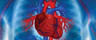
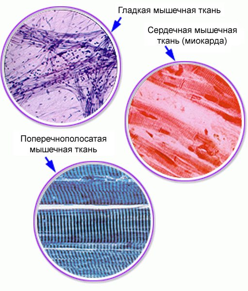
(Latin myocardium from ancient Greek μῦς “muscle” + καρδία “heart”) - muscular tissue of the cardiac type, the main histological element of which is the cardiomyocyte; corresponds to the middle layer of the heart and forms the thickness of the walls of the ventricles and atria.[B: 1][B: 2]
The muscle tissue of the heart consists of individual cells - myocytes. There are three types of cardiac myocytes:[1][B: 3][B: 4]
- conducting, or atypical
(obsolete), cardiomyocytes; - contractile, or typical
, cardiomyocytes, which are also called cells
of the working myocardium
; - secretory cardiomyocytes.
Other researchers[2] identify five types of cardiomyocytes, further dividing the group of conducting cardiomyocytes into sinus (pacemaker), transitional and conducting.
The fibers of the working myocardium of the atria and ventricles make up the bulk of the heart - 99%, providing its pumping function.[B: 5] The myocardium also includes supporting loose fibrous connective tissue and coronary vessels.[3]
Embryology
The myocardium, like the epicardium, is formed from the myoepicardial plate (visceral layer of the splachnotome of the embryonic neck), while the endocardium is formed from mesenchyme.[1] Sources of development of cardiac striated muscle tissue are symmetrical sections of the visceral sheet of the splachnotome in the cervical part of the embryo - myoepicardial plates
; epicardial mesothelial cells also differentiate from them.[2] After a series of mitotic divisions, G1 myoblasts begin the synthesis of contractile and auxiliary proteins and, through the G0 myoblast stage, differentiate into cardiomyocytes, acquiring an elongated shape.[1]
Unlike striated skeletal tissue, in cardiogenesis there is no separation of the cambial reserve, and all cardiophyocytes are irreversibly in the G0 phase of the cell cycle.[1] There are no stem cells or progenitor cells in cardiac muscle tissue, so dying cardiomyocytes are not restored.[2]
Striated cardiac muscle tissue
· Has a structure of myofibrils and protofibrils and a mechanism of muscle contraction similar to skeletal muscle tissue (there are few myofibrils, they are thin, weak transverse striations)
Features of cardiac striated muscle tissue:
o Muscle fiber consists of chains of individual cells - cardiomyocytes
(cells do not fuse)
o All heart cells are connected by membrane contacts (intercalated discs) into a single muscle fiber, which ensures contraction of the myocardium as a whole (separately the atrial myocardium and ventricular myocardium)
o Fibers have a small number of cores
· Cardiac muscle tissue is divided into two types:
o working muscle tissue
– makes up 99% of the mass of the heart myocardium (provides heart contraction)
o conductive muscle tissue
– consists of modified, incapable of contraction,
atypical
cells
- forms nodes in the myocardium, where electrical impulses for heart contractions are generated and from where they are distributed - the conduction system of the heart
Functions of cardiac striated muscle tissue
1. Generation and propagation of electrical impulses to contract the myocardium of the heart
2. Involuntary
rhythmic contractions of the myocardium of the heart to push blood (myocardial automation)
Smooth muscle tissue
· Localized only in internal organs (walls of the digestive tract, walls of the respiratory tract, blood and lymphatic vessels, bladder, uterus, oblique muscles of the hair of the skin, muscles surrounding the pupil)
· Cells are solitary, long, spindle-shaped, mononuclear, dividing throughout life
· The internal structure of the cell is the same as that of muscle fibers of striated tissue (myofibrils, consisting of protofibrils and the proteins actin and myosin)
· Light areas of actin and dark areas of myosin of different myofibrils lie disorderly, which leads to the absence of cross-striations of smooth muscle cells
· Form ribbons, layers, cords in the walls of internal organs (do not form separate muscles)
Innervated by autonomic nerves
Smooth muscles of internal organs are weak and contract involuntarily
without the fate of consciousness, slowly, do not get tired, capable of being in a state of contraction for a very long time (hours, days) -
tonic
contractions (consume little energy for work)
Functions of smooth muscles
1. Work (motor function) of internal organs (peristalsis, urine excretion, childbirth, etc.)
2. Tone of blood and lymphatic vessels (changes in the diameter of blood vessels lead to changes in blood pressure and velocity)
Nervous tissue
· During embryogenesis, it is formed by division of ectoderm cells
Properties of nervous tissue - excitability
and
conductivity
Organs formed by nervous tissue: brain, spinal cord, ganglia, nerves
· Consists of nerve cells (neurons)
– 15% of all cells and
neuroglia
(intercellular substance)
Neuroglia have cells (gliocytes) - 85% of all cells
Functions of neuroglia
1. Trophic (supply of neurons with everything necessary for life)
2. Supporting (skeleton of nervous tissue)
3. Insulating, protective (protection from adverse conditions and electrical insulation of neurons)
4. Regeneration of nerve cell processes
Nerve cells - neurons
- mononuclear, with processes, not dividing after birth (the total number of neurons in the human nervous system, according to various estimates, ranges from 100 billion to 1 trillion)
· Have a body
(contains granules, clumps) and
processes
Neurons have many mitochondria
, the Golgi complex and the system of support-transport microtubules -
neurofibrils
for the transport of substances (neurotransmitters)
· There are two types of shoots:
o Axon
– always one, long (up to 1.5 m), not branching (extends beyond the limits of the nervous system organ)
Axon functions
– carrying out a command (in the form of an electrical impulse) from a neuron to other neurons or to working tissues and organs
o Dendrites
– numerous (up to 15), short, branched (have sensitive nerve endings at the ends -
receptors
)
Functions of dendrites
– perception of irritation and conduction of an electrical impulse (information) from receptors to the body of a neuron (to the brain)
· Nerve fibers
– processes of nerve cells covered with connective tissue membranes
Structure of a neuron: Structure of a multipolar neuron: 1 - dendrites; 2 - neuron body; 3 - core; 4 - axon; 5 - myelin sheath; 6 – axon branches
· The gray matter of the brain is a collection of neuron cell bodies
- substance of the cerebral cortex, cerebellar cortex, horns of gray matter of the spinal cord and nerve nodes (ganglia)
· White matter of the brain -
a set of neuron processes (axons and dendrites)
Types of neurons (by number of processes)
o Unipolar
– have one process (axon)
o Bipolar
– have two processes (one axon and one dendrite)
o Multipolar –
have many processes (one axon and many dendrites) - neurons of the spinal cord and brain
Types of neurons (by function)
o Sensitive (centripetal, sensory, efferent) –
perceive irritations from receptors, form feelings, sensations (bipolar)
o Intercalated (associative)
– analysis, biological meaning of information received from receptors, development of a response command,
connection of sensory neurons with motor
and other neurons (one neuron can connect with 20 thousand other neurons); 60% of all neurons are multipolar
o Propulsive (centrifugal, motor, effector)
– transmission of the interneuron command to the working organs (muscles, glands); multipolar, with a very long axon
o Brake
o Some neurons are capable of synthesizing hormones: oxytocin and prolactin ( neurosecretory cells
hypothalamus of the diencephalon)
· Nerve fibers
– processes of nerve cells covered with connective tissue membranes
· There are two types of nerve fibers (depending on the structure of the membrane): pulpy and non-pulphate
| Myelinated nerve fibers | Unmyelinated nerve fibers |
| 1. Covered with a sheath of neuroglial cells (Schwann cells) to electrically insulate the fiber | 1. Also |
| 2. Schwann cell membranes contain a substance - myelin (significantly increases electrical insulation) | 2. Do not contain myelin (less effective electrical insulation) |
| 3. The fiber has areas without a sheath - nodes of Ranvier (accelerate the conduction of nerve impulses along the fiber) | 3. No |
| 4. Fat | 4. Thin |
| 5. The speed of nerve impulses is up to 120 m/sec | 5. The speed of nerve impulse is about 10 m/sec |
| 6. Form the nerves of the central nervous system | 6. Form nerves of the autonomic nervous system |
o Hundreds and thousands of pulpal and non-pulmonary nerve fibers extending beyond the central nervous system, covered with connective tissue, form nerves (nerve trunks)
Types of nerves
o Sensory nerves -
formed exclusively by dendrites, they serve to conduct sensitive information from the body’s receptors to the brain (into sensory neurons)
o Motor nerves
– formed from axons: they serve to carry out brain commands from the motor neuron to working tissues and organs (effectors)
o Mixed nerves
– consist of dendrites and axons; also serve to conduct sensitive information to the brain and brain commands to working organs (for example, 31 pairs of spinal nerves)
Communication and interaction between nerve cells is carried out using synapses
Synapse is the point of contact of an axon with another process or body of another cell (nerve or somatic), in which the transmission of a nerve (electrical) impulse occurs
o The transmission of nerve impulses at the synapse is carried out using chemicals - neurotransmitters
(adrenaline, norepinephrine, acetylcholine, serotonin, dopamine, etc.)
o Synapses are located on the branches of the axon terminal
o The number of synapses on one neuron can reach up to 10,000, so the total number of contacts in the nervous system is approaching an astronomical figure
o It is possible that the number of contacts and multipolar neurons in the nervous system is one of the indicators of a person’s mental development and labor specialization. With age, the number of contacts decreases significantly
Animal tissue (human tissue)
Reflex. Reflex arc
Reflex is the body’s response to irritation (change) in the external and internal environment, carried out with the participation of the nervous system
o the main form of activity of the central nervous system
v The founder of the idea of reflexes as unconscious automatic acts associated with the lower parts of the nervous system is the French philosopher and naturalist R. Descartes (XVII century). In the XVIII century. Czech anatomist and physiologist G. Prohaska introduced this term “reflex” to science
v I.P. Pavlov, Russian academician (XX century) divided the reflex into unconditioned ( congenital, specific, group) and conditioned (acquired, individual)
Previous3Next
Histology
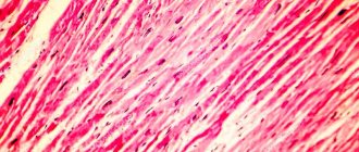
The myocardium is a tight junction of muscle cells - cardiomyocytes, which make up the main part of the myocardium. It differs from other types of muscle tissue (skeletal muscle, smooth muscle) in its special histological structure, which facilitates the propagation of the action potential between cardiomyocytes. A characteristic structural feature of cardiac muscle tissue is the presence in the area of intercalated discs of zones of tight fit of cardiomyocyte membranes - nexuses
.
Due to this, a low electrical resistance is created in the nexus area compared to other areas of the membrane, which ensures a rapid transition of excitation from one fiber to another. This pseudosyncytial structure of the cardiac muscle determines a number of its features.[4] In addition, the transverse portions of the projections of neighboring cells are connected to each other through interdigitations
and
desmosomes
;
a myofibril approaches each desmosome from the cytoplasm and is anchored in the desmoplakin complex, and thus, during contraction, the thrust of one cardiomyocyte is transferred to another.[2] This structural feature of the myocardium, which promotes faster propagation of action potentials in the myocardium, is designated as functional syncytium
to show that the heart is a functionally unified organ.[5]
Atrial and ventricular cardiomyocytes belong to different populations of working cardiomyocytes. Atrial cardiomyocytes are relatively small, 10 µm in diameter and 20 µm in length; in them the system of T-tubules is less developed, but in the zone of intercalary disks there are significantly more gap junctions. Ventricular cardiomyocytes are larger, 25 µm in diameter and up to 140 µm in length; they have a well-developed T-tubule system. The contractile apparatus of the myocytes of the atria and ventricles also differs in the composition of the isoforms of myosin, actin and other contractile proteins.[1] Unlike ventricular cardiomyocytes, whose shape is close to cylindrical, atrial cardiomyocytes often have a process-like shape and are smaller in size.[6]
The elementary contractile unit of a cardiomyocyte is the sarcomere - a section of myofibril between two so-called Z lines. The length of the sarcomere is 1.6-2.2 µm, depending on the degree of contraction. Light and dark stripes alternate in the sarcomere, which is why the myofibril appears transversely striated under light microscopy. In the center there is a dark strip of constant length (1.5 µm) - disk A, it is bounded by two lighter disks I of variable length. The myocardial sarcomere, like that of skeletal muscle, consists of intertwined filaments (myofilaments) of two types. Thick filaments are found only in disk A. They consist of the protein myosin, have a cigar shape, a diameter of 10 nm and a length of 1.5-1.6 µm. Thin filaments primarily include actin and extend from line Z through disk I to disk A. Their diameter is 5 nm and length is 1 μm. Thick and thin filaments overlap each other only in disk A; disk I contains only thin filaments. Under electron microscopy, cross bridges are visible between thick and thin threads.
Working cardiomyocytes are covered with a sacrolemma, consisting of a plasmolema and a basement membrane, into which thin collagen and elastic fibers are woven, forming a reliable external skeleton of these cells. The basement membrane of cardiomyocytes, which contains a large number of glycoproteins capable of binding Ca2+, can participate, along with the sarcotubular network and mitochondria, in the redistribution of Ca2+ in the contraction-relaxation cycle. The basement membrane of the lateral aspects of cardiomyocytes invaginates into the T-system tubules (unlike skeletal muscle).[6]
Some of the cardiomyocytes of the atria (especially the right one) have a pronounced secretory function (secretory cardiomyocytes): they contain a well-defined Golgi complex and secretory granules containing the hormone atriopeptin at the poles of the nuclei.[1]
Skeletal muscle anatomy
We gradually approached the question of what human muscles are made of. A muscle cell (myocyte) is the basic structural unit of muscle tissue. A distinctive feature of the myocyte is that it is hundreds of times longer than its cross section. It is also called muscle fiber. From 10 to 50 fibers are connected into a bundle, which actually forms the muscle. For example, the biceps consists of a million fibers.
The main substance contained in the muscle cell is sarcoplasm. It contains thin muscle filaments (myofibrils), due to which contractions occur. The myofibril, in turn, consists of elementary particles - sarcomeres. Their main feature is to contract under the influence of a nerve impulse.
Here are the fibers a muscle (muscle bundle) consists of:
- Cores.
- Contractile filaments.
- Cover membrane.
- The connective tissue membrane (fascia) is a muscle group that acts in one direction.
- Blood vessels.
Thanks to targeted strength training, you increase both the number of myofibrils and their cross-section. First, this process increases muscle strength, then its thickness. But the number of muscle fibers themselves does not change. It is determined by the genetic characteristics of the body and remains the same throughout life. From this we can conclude that sports anatomy is that athletes whose muscles contain more myocytes are more likely to increase muscle thickness during strength training than those whose muscles contain fewer fibers.
Thus, the strength of skeletal muscle depends on the cross-section, namely the thickness and number of myofibrils. It is noteworthy that the indicators of strength and muscle mass increase unequally: when muscle mass increases by 2 times, muscle strength increases by 3 times. Scientists cannot yet explain this phenomenon.
Biochemistry
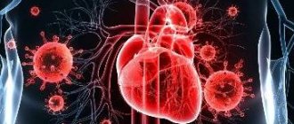
The main source of energy for the myocardium is the process of aerobic oxidation of non-carbohydrate substrates. These are free fatty acids and lactic acid (about 60%), pyruvic acid, ketone bodies and amino acids (less than 10%). During intense muscular work, lactic acid accumulates in the blood as a result of anaerobic glycolysis in the muscles. Lactate is an additional source of energy for the myocardium, and by breaking down lactic acid, the heart helps maintain a constant pH. About 30% of the energy consumed by the heart comes from glucose; During physical activity, the energy share of fatty and lactic acids increases while the energy share of glucose decreases. However, the greater dependence of the activity of the heart muscle on aerobic oxidation makes the heart very dependent on the supply of oxygen to cardiomyocytes. Therefore, when coronary blood flow deteriorates and there is insufficient oxygen supply to the heart muscle, pathological processes can develop in it, including a heart attack. The protective role for the heart is played by its myoglobin, which is contained in the heart muscle about 4 mg/g of tissue. It has a high affinity for O2, stores it during diastole of the heart and releases it during systole, when blood flow in the coronary arteries of the left ventricle almost stops (15% is retained); in the right ventricle and atria the blood flow is constant.[7]
Features of the structure of the heart
The heart of an adult weighs approximately 250-330 g. In women, the size of this organ is smaller, as is the volume of blood pumped.
It consists of 4 chambers:
- Two atria;
- Two ventricles.
The pulmonary circulation often passes through the right heart, and the large circulation through the left. Therefore, the walls of the left ventricle are usually larger: so that the heart can push out a larger volume of blood in one contraction.
The direction and volume of blood expelled is controlled by valves:
- Bicuspid (mitral) - on the left side, between the left ventricle and the atrium;
- Tricuspid - on the right side;
- Aortic;
- Pulmonary.
Physiology
The sequential contraction and relaxation of various parts of the heart is associated with its structure and the presence of the cardiac conduction system through which the impulse propagates. The myocardium of the atria and ventricles is separated by a fibrous septum, which allows them to contract independently of each other, since excitation cannot spread through the fibrous tissue. Excitation from the atria to the ventricles is carried out only through the atrioventricular bundle, extending from the atrioventricular node [B: 6].
Secretory cardiomyocytes of the atria, when they are strongly stretched due to increased blood pressure (BP), synthesize and secrete atriopeptin, which causes a decrease in blood pressure. [1]
Atypical cardiomyocytes
form the conduction system of the heart, consisting of:
sinoatrial node;
atrioventricular node;
atrioventricular bundle (bundle of His) trunk, right and left legs;
the terminal branches of the legs are Punkinje fibers.
Atypical cardiomyocytes ensure the generation of biopotentials, their conduction and transmission to contractile cardiomyocytes.
In their morphology, atypical cardiomyocytes differ from typical ones in a number of features:
they are larger (length 100 µm, thickness 50 µm);
the cytoplasm contains few myofibrils, which are arranged in a disorderly manner and therefore atypical cardiomyocytes do not have cross-striations;
the plasmalemma does not form T-tubules;
in the intercalary discs between these cells there are no desmosomes or gap junctions.
Atypical cardiomyocytes of different parts of the conduction system differ from each other in structure and function and are divided into three main types:
P-cells (pacemakers) pacemakers (type I);
transitional cells (type II);
His bundle cells and Purkinje fibers (type III).
Type I cells (P cells) form the basis of the sinoatrial node and are also found in small numbers in the atrioventricular node. These cells are capable of independently generating biopotentials at a certain frequency and transmitting them to transitional cells (type II), and the latter transmit impulses to type III cells, from which the biopotentials are transmitted to contractile cardiomyocytes.
The sources of development of cardiomyocytes are myoepithelial plates, which are certain areas of the visceral layers of the splanchnotome, and more specifically, from the coelomic epithelium of these areas.
Notes
- ↑ 1234567
Histology, 2002, Cardiac muscle tissue, p. 180-184. - ↑ 1234
Histology, 1998, Cardiac muscle tissue, p. 263-264. - Histology, 2002, Chapter 10. Cardiovascular system, p. 288-310.
- Sudakov, 2000, Physiology of the heart, p. 319-337.
- Tkachenko, 2005, § 2.8. Functions of cardiac muscle cells, p. 113-122.
- ↑ 12
Histology, 1998, Myocardium, p. 416-418. - Agadzhanyan, 2009, Chapter 11 Cardiovascular system, p. 260-310.
What tissue is the heart made of?
The heart is a hollow organ approximately the size of a human fist. It is almost entirely formed by muscle tissue, so many doubt: is the heart a muscle or an organ? The correct answer to this question is an organ formed by muscle tissue.
The heart muscle is called the myocardium, its structure is significantly different from the rest of the muscle tissue: it is formed by cardiomyocyte cells. Cardiac muscle tissue has a striated structure. It contains thin and thick fibers. Microfibrils are clusters of cells that form muscle fibers, collected in bundles of different lengths.
Properties of the heart muscle - ensuring the contraction of the heart and pumping blood
.
Where is the heart muscle located? In the middle, between two thin shells:
- Epicardium;
- Endocardium.
The myocardium accounts for the maximum amount of heart mass.
Mechanisms that provide reduction:
There are two phases in the heart cycle:
- Relative, in which cells respond to strong stimuli;
- Absolute – when, over a certain period of time, muscle tissue does not react even to very strong stimuli.
Literature
- Histology / ed. E. G. Ulumbekova, Yu. A. Chalysheva. — 2nd ed., revised. and additional.. - M.: GEOTAR-MED, 2002. - 672 p. — 3000 copies. — ISBN 5-9231-0228-5.
- Histology / ed. Yu. I. Afanasyeva, N. A. Yurina. - M.: Medicine, 1998. - 15,000 copies.
- Physiology. Fundamentals and functional systems / ed. K.V. Sudakova. - M.: Medicine, 2000. - 784 p. — ISBN 5-225-04548-0.
- Tkachenko B.I.
Normal human physiology. - M.: Medicine, 2005. - 928 p. - Agadzhanyan HA, Smirnov VM
Normal physiology. - M.: Publishing House "Medical Information Agency" LLC, 2009. - 520 p. — 5000 copies. — ISBN ISBN 978-5-9986-0001-2. - Guyton A.K., Hall D.E.
Medical physiology = Textbook of Medical Physiology / ed. IN AND. Kobrina. - M.: Logosphere, 2008. - P. 112. - 1296 p. — ISBN 978-5-98657-013-6.