Anatomical and physiological features of the cardiovascular system in children
The most important functions of the cardiovascular system are:
1) maintaining a constant internal environment of the body;
2) delivery of oxygen and nutrients to all organs and tissues;
3) removal of metabolic products from the body.
The cardiovascular system can provide these functions only in close interaction with the respiratory, digestive and urinary organs. Improvement in the functioning of the circulatory system occurs unevenly throughout childhood.
Features of intrauterine circulation in children
The formation of the heart begins in the 2nd week of intrauterine life. Within 3 weeks, the heart with all its parts is formed from the plate located on the border of the head and torso. In the first 6 weeks, the heart consists of three chambers, then four are formed due to the division of the atria. At this time, the process of dividing the heart into the right and left halves and the formation of heart valves occurs. The formation of the main arterial trunks begins from the 2nd week of life. The conduction system of the heart is formed very early.
Intrauterine blood circulation of the fetus
Oxygenated blood passes through the placenta through the umbilical vein to the fetus. A smaller part of this blood is absorbed into the liver, a larger part into the inferior vena cava. Then this blood, mixed with blood from the right half of the fetus, enters the right atrium. Blood from the superior vena cava also flows here. However, these two blood columns hardly mix with each other. Blood from the inferior vena cava enters the left heart and aorta through the oval window. Oxygen-poor blood from the superior vena cava passes into the right atrium, right ventricle and the initial part of the pulmonary artery, from here through the ductus arteriosus it enters the aorta and is mixed with blood coming from the left ventricle. Only a small part of the blood enters the lungs, from there into the left atrium, where it mixes with the blood entering through the oval window. A small amount of blood circulates in the pulmonary circulation before the first breath. Thus, the brain and liver receive the most oxygen-rich blood, and the lower extremities receive the least oxygen-rich blood.
After the birth of a child, the venous duct and umbilical vessels become empty, overgrow and turn into the round ligament of the liver.
All physiological life support systems are involved in the action.
Anatomical and physiological features of the heart and blood vessels in children
Children experience continuous growth and functional improvement of the cardiovascular system. The heart grows and improves especially vigorously in children from 2 to 6 years of age, as well as during puberty.
The heart of a newborn has a flattened cone-shaped, oval or spherical shape due to insufficient development of the ventricles and the relatively large size of the atria. Only by the age of 10-14 years does the heart acquire the same shape as that of an adult.
Due to the high position of the diaphragm, the newborn’s heart is located horizontally. The heart assumes an oblique position by the first year of life.
The weight of a newborn's heart is 0.8% of the total body weight, which is relatively larger than that of an adult. The right and left ventricles are equal in thickness, their walls are 5 mm. The atrium and great vessels are relatively large in size. By the end of the first year, the weight of the heart doubles, and by 3 years it triples. In preschool and primary school age, heart growth slows down and increases again during puberty. By the age of 17, the mass of the heart increases 10 times.
The parts of the heart also grow unevenly. The left ventricle significantly increases its volume; by 4 months it is twice as heavy as the right one. The thickness of the walls of the ventricles in a newborn is 5.5 mm, later the thickness of the left ventricle increases to 12 mm, the right - to 6-7 mm.
The volume of the heart at birth is about 22 cm3, during the first year it increases by 20 cm3, and subsequently by 6-10 cm3 annually. At the same time, the diameter of the valve openings increases.
In children, the heart is located higher than in adults. The volume of the heart in children is larger relative to the volume of the chest than in adults. In a newborn, the apex of the heart is formed on both ventricles, by 6 months - only on the left. By the age of 1.5 years, the projection of the heart from the 4th intercostal space descends into the 5th intercostal space.
In childhood, a qualitative restructuring of the heart muscle occurs. In young children, the heart muscle is undifferentiated and consists of thin, poorly separated myofibrils, which contain a large number of oval nuclei. There is no transverse striation. Connective tissue begins to develop. There are very few elastic elements; in early childhood, muscle fibers are close to each other. As the child grows, the muscle fibers thicken and rough connective tissue appears. The shape of the nuclei becomes rod-shaped, transverse striations of the muscles appear, and by 2-3 years of age the histological differentiation of the myocardium is completed. Other parts of the heart are also improving.
As the child grows, the conduction system of the heart improves. In early childhood it is massive, its fibers are not clearly contoured. In older children, overmodulation of the conduction system of the heart occurs, so cardiac arrhythmias are common in children.
The work of the heart is carried out due to the superficial and deep plexuses formed by the fibers of the vagus nerve and cervical sympathetic nodes, in contact with the ganglia of the sinus and atrioventricular nodes in the walls of the right atrium. The branches of the vagus nerve complete their development by 3-4 years. Until this age, cardiac activity is regulated by the sympathetic system. This explains the physiological increase in heart rate in children in the first 3 years of life. Under the influence of the vagus nerve, the heart rate slows down and respiratory-type arrhythmia appears, and the intervals between heartbeats lengthen. Myocardial functions in children, such as automatism, conduction, contractility, are carried out in the same way as in adults.
Features of blood vessels in children
The vessels supply and distribute blood to the child’s organs and tissues. Their clearance in young children is wide. Arteries are not equal in width to veins. The ratio of their lumen is
1:1, then the venous bed becomes wider, by the age of 16 their ratio is 1:2. The growth of arteries and veins often does not correspond to the growth of the heart. The walls of arteries are more elastic than the walls of veins. This is associated with lower levels of peripheral resistance, blood pressure, and blood flow velocity than in adults.
The structure of the arteries also changes. In newborns, the walls of blood vessels are thin, and their muscle and elastic fibers are poorly developed. Up to 5 years of age, the muscle layer grows rapidly; at 5–8 years of age, all vascular membranes are evenly developed; by 12 years of age, the structure of blood vessels in children is the same as in adults.
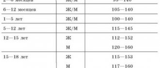
The heart rate in children depends on age. In a newborn it is 160-140 beats per minute, in 1 year - 110-140, in 5 years - 100, in 10 years - 80-90, in 15 years - 80.
With age, systolic blood pressure increases, and there is a tendency for diastolic pressure to increase.
Arterial systolic pressure is 90 + 2 xn, diastolic pressure is 60 + 2 xn, where n is the child’s age in years. For children under 1 year of age, systolic pressure is 75 + n, where n is the child’s age in months. Diastolic blood pressure is equal to systolic pressure minus 10 mm Hg. Art.
Heart and blood vessels during puberty
During puberty, intensive growth of various organs and systems occurs. During this period, disturbances in their functioning occur due to violations of their relationships and coordination of functions. In adolescents, due to the growth characteristics of both the heart and the whole body, relatively small mass and volume of the heart are observed compared to the mass and volume of the body. The ratio of body volume to heart volume in children is 50%, in adults - 60%, and in the puberty period it is 90%. In addition, there are anatomical features of the cardiovascular system in adolescents that are associated with the ratio of the volume of the heart and blood vessels.
In adolescents, the volume of the heart increases faster than the capacity of the vascular network, this increases peripheral resistance, which leads to a hypertrophic variant of the subadult heart.
In adolescents with deviations in the age-related evolution of the heart, sympathetic regulation predominates.
Thus, children have functional characteristics of the circulatory organs, which are characterized by:
1) 1) high level of endurance of the child’s heart due to its fairly large mass and good blood supply;
2) physiological tachycardia, caused by the small volume of the heart with a high need for oxygen in the child’s body, as well as sympathotomy;
3) low blood pressure with a small volume of blood flowing with each heartbeat, as well as low peripheral vascular resistance;
4) uneven growth of the heart and associated functional disorders.
Functions of the cardiovascular system
Main functions of the heart:
- delivery of oxygen to all organs and cells of the body;
- removal of metabolic products from cells;
- ensuring the invariability of the internal environment of the body.
The heart can perform all of these functions only in close cooperation with the respiratory, digestive and urinary organs. Failure in any of these organs over time begins to have a negative impact on the activity of the heart muscle. And when the functions of the heart muscle are suppressed due to any disease, the lungs, liver and kidneys begin to suffer first.
Treatment
VSD 4 mm, sometimes up to 6 mm - small in size - in the absence of disturbances in respiratory or heart rhythm and the normal development of the child, in some cases it allows not to use surgical treatment.
If the general clinical picture worsens or complications arise, surgery may be prescribed in 2–3 years.
Surgery is performed with the patient connected to a heart-lung machine. If the defect is less than 5 mm, it is closed with U-shaped sutures. If the hole is larger than 5 mm, it is covered with patches made of artificial or specially prepared bio-material, which is subsequently overgrown with the body’s own cells.
If surgical treatment is necessary for a child in the first weeks of life, but it is impossible due to some indicators of the baby’s health and condition, a temporary cuff is placed on the pulmonary artery. It helps equalize the pressure in the ventricles of the heart and alleviates the patient's condition. After a few months, the cuff is removed and surgery is performed to close the defects.
The mechanism of contraction of the heart muscle
Contraction of the chambers of the heart occurs in an orderly manner, since it is coordinated with the help of specialized cells that generate impulses and fibers through which impulses are carried through the myocardium.
The normal activity of the heart at rest and an increase in the number of contractions during exercise is ensured by the autonomic nervous system, consisting of the parasympathetic and sympathetic nervous systems. The parasympathetic nervous system reduces the frequency of contractions of the heart muscle and reduces the speed of impulses passing through the conductive fibers. The sympathetic system, on the contrary, ensures an increase in the frequency of contractions of the heart muscle. The center for regulating cardiac activity is located in the central nervous system. The nervous regulation of the activity of the heart does not depend on the will of a person.
With each contraction of the heart muscle, a new cardiac cycle begins, which consists of three phases: atrial contraction, ventricular contraction, and relaxation. The rhythm and frequency of heart contractions often change depending on whether a person is at rest or experiencing physical activity, resting or in a stressful situation.
Coronary arteries
The heart constantly needs nutrients and oxygen, which are supplied to it with blood through the coronary arteries. The diameter of these arteries allows the heart to supply everything it needs both at rest and during heavy physical exertion. In addition, the coronary arteries are capable of expansion, which may be necessary with constant additional stress on the heart muscle.
Narrowing of the lumens in the vessels that feed the heart leads to coronary heart disease. Impaired delivery of nutrients to the heart leads to myocardial infarction.
The cardiovascular system
The cardiovascular system consists of the heart muscle, aorta, arterial and venous vessels. The main purpose of the heart is to constantly pump blood, carrying oxygen and nutrients to the cells, and from them carbon dioxide and metabolic products.
Oxygen-rich blood moves through the arteries, and carbon dioxide-carrying blood moves through the veins. In a healthy heart, venous and arterial blood never mix, since there are special valves between the atria and ventricles. In diseases such as rheumatism, valves can become deformed (valve insufficiency) or become fused (valve stenosis), which leads to quite serious disturbances in the functioning of the heart.
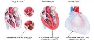
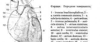
The heart itself consists of three layers: endocardium, myocardium and epicardium. The muscular layer of the left ventricle is several times more powerful than the muscular layer of the right ventricle, since it is on it that the work of pumping blood through the systemic circulation lies.
The endocardium is the inner layer that facilitates intravascular hemodynamics. The myocardium is the muscular layer of the heart. The epicardium performs the protective function of the heart muscle from mechanical injuries and overstretching; it consists of two layers: fibrous (surrounding the heart from the outside and performing a protective function) and cardiac, consisting of visceral and parietal (which fuses with the fibrous layer) layers. Between the visceral and parietal layers there is a fluid-filled cavity (to protect the heart muscle from injury).
If we assume that the heart pumps 5 liters of blood per minute, then in an hour this is 300 liters, in a day – 7200 liters, and in a year – 2 million 628 thousand liters. During an average human life, this organ has to pump more than 100 million liters of blood.
Prognosis for living with a patent foramen ovale
For children with open window symptoms, the prognosis for life is largely positive, although with certain limitations. They are prohibited from putting a high load on the heart. In medicine, a patent foramen ovale refers to abnormal features of the heart, and not to malformations of its development, since the functional load in almost all cases is within normal limits. If the child’s heart, in addition to the abnormal window, has no defects, if there are no chronic diseases of the pulmonary system, if the blood circulation of the heart is not impaired, then there is no need to worry and treatment is not carried out. It is enough to periodically visit a cardiologist, do an ultrasound of the heart (every six months) and avoid heavy exertion. It is not advisable to conduct such examinations more often, since the results will not be indicative, which will cause unnecessary stress in the child.
Our readers recommend!
Pinworms, Giardia, tapeworm, helminths, tapeworm... The list goes on for a long time, but how long are you going to tolerate parasites in your body?
But parasites are the main cause of most diseases, ranging from blood problems to cancer. But parasitologist Sergei Agapkin assures that it’s easy to cleanse your body even at home, you just need to drink...
.. »
We recommend reading:
How is it recognized?
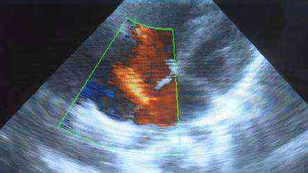
Ultrasound diagnostics will reveal the anomaly.
In almost all cases, an open window is discovered by chance, for example, another treatment is carried out with examination during an inpatient medical examination. However, there are certain symptoms that may indicate such an anomaly:
- The area of the nasolabial triangle turns blue. A newborn may appear blue with frequent coughing, screaming, crying, that is, when the child is tense. In a calm state, the blueness disappears;
- heart murmurs are heard;
- Weak endurance, frequent fatigue, fainting, frequent colds, and pneumonia are observed.
If you notice the above symptoms in newborns, you should contact your pediatrician. In this situation, the doctor will refer you for an ultrasound. Using this study, you can recognize the foramen ovale, its size (usually from 2 to 5 mm), the presence of an opening valve in it, determine whether the window is a defect, as well as how much blood flows through it and the presence of other heart anomalies.
An open oval window in children has the following symptoms:
- size from 2 mm;
- presence of a valve;
- thinning of the edges of the septum in the area of the oval fossa (thickening of the edges is a sign of a defect).