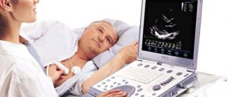
Is it possible for a person with a pacemaker to work on a computer, make cell phone calls, and do ultrasounds?
Modern stimulants impose very few restrictions on the patient's life.
The implantation of a pacemaker in itself does not prevent a person from living a normal, ordinary life. The only thing is that in the first 3-4 months you need to reduce the load on your hand (usually the left) in order for the wound to heal. And then any load is possible, naturally, within reasonable limits. There is no need to perform feats, but going to the store, cleaning, washing, sweeping the floor - all this is within the capabilities of a person with an artificial pacemaker.
A special note for fanatical gardeners: do not grab a shovel immediately after the operation. But in six months - just right. But again: the work should be moderate. If a person is tired, he should rest. Not because the stimulator is overworked - nothing will happen to it, but excessive load on a diseased heart can lead to bad consequences.
After all, the electrical device doesn’t care what a person is doing: lying, sitting, running. The heart is primarily responsible for overall blood circulation. And the amount of load permissible for the patient depends, firstly, on the underlying heart disease, and secondly, on how advanced the device was installed.
Indications for installation of a cardiac pacemaker
Indications for installation of a cardiac pacemaker are:
- the presence of bradycardia with clinical manifestations when the pulse is less than 40 beats/min;
- the occurrence of bradycardia accompanied by Morgagni-Adam-Stokes syndrome;
- development of severe disorders of myocardial contractile function during physical activity;
- the presence of a combination of increased and decreased heart contractions;
- the occurrence of an insufficient increase in heart rate during exercise and sufficient myocardial contraction under resting conditions;
- presence of carotid sinus syndrome;
- the occurrence of atrial fibrillation;
- presence of 2-3 degree A-V block;
- development of incomplete blockade;
- presence of sick sinus syndrome.
For example, the indication for installing a single-chamber stimulator is the presence of a chronic form of atrial fibrillation, as well as for the purpose of stimulating the right atrium when sick sinus syndrome occurs. In other cases, implantation of a dual-chamber pacemaker is preferable. Implantation of cardioverter-defibrillators is carried out in the treatment of the most dangerous arrhythmias, for example, ventricular fibrillation and ventricular tachycardia, as well as for the prevention of sudden cardiac death.
An endocardial electrode is installed in the presence of any bradycardia refractory to drug therapy and leading to hemodynamic failure. A pacemaker can be installed in acute myocardial infarction if there is a risk of high-degree blockade. An indication for the installation of a pacemaker is also the presence of threatening ventricular tachycardia, which occurs against the background of sinus bradycardia, as well as in the presence of tachybradyarrhythmic syndrome.
Among the indications for installing a pacemaker is the presence of very slow or suddenly stopping heart activity. A pacemaker should also be installed for certain types of heartbeat that do not respond to conventional treatment. The main indications for installing a heart pacemaker are the presence of blockades, various types of heartbeats, and also, in some cases, heart failure.
Medicine does not stand still; new medicines and devices are constantly appearing that can extend human life. Heart disease remained incurable for several decades. But now cardiologists have the opportunity not only to “look” into the heart, to see how it works inside, but to make it work. The heart pacemaker became a real salvation; doctors and patients always received only positive feedback.
The device seems to give people a “second chance” to live a full life again. The operation is not considered complicated; it takes only a few minutes. But do not forget that at first, for the first time after the operation, you need to listen to your condition and not overwork. To avoid problems in the future, you must follow the recommendations of doctors.
There are many horror stories about pacemakers. How true are they?
Yes, there are many unconfirmed tales about the dangers and limited scope of application of these devices. As if patients with an implanted device cannot ride in electric transport - in a trolleybus, tram. This is complete nonsense .
Some patients are afraid to use cell phones, work on a computer, or even watch TV. Concerns about television and computers do not hold water.
As for mobile phones , you should be careful here. For example, cell phones should not be placed in the chest pocket; they are allowed to be kept no closer than 20 centimeters to the stimulator. That is, if you have a stimulator on your left side, then when talking on a mobile phone, you should bring the receiver to your right ear.
The prejudice that has persisted longer than others is about the dangers of magnetic radiation from microwave ovens . But if a person was given a stimulant in the last 10-15 years, then he can be near a working microwave without fear. The old generation of stimulators may not have worked well with microwave ovens, but now more advanced models are used.
The pacemaker may fail due to electric shock.
Indications for installing a pacemaker
A device that supports the functioning of the heart is indispensable in case of arrhythmia if the heart rate remains at a sufficiently low level. With rare contractions of the heart muscle, the threat of acute heart failure remains. A sharp deterioration in the condition can occur at any time and lead to cardiac arrest.
The absolute indications for installing a pacemaker are:
- pulse less than 40 beats per minute during physical activity;
- bradycardia, which manifests itself in the form of dizziness and fainting;
- AV block with severe symptoms;
- sick sinus syndrome;
- transverse heart block.
If absolute indications are confirmed, the operation is performed urgently or planned.
Relative readings do not require urgent installation of the device. These include the following signs:
- Second or third degree AV block without symptoms;
- loss of consciousness, cardiac arrest.
What to do in this case?
You should definitely see a doctor , although short-term exposure to electric current, as a rule, does not disable the stimulator.
Modern stimulators have such a high level of protection that even a defibrillator, an electrical device that is used in medicine to start a stopped heart with an electrical impulse with a power of 10 kilovolts, cannot damage them.
But patients are not allowed to do electric welding. So if a patient with ECS works as an electric welder, he will have to say goodbye to the profession.
Installation of the device
Many people are interested in how the device installation process occurs. The operation is considered simple. The patient is prepared in advance, the necessary examinations are carried out. The process does not take long.
The essence of the procedure:
- insert a special device into the subcutaneous fat tissue
- place electrodes in various areas of the heart muscle
The operation is performed under local anesthesia. The following is done:
- The patient makes an incision in the collarbone area.
- Electrodes are inserted through a thin vein.
- The device is connected to the heart.
Important! Despite the fact that the procedure is simple, all work on installing a pacemaker is carried out using special X-ray equipment.
After installing the device, people's lives change, new requirements appear, and some restrictions arise. But you can get used to everything. We must remember that the heart remains the same and must be protected.
Frequently asked questions about x-rays
“Take a deep breath and don’t breathe!”
This team is probably remembered by those who once underwent an X-ray examination of the chest organs.
“The brainchild” of Wilhelm Conrad Roentgen is still relevant today. Without X-rays, it is impossible to detect a number of serious diseases.
Together with Yulia Aleksandrovna Rutskaya, a radiologist, deputy chief physician for radiation diagnostics at the Expert Kursk Clinic, we are looking for answers to frequently asked questions about x-rays.
— Yulia Aleksandrovna, how often can X-rays be taken per month and year?
The question of how many times an X-ray can be taken in a given period of time cannot always be answered unambiguously. In practice, it is performed as often as required by the doctor. Many diseases require monitoring over time, so studies can be performed at certain intervals. Of course, they are done no more often than necessary.
It is also important that the total dose does not reach the maximum permissible - 50 millisieverts/year. Usually in practice this dose is not achieved.
— How many days after fluoroscopy can a repeat x-ray be taken?
Fluoroscopy is an x-ray video examination, and the dose accordingly depends on the time of the procedure. If the patient's condition does not require frequent monitoring, repeated X-ray diagnostics are performed after about 2-4 weeks. However, again, this is largely determined by the specific clinical situation.
— Why can’t X-rays be done for pregnant women?
X-ray radiation is classified as ionizing radiation. It is harmful to the tissues and organs of the fetus, the genetic material of its cells. As a result of irradiation, various pathologies in the development of a child can occur.
Read the material on the topic: Is it possible to do an MRI during pregnancy?
Sometimes X-ray examinations are performed for pregnant women, but usually for health reasons, when the benefit outweighs the risk.
— Is it possible to take x-rays while breastfeeding?
If there is no urgent need to conduct this study in a nursing mother, it is better to postpone it. However, in any case, it is important to remember that the solution to this issue is within the competence of the attending physician.
— What happens if you feed your baby breast milk after fluorography?
A negative effect on the growing tissues of a small organism cannot be ruled out, so it is better not to take risks.
X-RAY STUDIES ARE CARRIED OUT FOR PREGNANT WOMEN, BUT USUALLY FOR VITAL INDICATORS, WHEN THE BENEFITS EXCEED THE RISK
After the examination, it is necessary to express milk for 24 hours and not feed it to the baby. The issue of selecting the appropriate mixture is decided together with the pediatrician.
— Why does the image of bones appear white on an x-ray?
The images appear different because tissues and organs absorb X-ray radiation to varying degrees. The bones absorb it well, without letting it pass through to the film to a significant extent. For this reason they appear white.
Read material on the topic: Nobel laureate without a high school diploma: X-ray as a proper name
— Is it permissible to do x-rays and fluorography on the same day?
Yes. This happens when fluorography reveals some changes in the lungs. In this case, further examination is necessary, so an x-ray is taken after it. An overview image of the chest is taken in two projections.
— Is it possible to take an x-ray during menstruation or is it better to wait?
In general, yes, however, depending on the specific situation, there may be time restrictions for the procedure.
— Why can’t you do x-rays and physical procedures on the same day?
X-ray radiation negatively affects body tissues. Physiotherapeutic procedures have an activating effect on them. It turns out that there is a kind of double burden on a person.
Therefore, it is advisable to separate them: on one day - an x-ray, and on the next - a physical procedure.
— Is it possible to do an ultrasound and an x-ray on the same day?
Yes. These studies are based on various physical phenomena. Ultrasound is not classified as ionizing radiation.
Often one area of the body is examined with one method, and another with another.
Read the material on the topic: How to prepare for an abdominal ultrasound?
It must be remembered that the sequence of examinations may matter. Therefore, you need to clearly discuss these points with your doctor so that during a particular study, “artifacts” (“shadows”) do not appear on the images that can complicate the interpretation of the results obtained.
— Why are they asked to remove jewelry before performing fluoroscopy?
Being made of metal, they do not transmit X-rays well and distort the image. At the same time, “artifacts” appear on the film, which cover one or another area of the examination.
You can find out the cost of the study and sign up for an x-ray here
Please note: the service is not available in all cities
— What are the contraindications for x-rays?
The main and absolute thing is pregnancy.
It is also not recommended for children: for them, the test is performed only when necessary and strictly as prescribed by the doctor.
The issue of conducting it in seriously ill patients is also decided together with the attending physician.
Other questions:
Why are x-rays dangerous?
What is the difference between MRI and X-ray?
When is MRI contraindicated?
For reference:
Rutskaya Yulia Alexandrovna
In 2002 she graduated from Kursk State Medical University.
In 2004, she completed an internship course in Radiology.
For more than six years she worked as a radiologist and ultrasound diagnostic doctor in a children's clinic.
Currently he holds the position of Deputy Chief Physician for Radiation Diagnostics at the Expert Kursk Clinic. Radiologist of the highest qualification category (MRI, CT, X-ray).
Receives at the address: Karl Liebknecht St., 7.
How is the operation performed?
The main indications for the use of such a measure to restore normal heart function are: bradycardia (its various forms), serious impairment of the ability of the myocardium to contract, frequent changes from a rapid heartbeat to a slow one, an incorrect ratio of contractions (increases at rest and decreases during exercise). Carotid sinus syndrome, A-V block, weakened sinus node, incomplete block - all these are also direct indications for surgery.
Despite the impressions that one gets about the operation to install a pacemaker, the process itself is not complicated. In general terms, the intervention consists of the following:
- a special-purpose device is inserted into the subcutaneous fatty tissue;
- the electrodes that come from it are placed in various parts of the organ.
Important! For the manipulation, general anesthesia is not required; everything is done under local anesthesia.
In order to carry out the above steps, an incision is made in the collarbone area. After this, using a thin vein, the electrodes are connected to the organ. The entire procedure is carried out using X-ray equipment for control purposes. When the electrodes are at their destination, their parts on the other side are connected to the device. It is placed in the area of the collarbone in the subcutaneous tissue, and the operation is considered complete. In the video of the Heart Pacemaker operation we see that the intervention is very simple. The most important condition for this is the literacy and experience of the surgeon.
The influence of braces and retainers on the results of the study
Metal elements distort the examination results and illuminate the installation site (skull area). This leads to the fact that the result will be incorrect, and a lot of money on the procedure will be wasted. If you have braces in the oral cavity, you can examine the limbs and cervical spine. However, MRI of jaw and brain tissue is questionable.
Before the procedure, it is important to ask your dentist what the braces are made of. Titanium and ceramic systems are not subject to heating under the influence of a magnetic field. Experts conducted an experiment with the participation of 10 patients with the following orthodontic devices: stainless steel braces, ceramics, and a stainless steel mandibular retainer.
INTERESTING: retainers: what are they and how are they placed?
According to the survey results, the distortion depended on the material of the systems. The worst readings were obtained when using steel structures and tubes, the best - when using ceramic braces. Thus, the material affects the course of the examination, and it is important to warn the doctor about the presence of orthodontic structures in the oral cavity.
Indications and contraindications for installation
The pacemaker has various indications for installation. These can be either congenital or acquired diseases.
These include:
When choosing a specific type of stimulator, the doctor pays attention to all the pros and cons of a particular device and the characteristics of the patient’s disease.
The operation to install a pacemaker is quite safe for the patient; there are no absolute contraindications. In some acute conditions, the operation is postponed while they are being relieved.
The most striking examples of such conditions are: acute abdomen (exacerbation of gastrointestinal ulcer, appendicitis, acute pancreatitis), acute inflammatory diseases, psychiatric diseases, due to which the patient is not in contact. These contraindications are relative, that is, temporary.
