Heart
is a muscular organ that provides blood supply to all organs and systems of the human body.
Holotopia
- This is a determination of the position of the heart relative to neighboring areas, located in the anterior mediastinum.
Skeletotopia
– the cardiac border with relative dullness, the cardiac upper border is along the third intercostal space on the left side of the left and right parasternal lines, on the left, the border of the heart will be from the left parasternal line to the apex beat.
Right border of the heart
will appear from the right parasternal line from the third intercostal space to the fifth costal cartilage, and the lower border of the heart will appear from the fifth costal cartilage along the parasternal line to the apex of the cardiac impulse.
Syntopy
– when the heart is surrounded by the cardiac sac.
- The front side of the heart is adjacent to the chest cavity.
- Along the bottom side directly to the diaphragmatic part.
- The lateral side is covered with mediastinal pleura.
- The esophagus, vagus nerve, bronchi, trachea, thyroid gland and others are adjacent to the back.
- Important cardiac vessels are attached along the upper side.
Heart structure:
the internal structure has an endocardium - the inner layer of the heart covers the chambers from the inside and forms the valve apparatus of the heart, which includes the pulmonary valve, extending from the right ventricle, the aortic valve, extending from the left ventricle, the aortic valve, extending from the left ventricle.
The tricuspid valve is located between the right atrium and the ventricle, the bicuspid valve is located between the left ventricle and the left atrium.
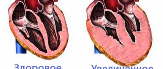
The middle layer of the myocardium consists of cardiomycytes. The size of the myocardium in the left ventricle is about one centimeter; an increase in its thickness by two or more millimeters indicates its hypertrophy. The papillary muscles extend from the myocardium; they connect to the valves through tendon filaments, the epicardium, pericardium or pericardium.
Properties of the heart muscle
- One hundred percent properties of cardiac tissue include conductivity, contractility, and excitability. An important property is the automaticity of the heart muscle.
- The volume of excitability of the heart muscle is slightly lower than that of skeletal muscle and has a higher threshold of irritability. The resting state value is 85 – 90 mV.
- Automaticity is the cardiac performance of an organ, a cell that contracts rhythmically under impulse influence.
Types of Automation:
- The sinoatrial node is located behind the right edge of the atrium.
- Internodal tracts.
- The atrioventricular node is located lower between the atria under the endocardium of the right atrium.
- The bundle of His emerges from the Ashov-Tavara node.
Cardiac conductivity is lower than striated skeletal muscle. The speed of excited conduction is about a meter per second. The spread of excitation occurs in all directions due to the special structure of the heart muscle.
Refractoriness
is a lack of sensitivity during a certain period. In cardiac muscle, refractoriness is much more pronounced than in skeletal muscles. At the initial time of contraction and relaxation, sensitivity completely disappears and has a period of 0.1-0.3 seconds. With complete relaxation of the heart, excitability cells are gradually restored.
Risk factors for developing cardiac arrhythmia
Risk factors for developing cardiac arrhythmias include:
- ✔ Age.
With age, the heart muscle becomes depleted, weakens and loses some of its nutrition. This can affect the formation and conduction of electrical impulses. - ✔ Genetics.
In people with congenital abnormalities of the heart, arrhythmias occur more often. Moreover, a number of arrhythmias (eg, Wolff-Parkinson-White syndrome, some, some forms of long QT syndrome) are congenital. - ✔ Coronary heart disease
, other heart diseases, open heart surgery. Narrowing and occlusion (complete closure) of the lumen of the coronary arteries (arteries that supply the heart muscle), pathology of the heart valves, previous open-heart surgery, cardiomyopathies and other factors damaging the heart are a serious risk factor for the development of almost all types of arrhythmias. - ✔ Thyroid diseases.
With increased thyroid function, increased production of hormones occurs, overall metabolism increases, and heart contractions become more frequent and irregular. Atrial fibrillation most often develops. With insufficient thyroid function, metabolism decreases, which causes, and in some cases, extrasystole.
- ✔ Medicines and biologically active food supplements.
Most often, arrhythmia is caused by uncontrolled and excessive use of cold medications containing ephedrine and pseudoepinephrine, as well as a number of other drugs, for example, diuretics, laxatives or anti-asthmatics. - ✔ High blood pressure.
This increases the risk of developing coronary heart disease. High blood pressure also causes the wall of the left ventricle to thicken, which can change the way impulses are conducted through it. - ✔ Obesity.
Being a risk factor for the development of coronary heart disease, obesity also increases the risk of developing arrhythmias. - ✔ Diabetes mellitus.
Diabetes mellitus in the stage of decompensation (uncontrolled blood sugar levels) greatly increases the risk of developing coronary heart disease and arterial hypertension. In addition, (low blood sugar) can be a trigger for the development of cardiac arrhythmia.
✔ Pathological sleep apnea syndrome.
This pathology may be accompanied by bradycardia and atrial fibrillation.
✔ Electrolyte disturbances.
Electrolytes such as potassium, magnesium, sodium and calcium form the basis for the formation, maintenance and conduction of electrical impulses in the heart. Too high or too low concentrations of electrolytes in the blood and in heart cells affect the electrical activity of the heart and can cause the development of arrhythmias.
✔ Alcohol consumption.
Drinking large doses of alcohol increases the risk of developing atrial fibrillation. Sometimes, the development of atrial fibrillation after excessive alcohol consumption is called “holiday heart syndrome”. Chronic alcohol abuse has a detrimental effect on heart cells and can lead to cardiomyopathy, which is also the basis for the development of cardiac arrhythmias.
✔ Use of stimulants.
Psychostimulants such as caffeine, nicotine, etc. are a cause of development and can also lead to the development of more severe heart rhythm disturbances over time. The use of amphetamines and cocaine can damage the heart muscle with the development of any of the existing arrhythmias and even lead to sudden cardiac death due to the development of ventricular fibrillation.
Heart function and work
Contractile function
– rapid contractility of the heart, controls the level of hypertension.
Endocrine function
– the ability to produce natriuretic factor occurs.
Information function - information is transmitted in the form of blood flow speed in the tissue with a change in its metabolism.
Blood feeds the body with the help of the heart with useful elements and oxygen. Endocrine cells (distribute cardiodilantin - hormones that enhance the work of the heart - throughout many organs) are formed in the myocardium - the heart muscle and in the atria.
There is the following function that can remove carbon dioxide and used substances from the body.
Symptoms of a slow heart rate
A slow heart rate, or bradycardia, can cause the following symptoms:
- Weakness
- Difficulty walking, climbing stairs, or exercising
- Fatigue
- Irregular breathing
- Dizziness
- Fainting
How to increase your heart rate naturally?
Any physical movement makes the heart beat faster, but among the simplest in terms of execution and load, you can try these:
- Aerobic exercise : Aerobic exercise safely increases your heart rate. These include walking, water aerobics, cycling, swimming, running, kickboxing and dancing. Choosing the right aerobic exercise should be based on your personal needs, abilities and preferences. If you're just starting to exercise, it's best to start with lower-impact, gentle-movement activities like walking and swimming. Once your heart starts to get stronger, you can move up a level to longer activities like dancing or cycling.
- Strength exercises : These involve the use of kettlebells or any other mechanism that adds weight. When muscles meet resistance, they demand more oxygenated blood, which in turn increases your heart rate. At first you can use your own body as a weight by doing push-ups or squats, but over time you can add additional weight.
- Stretching : What many people don't know is that stretching can get your heart pumping and it certainly can and should be done for good heart health. Stretching increases flexibility and improves blood flow, and is not very difficult to do.
Regulation of the heart
- There are two types
of regulatory capacity for the heart - humoral and nervous regulation. - Some electrolytes, hormones
, are highly active substances that have humoral regulation on the heart. The mediator adrenaline has a stimulating effect (increases cardiac activity) in both small and small doses. - Many electrolytes affect the automaticity of the heart
, namely the heart muscle and the cardiac conduction system. A deficiency of ions such as potassium contributes to a decrease in the strength of heart contractions and, accordingly, their frequency, the rate of decrease in the distribution of excitation throughout the conduction system, but leads to a relaxation phase. - The characteristic features of
excess potassium ions are similar to the vagal effect. An excess of potassium ions has the opposite effect. In the future, this can lead to the death of the patient as a result of cardiac arrest with insufficiency of myocardial diastole; in general, ions that enhance cardiac activity have a certain ratio in the peripheral blood. - Temperature also has a stimulating effect
on cardiac tissue, so when exposed to heat, the sinocarotid zone leads to an increase in the work of the heart, and when exposed to cold, a decrease. - Nervous regulation is provided by the vagus
- this is the parasympathetic nervous system, and the sympathetic nervous system, respectively, the vagus or vagus nerve helps to slow down cardiac activity (force, frequency, rhythm, excitation, conduction), and the sympathetic nervous system, on the contrary, increases cardiac activity.
The influence of hormones on the heart in diseases
In diseases accompanied by an increase in the tone of the parasympathetic department (release of acetylcholine) and a decrease in sympathetic activity (lack of adrenaline), the work of the heart decreases. In such cases, patients are diagnosed with bradycardia, myocardial dystrophy, and hypotension occurs due to insufficient cardiac output. Such pathological conditions include:
- pancreatitis;
- peptic ulcer of the stomach and duodenum;
- cholelithiasis;
- insufficient adrenal function;
- hypothyroidism;
- severe liver disease;
- traumatic brain injuries.
There are also diseases in which the heart works harder and wears out quickly. Stress hormones play a leading role in the mechanism of development of tachycardia, hypertension and heart failure in such pathologies. Diseases with excessive hormonal activity stimulating the heart muscle:
- thyrotoxicosis;
- pheochromocytoma;
- tumors of the pituitary gland, adrenal glands;
- hyperaldosteronism;
- Itsenko-Cushing syndrome;
- polycystic ovary syndrome.
And here is more information about dysmetabolic myocardial dystrophy.
Heart cells have the ability to synthesize and release hormones into the blood. Atrial natriuretic factor is formed when pressure increases and stretches the muscle fibers of the atrium. It promotes the release of sodium and water from the body. Atriopeptins are responsible for the sensation of thirst and salt intake. Relaxil facilitates labor.
The work of the heart muscle is controlled by hormones produced by the endocrine system. Acetylcholine inhibits the rhythm and strength of contractions, and hormones of the adrenal glands, thyroid gland and pituitary gland activate them.
What makes the heart work better?
At the points where arterioles pass into capillaries there are circular smooth muscle fibers. In the passive form of the organ, two-thirds of the capillaries are closed. In the active state, many capillaries are open and blood circulation in the organ occurs.
In stressful situations, abundant blood supply occurs, affecting all organs at once, capillaries open. If there is a sharp drop in blood pressure, then blood will not flow into the open capillaries.
To prevent pressure drop:
- Vascular stenosis;
- Increased cardiac activity;
- Blood circulation thanks to the veins at rest at 70 - 80%.
REVIEW FROM OUR READER!
The sympathetic nervous system and the hormone adrenaline increase the functional activity of the heart and vascular stenosis.
The parasympathetic nervous system and the mediator acetylcholine inhibit cardiac function.
How to strengthen the work of the heart?
There are many ways to enhance cardiac activity. Substances that enhance the work of the heart are adrenaline and sympathetic nerves.
Methods to increase heart function:
- Activity
. The more a person moves, the better the heart will work. - Regular blood pressure measurement.
As people age, they should visit their doctor twice a year if possible, or even more. The level not exceeding the norm is 140/90. - Limiting salty foods
. Limiting salt does not mean giving up, but normally it’s best to consume a teaspoon or 5 g of salt per day. - Give up bad habits completely.
You can quit smoking at any age; it will not affect your body’s metabolism in any way.
With a poorly functioning heart, there is a risk of heart failure or worsening of the person’s condition, weakness, fatigue and frequent dizziness. To do this, you can strengthen your heart by doing physical exercise every day and following a healthy diet.
You should not eat foods that contain trans fats, they can lead to the spread of chronic diseases; trans fats are found in margarine and baked goods.
Another method that is necessary to improve heart activity is physical activity:
- Stretching.
- Aerobics.
- Running exercises.
- Swimming.
- Skiing, cycling or skating.
Result:
the heart is the central organ of the cardiovascular system, providing blood to many systems and organs in general; the properties of the heart are conductivity, excitability, refractoriness, contractility; the work of the heart is regulated by various complex systems, both humoral and nervous.
The work of the heart plays a subordinate role, since changes in metabolism are caused through the nervous system. Shifts in the content of various substances in the blood, in turn, affect the reflex regulation of the cardiovascular system.
Heart function is affected by changes in potassium and calcium levels in the blood. An increase in potassium content has negative chronotropic, negative inotropic, negative dromotropic, negative bathmotropic and negative tonotropic effects. Increasing calcium levels does the opposite.
For normal heart function, a known ratio of both ions is necessary, which act similarly to the vagus (potassium) and sympathetic (calcium) nerves.
It is assumed that when the membranes of the muscle fibers of the heart are depolarized, potassium ions and ions quickly leave them, which contributes to their contraction. Therefore, the blood reaction is important for the contraction of the muscle fibers of the heart.
When the vagus nerves are irritated, acetylcholine enters the blood, and when the sympathetic nerves are irritated, a substance similar in composition to adrenaline (O. Levi, 1912, 1921) - norepinephrine. The main transmitter of the sympathetic nerves of the heart of mammals is norepinephrine (Euler, 1956). The adrenaline content in the heart is approximately 4 times less. The heart accumulates adrenaline introduced into the body more than other organs (40 times more than skeletal muscle).
Acetylcholine is quickly destroyed. Therefore, it acts only locally, where it is released, that is, at the endings of the vagus nerves in the heart. Small doses of acetylcholine stimulate the automaticity of the heart, and large doses inhibit the frequency and strength of heart contractions. Norepinephrine is also destroyed in the blood, but it is more persistent than acetylcholine.
When the common trunk of the vagus and sympathetic nerves of the heart is irritated, both substances are formed, but the effect of acetylcholine appears first, and then norepinephrine.
The introduction of adrenaline and norepinephrine into the body increases the release of acetylcholine, and, conversely, the introduction of acetylcholine increases the formation of adrenaline and norepinephrine. Norepinephrine increases systolic and diastolic blood pressure, while epinephrine increases only systolic blood pressure.
In the kidneys, under normal conditions and especially when their blood supply decreases, rhenium is formed, which acts on hypertensinogen and converts it into hypertensin, causing vasoconstriction and a rise in blood pressure.
Local vasodilation is caused by the accumulation of acidic metabolic products, especially carbon dioxide, lactic and adenylic acids.
Acetylcholine and histamine also play a major role in the dilation of blood vessels. Acetylcholine and its derivatives irritate the endings of the parasympathetic nerves and cause local dilation of small arteries. Histamine, a product of protein breakdown, is formed in the wall of the stomach and intestines, in muscles and other organs. Histamine, when it enters the bloodstream, causes dilation of the capillaries. Under normal physiological conditions, histamine in small doses improves blood supply to organs. In muscles during work, histamine dilates capillaries along with carbon dioxide, lactic and adenylic acids and other substances that are formed during contraction. Histamine also causes dilation of skin capillaries when exposed to sunlight (ultraviolet part of the spectrum), when the skin is exposed to hydrogen sulfide, heat, or when rubbed.
An increase in the amount of histamine entering the blood leads to a general dilation of the capillaries and a sharp drop in blood pressure - circulatory shock.
summary of other presentations
“Information about blood” - Explain the picture. We conduct training. Blood flow speed. Movement of blood through blood vessels. Type of bleeding. Blood movement. Heart attack. Blood. Reception at the emergency room. What is shown in the picture. Vaccine.
“Myopia and farsightedness” - Farsightedness. Surgery to restore vision. Myopia. Refraction of light rays. Cornea and lens. Focus. Refraction of light rays during farsightedness. Myopia and farsightedness. Symptoms of myopia. Symptoms of farsightedness.
“Plants listed in the Red Book” - Pyramidal tenacious. Marine pancrates. Bear bow. Japanese beard. Changing of the climate. Perennial herbaceous plant. Spectacular flower. Lunar coming to life. Rare plants listed in the Red Book. Swamp waxweed. Arctic sunflower. Protect and take care of nature.
“Flora world of Russia” - Mixed forests. Steppe plants. Steppe. Flora of Russia. Flora of Russia. Tundra plants. Desert plants. Taiga. Plants of mixed forests. Meadows. Vegetable world. Taiga plants. Tundra. The flora and fauna of our country. Swamps. Deserts. Vegetation types.
“General characteristics of the class of Mammals” - Habitat and external structure of mammals. The baby platypus hatches from eggs. When a dog is hot, it sticks out its tongue. Remember what types of life exist on Earth. Study the features of the external structure of mammals. Determine what sense organs are developed in mammals. External structure of the dog. Presence of horny scales. Ecological groups of mammals. The structure of mammalian skin. Mammary glands.
“Healthy food” - A small debate. “A man is what he eats” by G. Heine. Sources of protein. Amount of water. Vegetables are a source of vitamins on your table. 99% of fast food consists of chemicals. You can do a few days as fasting days. School age is a very important period of life. Proper nutrition. How often do you eat fast food? Food is the main source of human existence. Basics of rational nutrition.
Test No. 4. Topic “Circulatory system”
Option 1.
1. The blood vessels through which blood moves from the heart are:
1) veins of the pulmonary circulation; 2) veins of the systemic circulation;
3) arteries of the pulmonary and systemic circulation; 4) capillaries of the pulmonary and systemic circulation.
2. What kind of blood flows in the veins of the systemic circulation:
1) venous; 2) arterial; 3) oxygenated; 4) mixed.
3. The pulmonary circulation includes veins:
1) liver; 2) lungs; 3) upper limbs; 4) lower extremities.
4. The human heart has a chamber structure. Number of cameras:
1) 3; 2) 2; 3) 4; 4) 5.
5. The pericardial sac is called:
1) epicardium: 2) endocardium; 3) myocardium; 4) pericardium.
6. The heart valve that prevents the movement of blood from the right ventricle to the right atrium is called:
1) bivalve; 2) tricuspid; 3) semilunar; 4) pocket-shaped.
7. In a person’s calm state, the duration of cardiac diastole is:
1) 0.8 sec; 2) 0.4 sec; 3) 0.3 sec; 4) 0.1 sec.
8. Substance that enhances heart function:
D) capillaries of a large circle; D) left ventricle; E) right atrium.
Answers. Topic: “Circulatory and lymphatic systems”
Option 1.
Option 2.
To use presentation previews, create a Google account and log in to it: https://accounts.google.com
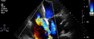
Cardiac output (CO)
Cardiac output is defined as the volume of blood that the heart pumps out in a given period of time. Minute volume refers to the amount of blood the heart pumps in a minute. The same volumes of blood always pass through the right and left halves of the heart, since otherwise there would be an overflow of one circle of blood circulation, and a lack of blood would be felt in the other. If at rest the heart contracts at a speed of 70 beats/min (pulse rate) and with each contraction about 70 ml of blood is released into the systemic circulation (stroke volume), then the estimated minute volume will be about 5 liters (70 beats/min x 70 ml = 4900 ml). This amount of blood roughly corresponds to that contained in the body of a person weighing 70 kg.
During physical activity, blood supply to muscles and other organs increases. Consequently, blood circulation and blood pressure should increase. As circulation increases, heart rate and stroke volume may increase. During severe physical exertion, cardiac output can increase to 25 liters per minute, i.e., five times the total blood volume in the body. This increase in cardiac output can be achieved if, for example, stroke volume increases from 70 to 140 ml and heart rate increases to 180 beats/min (180 beats/min x 140 ml = 25 200 ml/min = 25. 2 l/min).
Slide captions:
For home: § 11 Topic: The structure and work of the heart Objectives: To study the structure, work and regulation of the heart
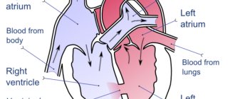
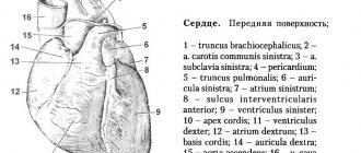
Structure of the heart The human heart is located approximately in the middle of the chest. This is a four-chambered muscular organ that works continuously throughout life. The shape of the heart resembles a flattened cone and consists of two parts - right and left. Each part includes an atrium and a ventricle. The size of the heart is approximately the size of a fist. The average heart weight is about 300 g in men, and about 220 g in women. The heart is covered with a thin and dense membrane, forming a closed sac - the pericardium, the pericardium. Between the heart and the pericardial sac there is a fluid that moisturizes the heart and reduces friction during its contractions. The walls of the ventricles have more developed walls than the atria.
The structure of the heart The muscular wall of the left ventricle is especially thick, which, when contracting, pushes blood through the vessels of the systemic circulation. The atria and ventricles are connected by openings.
Structure of the heart The leaflet valves of the heart are located at the edges of the openings. On the side of the valves facing the ventricular cavity there are special tendon threads. These threads keep the valves from bending. Between the left atrium and the left ventricle the valve has two leaflets and is called bicuspid, between the right atrium and the right ventricle there is a tricuspid valve. The bicuspid and tricuspid valves allow blood to flow in one direction - from the atria to the ventricles. The wall of the heart consists of three membranes: 1. Inner membrane (endocardium), 2. Middle membrane (myocardium), 3. Outer membrane (epicardium).
Structure of the heart Each semilunar valve consists of three leaves that resemble pockets. Semilunar valves allow blood to flow in only one direction - from the ventricles to the aorta and pulmonary artery. There are also valves between the left ventricle and the aorta extending from it, as well as between the right ventricle and the pulmonary artery extending from it. Because of the peculiar shape of the valves, they are called semilunar.
Laboratory work “Structure of the heart” 1. Find the right and left atria, right and left ventricles. 2.Compare the thickness of the walls of the atria and ventricles. 3.Why are the muscular walls of the left ventricle the thickest? Explain. 4.Locate the semilunar and leaflet valves on the model. Explain their functions.
Work of the heart. Cardiac cycle The atria and ventricles can be in two states: contracted and relaxed. Contraction and relaxation of the atria and ventricles of the heart occur in a certain sequence and are strictly coordinated in time. The cardiac cycle consists of contraction of the atria, contraction of the ventricles, relaxation of the ventricles and atria (general relaxation). The duration of the cardiac cycle depends on the heart rate.
Cardiac cycle In a healthy person at rest, the heart contracts 60-80 times per minute. Therefore, the time of one cardiac cycle is less than 1 s. The cardiac cycle begins with contraction of the atria, systole, which lasts 0.1 s. At this moment, the ventricles are relaxed, the leaflet valves are open, and the semilunar valves are closed. During contraction of the atria, all the blood from them enters the ventricles. Contraction of the atria is replaced by their relaxation, diastole.
Cardiac cycle Then ventricular systole begins, which lasts 0.3 s. At the onset of ventricular contraction, the semilunar and tricuspid valves remain closed. Contraction of the muscles of the ventricles leads to an increase in pressure inside them. The pressure in the cavities of the ventricles becomes higher than the pressure in the cavities of the atria. Blood moving towards the atria meets the valve leaflets on its way. The valves cannot turn inside the atria; they are held in place by tendon threads.
Cardiac cycle Blood enclosed in the closed cavities of the ventricles has only one path left - to the aorta and pulmonary artery. Ventricular systole is replaced by general diastole, relaxation, which lasts 0.4 s. At this moment, blood flows freely from the atria and veins into the cavity of the ventricles. The semilunar valves are closed. The peculiarities of the cardiac cycle include the ability to maintain the working activity of the heart throughout life.
Cardiac cycle Of the total duration of the cardiac cycle of 0.8 s, the cardiac pause accounts for 0.4 s. This interval between contractions is sufficient to fully restore the heart’s performance. During each contraction of the ventricles, a certain portion of blood is pushed into the vessels. Its volume is 70-80 ml. In 1 minute, the heart of an adult at rest pumps 5-5.5 liters of blood. The heart pumps about 10,000 liters of blood per day.
Cardiac cycle During physical activity, the amount of blood pumped by the heart in 1 minute in a healthy, untrained person increases to 15-20 liters. For athletes, this value reaches 30-40 l/min. Systematic training leads to an increase in the mass and size of the heart and increases its power.
Automaticity of the heart In the human heart, the source of automaticity is special muscle cells. They are located in its various departments. The main center for the generation of automatic impulses are muscle cells located in the right atrium. The beating heart creates weak bioelectrical signals that are carried throughout the body. These signals recorded from the skin of the arms and legs and from the surface of the chest are called an electrocardiogram. An electrocardiogram reflects the state of the heart muscle and serves as the most important indicator of its activity. The isolated heart of animals can work rhythmically for a long time if nutrient solutions saturated with oxygen are passed through the vessels feeding the heart. Automaticity of the heart is the ability of the heart to contract rhythmically without external stimulation under the influence of impulses arising within itself.
Regulation of heart function. Nervous regulation. Impulses are transmitted to the heart along nerve fibers from nerve centers located in the medulla oblongata and spinal cord. Influences that weaken the work of the heart are transmitted through the parasympathetic nerves, and those that enhance its work are transmitted through the sympathetic nerves. The central nervous system constantly monitors the functioning of the heart. Inside the cavities of the heart itself and in the walls of large vessels there are nerve endings - receptors that perceive pressure fluctuations in the heart and blood vessels. There are two types of nervous influences on the heart: some are inhibitory, i.e. those that reduce the heart rate, others that accelerate.
Nervous regulation of work Under the influence of positive emotions, people can perform colossal work, lift weights, and run long distances. For example, a person’s heart rate increases when he quickly gets up from a lying position.
Humoral regulation of work Adrenaline has the opposite effect. Even in very small doses it enhances heart function. In medical practice, adrenaline is sometimes injected directly into a stopped heart to force it to contract again. An increase in the content of potassium salts in the blood depresses, and calcium increases the work of the heart. Along with nervous control, the activity of the heart is regulated by chemicals constantly entering the blood. This method of regulation is called humoral regulation. A substance that inhibits the work of the heart is acetylcholine. The sensitivity of the heart to this substance is so great that in a dose of 0.0000001 mg acetylcholine clearly slows down its rhythm.
Repetition: **Test 1. Which valves are located on the right side of the heart? Where? Between the atrium and the ventricle - bicuspid. Between the atrium and the ventricle is the tricuspid. Semilunar valves at the root of the aorta. Semilunar valves at the root of the pulmonary artery. **Test 2. Which valves are located in the left side of the heart? Where? Between the atrium and the ventricle - bicuspid. Between the atrium and the ventricle is the tricuspid. Semilunar valves at the root of the aorta. Semilunar valves at the root of the pulmonary artery.
Repetition: Test 3. Correct judgment for a cardiac cycle lasting 0.8 sec: Atrial systole – 0.1 sec, ventricular systole – 0.3 sec, total diastole 0.4 sec. Atrial systole – 0.2 sec, ventricular systole – 0.2 sec, total diastole 0.4 sec. Atrial systole – 0.3 sec, ventricular systole – 0.3 sec, total diastole 0.2 sec. Atrial systole – 0.1 sec, ventricular systole – 0.4 sec, total diastole 0.3 sec.
Repetition: Test 4. Which nerves strengthen and which inhibit the work of the heart? Sympathetic enhances, parasympathetic inhibits. Sympathetic ones inhibit, parasympathetic ones strengthen. Both sympathetic and parasympathetic inhibit. Both sympathetic and parasympathetic strengthen.
Repetition: Test 5. What substance inhibits the heart? Adrenalin. Acetylcholine. Norepinephrine. Vasopressin. **Test 6. What hormones increase heart function? Adrenalin. Acetylcholine. Norepinephrine. Vasopressin. Test 7. Which ions enhance and which inhibit the work of the heart? Potassium ions enhance, calcium ions inhibit. Calcium ions enhance, potassium ions inhibit. Iron ions enhance, magnesium ions inhibit. Magnesium ions strengthen, iron ions inhibit.
Conduction system of the heart
The processes of excitation in the heart, impulse conduction and contraction of the heart muscle are unique.
For example, in the presence of adequate nutrients and oxygen, an isolated heart can contract for a long period of time without an external nerve impulse. The heart has an autonomous system for generating nerve impulses, the sinoatrial node, which is located in the right atrium at the level of the confluence of the superior vena cava. This so-called pacemaker (pacemaker) generates approximately 60-70 impulses per minute. The impulse causes contraction of the atrium muscles, through which it is transmitted to the atrioventricular node (AV node). From there, the impulse reaches the His bundle, which is a fibrous muscle structure, and is transmitted further to the ventricular myocardium. There, it first passes through the branching node (Tovar's node) along the interventricular septum to the apex of the heart and is distributed along the Purkinje fibers throughout the entire ventricular myocardium. Thus, the heart muscle is excited and, in accordance with the impulse conduction system, contracts rhythmically. The frequency, speed of impulse conduction and degree of contraction depend on the autonomic nervous system. This brings the activity of the heart into line with the body’s demands (for example, an increase in minute volume of blood flow during physical activity).
Complications of arrhythmia
A number of heart rhythm disorders can increase the risk of developing conditions and diseases such as:
- Stroke
. When the atria fibrillate, they are unable to adequately pump blood to the ventricles. Slowing blood flow in the atria leads to the formation of clots. If a small piece breaks off from the clot, it can enter the bloodstream, spread throughout the body and clog the cerebral arteries, causing the development of ischemic stroke, i.e. damage or death of part of the brain, and sometimes leads to death. - Congestive heart failure
. Due to a prolonged period of bradycardia or tachycardia, such as atrial fibrillation, the heart may not pump efficiently. By controlling your heart rate, you can improve left ventricular contractility and reduce signs of heart failure.
These conditions are not dangerous; they arise under the influence of physiological processes and pass quickly. But cardiac arrhythmias that occur for no apparent reason require consulting a doctor.