What diseases does the examination reveal?
Echocardioscopy is used to identify the following diseases:
- heart defects
- thoracic aortic aneurysm
- heart tumors
- cardiac aneurysm
- intracardiac thrombi
- coronary heart disease, including myocardial infarction
- cardiomyopathy
- endo-, myo-, pericarditis
- some other pathology.
The study does not analyze the nature of the heart rhythm (only the order of contraction of the heart chambers and the frequency of contractions are determined) - for this, an electrocardiogram is used in a comprehensive examination.
Types of research
EchoCS is conventionally divided into three groups:
- methods for imaging cardiac structures: one-dimensional and two-dimensional studies
- methods for assessing blood circulation in the heart and large vessels extending from it: Doppler examination (it can be pulsed, continuous and color two-dimensional, each has its own indications)
- additional techniques: transesophageal, contrast and stress echocardiography (they are carried out only as prescribed by a cardiologist, in a clinic equipped with a cardiac intensive care unit).
What is the difference between echocardioscopy and echocardiography? It doesn’t matter what you call this study to the medical staff, you will be clearly understood.
By the term “echocardiography”, doctors understand either ultrasound of the heart as a science, or ultrasound of the heart with a printed graphic image of the heart. “Echocardioscopy” - observation, visualization of the heart in real time on the monitor screen, without printing the image.
Who needs to undergo the study
Heart ultrasound for children and adults is performed in the following cases:
- when listening to noises by a doctor using a phonendoscope
- with noted changes on the ECG
- if there are complaints about interruptions in heart rhythm
- shortness of breath appeared when doing physical work or at rest
- chest pain
- if there is an increase in blood pressure
- after a heart attack (the diagnosis itself is made by ECG and blood test for troponins)
- for rheumatic diseases
- with flu or sore throat, if there are complaints of heart pain, arrhythmias or shortness of breath
- with varicose veins of the lower extremities.
Fetal echocardioscopy is performed during pregnancy (usually at 18-22 weeks) in perinatal centers in the following cases:
- pregnant woman suffers from heart defect
- children have already been born with heart defects
- pregnant woman suffers from diabetes
- a woman takes certain medications (for example, anticonvulsants) for health reasons during pregnancy
- during the first screening, deviations in the thickness of the nuchal translucency were noticed, but amnio- or cordocentesis did not show any deviations (the nuchal translucency may increase due to the fact that the heart does not cope well with the load)
- the second screening ultrasound revealed abnormalities in the size or function of the heart
- with intrauterine growth restriction of the baby
- a woman suffered from infectious diseases during pregnancy
- Some malformations were noted on routine ultrasound (they can also be combined with a heart defect).
How to prepare for the procedure
No preparation is required for the study. For small children (newborns and infants), it is advisable that they sleep during the procedure. Such patients need to be fed one and a half to two hours before the ultrasound, and those who are falling asleep or sleeping should be brought in. Feeding immediately before the procedure is not recommended.
Adults with a pulse more than 90 and/or an increase in “upper” blood pressure above 160 mmHg. It is imperative to consult with a cardiologist about taking medications to eliminate these symptoms, otherwise the study will be inaccurate.
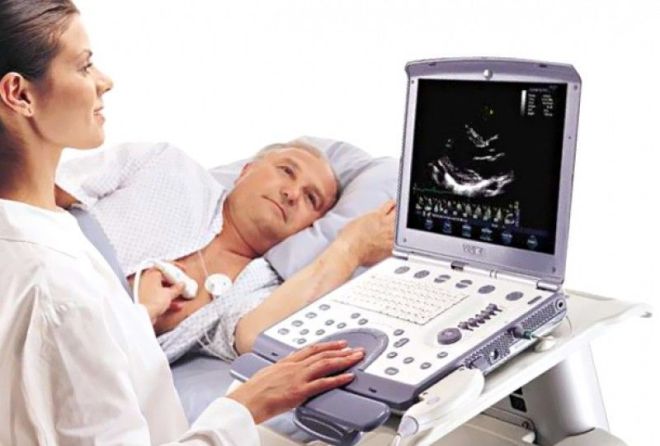
Executing the procedure
Let's talk about how echocardioscopy is done.
- The patient comes to the office, undresses to the waist so that the chest area is accessible to the researcher.
- Then you need to lie down.
- A gel is applied to the skin to prevent air from getting under the ultrasonic sensor.
- The sensor is placed in one of the intercostal spaces to the left of the sternum, and one ultrasound section of the heart is obtained.
- From this position, measurements are taken and the movement of valves, septa and contraction of the heart cavities are observed online (that is, in real time).
- Further, during the study, the sensor is moved along the intercostal spaces, placed under and above the sternum, its scanning plane is changed, making new measurements and observing heart contractions from different positions.
- Blood flow characteristics are also assessed from different positions using Doppler.
There should be no unpleasant sensations or discomfort during the examination. It lasts about 40 minutes, after which you almost immediately receive a conclusion from a sonologist.
How to decipher a study
Using echo kg of the heart you can:
- quantify systolic and diastolic ventricular function
- determine the size of the heart cavities
- find out the thickness of the walls in different parts of the heart
- assess the condition of the heart muscle
- measure pressure in the pulmonary trunk
- quantify the type and degree of changes in the heart valves.
Decoding of the obtained data is carried out by comparing the measured parameters with their standard values. Thus, for a comprehensive assessment of the structure and function of the heart, the following indicators are used:
- for valves – opening diameter and hole area
- for the cavities of the heart: anterior-posterior size, pressure in the cavity (meaning the ventricle) at the end of diastole, size of the cavity at the end of systole and diastole
- thickness of the interventricular septum (IVS)
- stroke volume (SV) of the left ventricle, cardiac index (CI) and minute volume (MV) of the heart (interrelated calculated indicators)
- peak diastolic filling rates
- maximum linear speed
- pressure gradient between the cavities of the heart
- fluid in the pericardial cavity.
The norm of the main indicators measured during echocardiography:
- Aorta: valve opening: 1.50-2.60 cm, opening area - more than 2 square meters. cm
- Left ventricle: EDD (end-diastolic size) – 3.70-5.60 cm, EDD (end-diastole diameter) – 5.8-154 ml; ESV (volume at end systole) – 25-54 ml, SV – 44-100 ml, SI – 2-4.1 l/sq.m. meter body area
- Pulmonary artery: diameter – up to 3 cm, ring – 1.81-2.50 cm
- Right ventricle: anteroposterior size – up to 32 mm
- interventricular septum – 0.6-1.1 cm.
In children and fetuses, the norms differ from those of adults, depend on age (gestational age), and are recorded in special tables, which the ultrasound doctor checks with.
Where to take the study
With a referral from a cardiologist, you can undergo echocardioscopy in a community clinic, a large hospital with a cardiology department, or at state cardiac clinics. The cost of the study in these cases is minimal (about 250 rubles), you can even undergo a heart ultrasound for free.
You can also undergo this type of research in multidisciplinary medical centers and specialized clinics. In this case, it is not even necessary to have a doctor’s referral. The average price of EchoCS in such institutions is about 2000 rubles, the range is from 1400 to 4000 thousand rubles.
Patients' opinions
Feedback from the study has been positive, with this precise and painless procedure providing patients with treatments that worked for them. In some cases, it was necessary to supplement echocardioscopy with other, more specific studies (for example, coronary angiography), but this does not indicate the shortcomings of the technique, but its specificity.
Thus, cardiac echocardioscopy is a simple, inexpensive and accurate diagnostic technique that allows you to clarify the nature of cardiac pathology and assess the degree of risk of the disease in the occurrence of life-threatening disorders. The method is widely used in clinical practice: today there is not a single area of cardiology in which the results of this study are not needed.
Share information with friends: What else to read
ATTENTION! The information on the site is for reference or popular information only. Correct treatment and prescription of medications can only be carried out by a qualified specialist, taking into account the diagnosis and medical history.
Successful diagnosis and treatment, health and well-being! Your uzilab.ru.
Why is Echo-CG performed?
The main objectives of the examination are always to assess the mechanical functioning of the heart and its morphological characteristics. With the help of cardiac ECHO it became possible:
- receive information about the size of the heart, the volume of its cavities;
- determine the condition of the organ membranes (pericardium);
- record information about the thickness of the walls of the heart;
- detect cicatricial changes in the myocardium;
- study the contractile function of the myocardium, that is, the ability to contract the ventricular muscles;
- analyze the operation and condition of the valves of the organ;
- assess intracardiac blood flow, determine the presence of pathological blood flow, measure blood pressure in the heart chambers;
- assess the condition of the largest vessels of the organ.
Using echocardiography, doctors identify a whole range of heart diseases and pathological conditions, including:
- ischemic disease;
- myocardial pericarditis, that is, an inflammatory process;
- aneurysms of any degree;
- hypertrophy and dilatation of the heart chambers;
- damage to the blood vessels of the organ;
- heart valve damage;
- the presence of intracardiac blood clots, heart tumors;
- identifying the level of pressure in the pulmonary artery.
Today, Echo-CG (ultrasound of the heart) is the only method for informative and accurate diagnosis of acquired or congenital heart defects. The examination is used not only in diagnosing functional organ disorders. It is also indispensable in preventive cardiology. Using this procedure, you can identify even the slightest deviations in the functioning of the heart, prevent a wide range of pathologies and prevent their further development. Using this procedure, you can identify even the slightest deviations in the functioning of the heart, prevent a wide range of pathologies and prevent their further development.
Advantages of cardiac ultrasound (cardiac echocardiography) at SM-Clinic
Heart ultrasound in Moscow at SM-Clinic is performed using the latest digital devices - expert-level echocardiographs from well-known manufacturers of medical equipment. Modern devices allow you to perform examinations at high speed and obtain impeccable data processing quality. That is why the study provides highly accurate results. Echocardiography at the SM-Clinic is performed by diagnosticians of the highest qualification category, who have been trained in ultrasound diagnostics in the cardiological field and have certificates confirming this specialization. Our specialists have extensive practical experience in conducting functional examinations. Features of ultrasound examination of the heart at SM-Clinic:
- echocardiographic devices used for research allow obtaining images in four mutually perpendicular planes, which guarantees maximum diagnostic accuracy;
- using Doppler echocardiography, the speed and direction of blood flow in the heart valves are determined, and the dynamics of changes in these parameters are monitored;
- the study is absolutely safe for the patient, there is no effect on the body;
- Heart echo has a price that is affordable for most of the clinic’s patients.
Videos on the topic
An echocardiogram (ultrasound of the heart) is a safe and effective examination method with virtually no contraindications. It is mandatory for professional athletes whose activities involve increased physical activity.
This research method is highly informative and is rightfully considered the leading method for diagnosing pathologies of the cardiovascular system. Compared to other types of hardware diagnostics, cardiac ultrasound has a fairly low cost, and this is its undoubted advantage.
ECG and EchoCG: what are the differences
There are four main differences between the procedures: An echocardiogram is performed using a transducer that is placed near the heart to the patient's chest. The transducer picks up the ultrasound waves that pass through the walls of the heart and then reflects them and receives the returned signals. They are processed by a computer. An ECG is performed according to a different principle: special sensors are attached to the patient’s chest. They measure the activity of the heart. Sensors (electrodes) are connected to a special device, which displays a graph indicating the nature and strength of the received electrical signals. Ultrasound examination of the heart determines how well the organ pumps blood. With the help of such diagnostics it is also possible to identify violations of this function, which indicate heart failure. Electrocardiography, in turn, only measures the signal level and checks whether the heart is sending stable impulses. The ECG result is presented on a graph, and the EchoCG result is presented in the form of photographs. An electrocardiogram can detect arrhythmia, tachycardia, abnormal heart rhythm, and bradycardia. Echocardiography evaluates the state of heart function after attacks, heart valves, possible localization of blood clots and other abnormalities in the functioning of the organ.
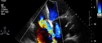
How is Dopplerography of the heart performed?
Doppler echocardiography is performed as a standard ultrasound and consists of the following stages:
- Undress, freeing your torso.
- Lie on your left side, the level of your head should coincide with the level of your heart.
- The diagnostician applies acoustic gel and touches the sensor to the chest.
- The sensors produce waves that are transformed into an electrical signal that is read by the device.
- The data is displayed on the screen.
The indicators are deciphered after the procedure. In some pathologies, the presence of characteristic signs can be determined immediately.
How safe is it?
The process is painless and safe, and is even indicated for pregnant women in cases of hereditary heart disease, diabetes and antibiotic use.
Types of EchoCG
The examination is almost always carried out through the chest. This method is called transthoracic. Transthoracic echocardiography, in turn, is divided into two-dimensional and one-dimensional. With one-dimensional diagnostics, information is displayed in the form of a graph on a computer monitor. With the help of such a study, you can obtain information about the size of the atrium and ventricles, and evaluate their performance. With a two-dimensional examination, information is provided in the form of an image of the organ. Two-dimensional echocardiography makes it possible to obtain an accurate picture of the heart’s functioning, determine its size, wall thickness, and chamber volume. There is also Doppler echocardiography - a study that checks how well the blood supply to the organ occurs. For example, during the procedure, the doctor observes the movement of blood in the vessels and parts of the heart. Normally, blood flow should move in one direction, but if the valves are malfunctioning, the blood may flow in the opposite direction. Doppler examination is usually prescribed in combination with one-dimensional or two-dimensional ultrasound examination.
Results of the diagnostic procedure
After completion of the manipulation, the SM-Clinic diagnostician analyzes the results. It determines the thickness of the heart septa, the size and condition of the heart, its topographic position within the anatomical structure. The specialist also evaluates the functioning of the heart valves and other functional structures, and the condition of the soft tissues. Based on the results obtained, the doctor will identify possible pathologies. After an ultrasound examination of the heart at SM-Clinic, the patient receives:
- echocardiogram - visualization of soft x-ray negative tissues on photographic paper or an ultrasound image of the heart;
- conclusion of a diagnostician.
Also, the EchoCG protocol must indicate the norms for people who correspond to a certain gender and age group. When writing a conclusion, these standards are taken into account and correlated with the results obtained. Diagnostics at SM-Clinic are carried out by qualified specialists with impressive practical experience. The availability of modern equipment, as well as highly qualified diagnosticians, guarantees the most accurate examination results. You can get an ECHO of the heart in Moscow inexpensively here at SM-Clinic. We perform tests at the best price and quickly provide diagnostic results to patients. You can find out all the details you are interested in, clarify the cost of cardiac ultrasound and other information, and also sign up for an examination from the Contact- operators.
What you need to know before having an echocardiogram
If you are referred for an echocardiogram, please bring a towel or diaper with you (which can be placed on the couch during the examination and used after the examination to remove any remaining gel). Try to arrive for the examination in advance (5-10 minutes) so that you have time to rest and the echocardiography is performed in a calm state. Be sure to take your ECG film (preferably no more than 1 month old). If you have several ECG films left, take them all. Previous EchoCG findings are also required if you have had this study previously; a report from your doctor (cardiologist or therapist), which may indicate the purpose of the echocardiography.