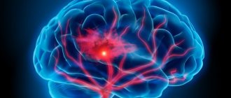
Cerebral angiography is a hardware diagnostic examination aimed at studying the state of cerebral vessels and circulatory processes. This technique makes it possible to promptly identify cerebrovascular disorders and prevent the further development of pathologies with extremely dangerous consequences.
How can you see the blood vessels of the brain?
Cerebral angiography is an x-ray method for visualizing cerebral vessels, which consists of staining the vascular bed with previously injected contrast. This is a highly effective and modern diagnostic method that allows you to make an accurate diagnosis. The method of visualizing blood vessels using a contrast agent has been known to medicine for about a century. Back in 1927, a neurologist from Portugal began using this method, and it came to Russia in 1954. Despite such a long use, cerebral vascular angiography has changed significantly over this time, becoming more sophisticated.
Methods of conducting research
There are several different types of cerebral angiography.
CT angiography of cerebral vessels
Angiography using computed tomography (CTA) provides a detailed image of the vessels and shows patterns of blood flow. In this case, intravenous contrast enhancement is used.
After CTA, image reconstruction is performed.
A definite positive side of this method is the reduced radiation exposure to the patient’s body.
CT angiography is often performed for stenosis, thrombosis, aneurysms, and vascular developmental defects.
Contraindications are allergies to the contrast agent, diabetes, pregnancy, obesity, thyroid problems, myeloma, heart disease, uncontrollable arrhythmia and tachycardia.
The study is carried out on an outpatient basis. Approximately 100 ml of contrast agent is injected into a venous catheter, which is inserted into the cubital vein. The patient lies on the CT scanner table.
X-rays scan the area under study in parallel with the introduction of a contrast agent.
MR angiography of cerebral vessels
Magnetic resonance angiography (MRA) allows you to study the functions of blood flow and its anatomical features.
The basis of magnetic resonance imaging is to track energy changes in tissues, their structure and chemical composition. Contrast agents are practically not used in MRA (occasionally gadolinium-based to obtain high-precision images).
MRI angiography of cerebral vessels is used to diagnose aneurysm dissection, congenital heart defects, and vasculitis.
Article on the topic: Exelon in capsules: indications, instructions and reviews
Contraindications include installed implants, pacemakers, nerve stimulators, blood-restoring clips, insulin pumps, prosthetic heart valves, heart failure, pregnancy, claustrophobia.
Cerebral angiography of cerebral vessels
Cerebral angiography is a kind of “gold standard” for studying cerebral vessels.
The author of this method is Egas Monitz, who performed angiography for the first time in 1927.
The method is of the highest value because it allows you to accurately detect aneurysms, narrowing of blood vessels or the site of their blockage, and brain tumors.
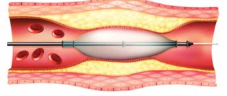
The catheter is inserted into the vessel through the femoral artery and directed to the carotid artery. A contrast agent is injected into the blood vessels and X-rays are taken to determine the state of the inflow and outflow of venous blood.
During cerebral angiography, surgical intervention is possible. The information content of the method is much superior to CTA and MRA.
Arteriography
Arteriography involves the introduction of a contrast agent into the lumen of the vessel, which makes it possible to determine the presence of tumors located close to blood vessels, arterial pathologies and other circulatory disorders.
Most often, this method is used to examine limbs .
Arteriography is relatively simple, performed on an outpatient basis, but is painful because the contrast moves quite quickly through the arteries.
X-ray contrast agents (about 30-40 ml) are injected through a catheter or directly into the artery under strong pressure in the direction of the blood flow (less often against the blood flow).
This method allows you to diagnose changes even in the deepest arteries, which are monitored using the screen of an X-ray machine.
Venography
Another name for venography is phlebography. The essence of the method corresponds to its name.
Venography allows you to see the distribution of veins ; it is actively used for varicose veins and thrombosis, as well as arrhythmia. The patient is advised to breathe calmly and relax during the procedure.
This is a simple and painless method, but in rare cases, your health may deteriorate after the procedure, and phlebitis may appear - inflammation at the site of contrast injection.
Venography involves the use of small amounts of a contrast agent that is injected directly into a vein (direct venography). After the procedure, an injection is made using 60 ml of saline to cleanse the vessels.
It is most justified to use venography before surgery on the veins.
Indirect venography can be performed in three ways:
- contrast is injected into the artery and then enters the veins through the capillaries;
- contrast is introduced into the tissue of the affected organ that needs to be examined, and the pictures show veins draining blood from the organ;
- contrast is injected directly into the medullary space.
What is the danger of post-traumatic encephalopathy of the brain - treatment and prevention of the disease.
What is the most common cause of a retrocerebellar arachnoid cyst of the brain and what to do if there is a suspicion of this formation.
Lymphography
Lymphography is a method of studying the lymphatic system also using a radiopaque agent.
The study is carried out in three projections and the data is studied immediately after the administration of contrast (early lymphogram) and after 1-2 days (late lymphogram).
Early lymphograms make it possible to examine the condition of the lymphatic vessels, late lymphograms - the lymph nodes.
Article on the topic: Ginkgo Biloba - instructions and reviews of use
This method allows you to identify changes in the external and common iliac, inguinal, supra- and subclavian, lumbar, axillary lymph nodes; identify the presence of tumor processes and optimize cancer treatment.
Types of cerebral angiography
According to the established classification, this diagnostic technique is divided into 2 types:
- Selective - targeted, local. When performing selective cerebral angiography, an iodine-containing contrast agent is injected into an arterial vessel that supplies one of the brain regions.
- Survey is an extended research method. A contrast agent is injected into the area of the main artery, which is responsible for the general cerebral blood supply and nutrition. Using this technique, a specialist can carefully examine all cerebral vessels.
The optimal type and method of implementation is determined by a specialist individually, taking into account the characteristics and severity of the clinical case.
When is an MRI of the vessels of the neck and head performed?
As a rule, a doctor gives a referral for magnetic resonance imaging of the vessels of the head and neck. The main indications for examining the vessels of the head and neck are:
- suspicion of circulatory pathologies in this area;
- prevention of health conditions after a stroke;
- assessing the effectiveness of the surgery or drug treatment;
- neurosurgical operation (preliminary examination is carried out to identify the exact location of the vessels);
- primary diagnosis of the pathological process;
- suspicion of cancer.
Indications
Indications for cerebral angiography are pathological conditions that cause disturbances in the functioning of the brain. Hemorrhagic circulatory disorders:
- aneurysms;
- diverticulum;
- angioma.
Ischemic circulatory disorders:
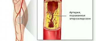
- cerebral atherosclerosis;
- blood clots;
- arterial deformation.
Tumors leading to changes in the vascular pattern, as well as the lack of results after other methods for diagnosing brain diseases in the presence of the following symptoms:
- constant dizziness not related to blood pressure;
- epileptic seizures;
- confusion of consciousness;
- increased intracranial pressure;
- previous stroke or suspected micro-stroke;
- intracranial hematomas caused by head trauma;
- chronic headache of unknown origin;
- nausea accompanied by dizziness and headaches;
- noise in ears.
It is also advisable to perform cerebral angiography to plan upcoming surgery and to monitor the patient’s recovery after brain surgery.
What pathologies can be detected by MRI of the vessels of the head and neck?
Magnetic resonance imaging makes it possible to identify the following pathological processes and abnormalities:
- atherosclerotic plaques and blood clots in the vessels of the head and neck;
- microstroke (minor hemorrhage);
- vascular aneurysm (specific expansion of the vascular wall);
- compression of blood vessels by neoplasms or abscesses;
- source of hemorrhage after head injury;
- areas of necrotic brain tissue;
- vascular defects as a consequence of osteochondrosis;
- compression of blood vessels by cervical vertebrae;
- salt deposits;
- disturbances of blood flow in the vessels.
Contraindications to the procedure
As with any other procedure, there are contraindications for cerebral angiography. They are associated both with the procedure itself and with the contrast agent that is injected into the bloodstream. Iodine compounds are used as the administered substance. The amount of the substance depends on the volume of the examination; it can be 5-10 ml.
Cerebral angiography is not done in the following cases:
- allergic reactions to iodine-containing contrast agents,
- individual intolerance,
- acute or chronic renal failure, which does not allow the use of a contrast agent,
- exacerbation of chronic diseases,
- pregnancy or lactation,
- diseases accompanied by blood clotting disorders,
- myocardial infarction,
- age up to 2 years,
- mental illness.
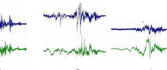
What do angiography indicators mean?
The amount of radiation that will penetrate the veins and other brain tissue is determined by their density. It is expressed in various color shades. The bone in the image will be white, and the cerebrospinal fluid will practically not appear on the resulting images. Other brain substances have different colors and densities. Using them, doctors evaluate the internal structure. The doctor will provide a detailed transcript of the received images.
Preparation for the procedure
Before the study, the patient should not eat for 10 hours and not drink for 4 hours. He needs to remove all metal objects. If surgical intervention is required to administer contrast, the following is prescribed:
- iodine allergy test;
- urine and blood tests;
- ECG;
- kidney function testing;
- consultations with an anesthesiologist and therapist.
Before performing an MRI or CT scan of the brain with angiography, you must adhere to a diet for several days. Eliminate from your diet carbonated drinks, refined sweets and sweet fruits, dishes made from legumes and other products that cause increased gas formation in the gastrointestinal tract.
Methodology for cerebral angiography
The entire procedure of this study can be divided into several stages:
- preparing the site for puncture - treating the skin with an antiseptic solution to destroy microorganisms and local anesthesia;
- direct puncture of the vessel with a special needle and insertion of a flexible catheter;
- introduction of a contrast agent into the vascular bed of the brain through a catheter;
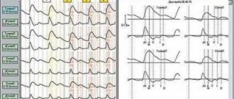
- performing a series of X-ray images of blood vessels; during CT cerebral angiography, a series of layer-by-layer images is taken;
- interpretation and interpretation of the received images with a conclusion - is carried out by a radiologist together with a vascular surgeon.
Considering the paramount importance of proper cerebral circulation, adequate and correct diagnosis of vascular problems is the basis for their further successful treatment.
Where can I get an MRI of the vessels of the head and neck?
Magnetic resonance imaging of the vessels of the head and neck can be done in a private multidisciplinary medical center located in Tushino, at Skhodnenskaya metro station. You can undergo the examination inexpensively and without queues.
The MRI center is equipped with modern equipment, which allows you to obtain reliable and informative results. You can sign up for the procedure either in person at the clinic or by phone. The medical center is open 24 hours a day, seven days a week, so you can visit the study at any convenient time. Additional information can be found by calling the phone number listed on the website.
Possible complications
Despite the high level of safety for patients of different ages, angiography may result in the development of negative consequences for the patient. The most commonly observed conditions are:
- release of a radiopaque substance from the vascular bed into the surrounding tissues. This situation can lead to inflammatory changes of varying severity;
- allergic reactions to the contrast agent or its individual intolerance. In such cases, the patient may experience itching, urticaria, Quincke's edema and other allergy-specific symptoms;
- acute renal dysfunction, as a complication of the examination, is observed in patients with their diseases.
To prevent complications of the procedure, it is necessary to ensure a comprehensive examination of the patient before the study.
Related posts:
- Sexual Aversion Disorders Aversion to sexual activity is a disorder defined as persistent or...
- Argyrosis or blue skin Argyrosis is a persistent bluish-gray discoloration of the skin caused by deposits in…
- Numbness of the hands - dangerous or not? Constantly chilly fingers, frequent feeling of numbness in the hands, especially...
- Morton's neuroma (foot pain) Morton's neuroma is a fairly common disease characterized by thickening of the nerve sheath...