Symptoms and diagnostic methods
Xanthelasma usually occurs in older people. Women get sick much more often than men.
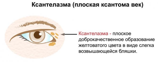
Yellow plaques characteristic of this disease form on the unchanged skin of the upper eyelid. They are characterized by a gradual and slow increase in size. Without proper treatment, they persist throughout life.
Skin lesions of the upper eyelids of both eyes are often observed. Xanthelasmas can be single or multiple. Sometimes the lesions can merge into a single strip of compacted tissue, which gives the eyelid a yellowish color.
The disease does not pose a true health hazard, since plaques never degenerate into malignant formations. In addition, xanthelasmas are absolutely painless and not prone to inflammation or ulceration. Patients experience concern only in connection with aesthetic problems, since the spots are yellow in color and protrude above the surface of the skin, therefore they are noticeable to others. In some cases, the lesions become very large, which further aggravates the aesthetic discomfort.
A routine visual examination is sufficient to diagnose xanthelasma. In addition to a dermatologist, the patient should visit an endocrinologist. In such patients, serum cholesterol levels and blood lipoprotein concentrations must be determined.
Xanthomas on the skin: what is it?
If there is a violation of fat metabolism in tissues and cells, then focal neoplasms called xanthomas appear on the human body. They look like small white or yellowish growths containing fat cells inside. Such neoplasms can be local, they appear only in one area of the body, and generalized, that is, those that spread throughout the entire skin.
It was found that one of the main reasons for the appearance of xanthomas is excess cholesterol in the blood; their appearance can also be affected by:
- weakened immune system;
- injuries;
- chronic diseases;
- diabetes.
Neoplasms appear when the blood is oversaturated with processed fat residues. This disease is dangerous because it is accompanied by damage to blood vessels and some internal organs.
Types of xanthoma
Let us examine in more detail individual types of xanthoma, their location and appearance.
This type of xanthoma looks like soft, slightly raised papules above the skin. They are flat and round in shape and are most often light yellow in color. Such formations are located on skin folds, palms and soles. Such xanthomas occur in older people, with liver disease, obesity and atherosclerosis.
Experts distinguish two types of flat xanthomas:
- Intertriginous. They look like small yellow-red lesions with clear boundaries.
- Diffuse. It appears as a cluster of papules and plaques of a yellowish tint, connecting with each other and turning into one large xanthoma.
This type forms on the eyelids and around the eyes, in the form of many small yellow plaques.
This is the most common type of xanthoma and can appear even in people who do not have high blood lipid levels. Multiple nodular xanthoma.
It has the shape of flat and semicircular yellow formations, they are soft to the touch and have clear boundaries. The peculiarity of multiple nodular xanthoma is that it appears suddenly and chaotically. For example, appearing on the thighs or buttocks, when merging, it forms extensive plaques. This species is characterized by brown nodes, reaching sizes from 1 to 5 cm. The contents of tuberous xanthomas are cholesterol. The main locations on the body are the legs, buttocks, arms and fingers. This type of xanthoma is dangerous because it can affect the coronary arteries, since the level of cholesterol in the blood is too high. The main sites of formation of such xanthomas are the extensor tendons of the fingers and the Achilles tendons. Outwardly, it looks like a small dense tumor-like formation, flesh-colored. They move with the tendon as the joint flexes and extends. Xanthomas are insidious neoplasms. They can appear not only on the body, but also on the internal organs of a person. The most common is gastric xanthoma. It can only be detected through diagnostics and fibrogastroscopy. This xanthoma looks like fat deposits on the mucous walls of the stomach. Most often, such plaques appear in elderly people, when atherosclerotic processes in the body are most active.
Removal methods
Surgical treatment is performed on an outpatient basis using local anesthesia. The following methods are used to remove xanthelasma:
- Mechanical excision.
- Laser destruction.
- Electrocoagulation.
- Radio wave method.
Electrocoagulation is usually used to treat small lesions. Extensive plaques are removed using surgical scissors and tweezers. The choice of the exact method for removing xanthelasma depends on the technical equipment of the clinic.
Forecast
The prognosis of the disorder directly depends on the factors that provoked it. This is due to the fact that in some cases xanthomas can disappear on their own, while in others they can lead to consequences such as atherosclerosis of the coronary or cerebral vessels, heart disease and an increase in liver size. However, the most common danger of the disease lies in its frequent relapses.
In addition, patients should not forget about the possible formation of complications of the underlying disease. Often, in addition to cosmetic inconveniences, xanthomas do not cause any other discomfort, which is why the prognosis of the disease is favorable.
Treatment of the underlying disease and prevention of relapses
If neoplasms develop against the background of one or another pathology, then additional treatment is necessary, which will be aimed at the cause of xanthelasma. In case of lipid metabolism disorders, a diet low in animal fats is prescribed. According to indications, lipotropic drugs (controlling fat metabolism in the body) and statins (lowering blood cholesterol levels) are prescribed.
Prevention of relapses of xanthelasma is achieved by following a special dairy-vegetable diet. At the same time, animal fats are almost completely excluded from the diet and carbohydrates are limited. The patient can meet the body's need for fats by eating any vegetable oils.
Reasons: why do xanthomas form?
The cause of this phenomenon is usually associated with changes in lipid metabolism, systemic or rarely local in nature.
- Xanthomas are common among patients with severe forms of hypercholesterolemia and hypertriglyceridemia, usually on a hereditary (primitive) basis, sometimes appearing large in homozygous individuals; these patients have very high levels of cholesterol and/or triglycerides in the blood .
- Xanthomas may appear less frequently even in patients with primary biliary cirrhosis, pancreatitis , diabetes mellitus , heart failure , and some forms of cancer and inflammatory diseases.
Xanthelasma in children
Diagnosis and treatment of these formations in children has a number of features.
- When a formation appears, you need to make sure that it is not a diabetic xanthoma caused by a dysfunction of the pancreas. Often, after correcting the level of carbohydrates in the blood, skin manifestations immediately disappear.
- With concomitant obesity, the child must follow a diet. Usually, after consultation with a pediatrician and endocrinologist, a dairy-vegetable diet is prescribed.
- Be sure to check the condition of the liver and pancreas, and treat any detected disorders.
Surgical removal of xanthomas in children is rarely required, since they disappear after properly prescribed drug treatment or during puberty. If removal of the lesion is necessary, diathermocoagulation and cryodestruction are used for this.
Source: BellaEstetica.ru
Treatment of xanthoma
The disease can be treated using conservative methods.
- Sequestrants are prescribed to activate the release of bile acids, which leads to a decrease in cholesterol in the blood.
- Statins work by binding to cholesterol-producing enzymes in the liver.
- Hepatoprotectors ensure restoration of liver cell membranes and protect against destruction.
Drug treatment methods are effective for minimal manifestations of xanthelasma. If the xanthoma is extensive, it cannot be cured without surgery; getting rid of xanthelasma is possible only with the help of surgery. This is dangerous, accompanied by negative consequences, and requires healing.
Therefore, plaque removal is prescribed only for the following indications:
- Inflammatory process of any etiology;
- External change in the appearance of xanthelasma: redness, sharp increase, reproduction;
- Very high cholesterol levels.
Let's look at removal methods in more detail.
Surgical removal
This long-standing method was used back in the middle of the 20th century. To remove a xanthoma, no special equipment is required. The main tool is a surgical scalpel. An incision is made at the site of the tumor, followed by excision of the xanthelasma. Used to remove multiple xanthomas or when a malignant tumor is suspected. The edges of the wound are cauterized with electric current. Disadvantages of the surgical method:
- The incision requires stitches, which leave scars. As a result, additional cosmetic surgery is required.
- During the operation, due to the close proximity of blood vessels, bleeding occurs, which leads to the formation of postoperative hematomas.
Laser removal
The difference from the surgical method is that the operation takes place under local anesthesia. This method demonstrates a number of advantages:
- Precise localization of the intervention. Laser devices operate using a beam or carbon dioxide. With the latter, the removal of the skin occurs in layers and painlessly, without damaging healthy tissue, exactly at the location of the plaque.
- Laser removal is a bloodless procedure and does not cause hematomas.
- Sterility reduces the degree of manifestation of inflammatory processes and rapid restoration of the skin. After the procedure, the wounded area is covered with a crust, which disappears within seven days.
- Does not leave any traces of intervention on the skin.
- Minimal risk of relapse.
The disadvantages of the method include the price - this is an expensive operation and is performed in a clinic.
There are contraindications for use:
- Diabetes;
- Malignant formation;
- Epilepsy;
- Inflammatory processes;
- Presence of a pacemaker.
Electrocoagulation
Burning of formations with a coagulator occurs by exposure to electric current; the neoplasm is fixed with tweezers and cut off with an electrode at the surface of the skin. Contraindications are similar to laser removal. The electrocoagulation method is cheaper.
Radio wave method
The radio wave method can be used to remove xanthelasma without contact. Excision is achieved due to the ability of the skin to resist the radiation of high-frequency waves; heating the surface leads to its dehydration and dissection. The radio wave is localized at the end of the device, which maximizes the concentration of the process on painful tissues.
Cryodestruction
The method allows you to remove xanthoma by exposure to liquid nitrogen at ultra-low temperatures; under its influence, the cell membrane freezes, cryonecrosis is formed, which crumbles.
Advantages of the method:
- The speed of the procedure is no more than 2 minutes.
- Painless.
- The skin is quickly restored.
- No bleeding.
- Reduced trauma.
- Minimal risk of relapse.
The only drawback of the method is the difficulty of localizing the border of unhealthy tissues.
Diagnosis of the disease
In 90% of diagnosed cases, xanthelasma develops asymptomatically, manifesting itself only in the presence of a plaque; the eye does not lose mobility and closes. As a rule, such neoplasms do not become inflamed. As senile xanthelasma grows, it spreads to the eyelids, bridge of the nose, and chin. The plaque creates a general unaesthetic appearance; in special cases, the disease can affect vision and eyelid mobility.
If the disease progresses, removing xanthelasma will become problematic. In the eye area, the vessels are located very close to the surface of the skin. Xanthelasma cells “fuse” with connective tissue, blood vessels and nerves. When removing a plaque, you can easily damage the vascular wall or nerve.
The appearance of xanthelasma as a systemic disease requires an integrated approach to diagnosis, which includes consultation with specialist doctors: ophthalmologist, dermatologist, endocrinologist.
Biochemical blood test (lipidogram) is the most common and effective diagnosis and monitoring of the course of diseases. The accuracy of the results depends on following these rules.
- The test must be taken on an empty stomach 12 hours before blood sampling.
- For three days, you need a diet: exclude fatty and fried foods from your diet.
- Stop taking dietary supplements and antibiotics.
The analysis is taken by drawing blood from a vein.
Lipidogram norms:
| Name | Lower indicator | Upper indicator |
| Total cholesterol content | 3.2 mmol/l | 5.2 mmol/l |
| Triglycerides | 0.14 mmol/l | 1.82 mmol/l |
| HDL | From 1 mmol/l |
With xanthelasma, cholesterol and triglyceride levels are exceeded.
A general blood test determines:
- The degree of body resistance;
- The presence of inflammatory processes;
- Hemoglobin level to rule out anemia;
- Blood sugar.
Diascopy - bleeding of the plaque by squeezing the xanthelasma with a spatula; the study is carried out to identify the color of the neoplasm. The mentioned disease is characterized by a yellow color.
Histology. When removing xanthelasma, it is recommended to take a histological analysis to identify pathological changes.