Leukemia (viral leukemia) in cats
Feline leukemia is one of the most dangerous incurable retroviral diseases of cats, which is dormant (hidden, sluggish) and does not manifest itself until the beginning of the terminal phase.
Together with infectious peritonitis and immunodeficiency in cats, leukemia is included in the ominous list of “tri-fatal” infections, characterized by high contagiousness and lethality. Other names for leukemia in cats are VLK, FeLV, blood cancer, leukemia, viral leukemia, Leucaemia viralis, lymphosarcoma, hemoblastosis.

Possible causes of leukemia development
If 60-40 years ago the diagnosis of leukemia was tantamount to a death sentence, then today the situation has changed radically - the problem with the disease has been solved, albeit not perfectly, but the results speak for themselves: 90% of patients return to normal activities.
Regarding the reasons that provoke the appearance of leukemia (leukemia), there is no exact data in medicine; there are a lot of assumptions and factors that provoke the appearance of the disease, but in essence, these are in most cases just guesses. Scientists suggest that environmental and genetic factors combine to trigger the mechanics of change, causing DNA mutations that cause cells to grow abnormally, losing the functional characteristics of the white blood particles in the cell. The causes of changes in the DNA of cells are attributed to chromosome translocation; these changes lead to the rupture of one of the chromosomes and attachment to another.
Another possible cause, experts say, is exposure to radiation, which increases the risk of developing the disease. To the same list, doctors add the abuse of bad habits, for example, smoking, or the consequences of taking chemotherapy drugs.
Why is it dangerous to neglect treatment for leukemia?
Lack of treatment is fraught with the development of the disease and its accompanying pathologies, which lead to death. An animal that does not receive veterinary care develops:
- anemia;
- spontaneous bleeding;
- chronic cystitis;
- constant low-grade fever;
- bacterial infections;
- viral respiratory diseases;
- peritonitis;
- ascites;
- toxoplasmosis;
- autoimmune glomerulonephritis;
- stillbirth;
- damage to the mammary glands in females;
- miscarriages;
- fetal resorption and fading kitten syndrome;
- sarcoma of the lymphatic system.
A pregnant cat with leukemia will give birth to already infected kittens, which usually do not survive due to the body’s lack of strength to fight the virus. Therefore, cattery owners should definitely examine their cats.
Which cats can get leukemia?
All cats are at risk. Stray animals, domestic animals, young and old, purebred and mixed-breed animals are susceptible to the disease.
Why, then, are they not all carriers of the virus? The fact is that the virus is quite unstable to external influences. In the external environment, it most often dies within a couple of days. It is dangerous from sunlight, ultraviolet rays, low and high temperatures. It is vulnerable to disinfectants and alcohol-containing products. Nevertheless, it is incorrect to even talk about the relative safety of animals.
Reference! Cats, due to their lifestyle, are more often susceptible to the disease (fights, including for the right to mate with a cat in heat).
Leukemia rashes
Changes in the skin under the influence of leukemia can manifest themselves in the form of nonspecific or specific rashes. Specific modifications manifest themselves in the form of extramedullary foci of hematopoiesis in the layers of the skin, which inherently turn into malignant proliferation. Nonspecific changes occur due to exposure to toxic substances on the skin.
With specific changes in the dermis, the appearance of plaque-infiltrative rashes, the presence of nodules, and papular formations are observed. Finding out the cause of the manifestations through research, the diagnosis is confirmed when infiltrates are detected, which may differ depending on the form of leukemia.
Nonspecific changes are characterized by itching, a symptom that can develop and progress much earlier than the disease itself. Along the way, various types of rashes may appear on the skin.
The disease can hide itself for a long time and develop without the manifestation of some symptoms, so often a rash, as one of the factors indicating the presence of leukemia, may appear in the presence of other companions: enlarged lymph nodes, changes in the blood. Whether it is a genetic feature or the influence of other factors that is to blame, doctors do not have exact explanations for why the disease appears; there are only a number of assumptions that partially explain this phenomenon.
The cause of the manifestation of leukemia on the skin in the form of specific neoplasms is the infiltration of areas of the dermis by hematopoietic cells of the malignant series. The lesion can have a different appearance: tumors, nodules on the surface, infiltrates. The localization of neoplasms can be located on any area of the skin; it can look like one or several elements, or grow like colonies. To accurately diagnose the causes and categories of formations, a study of immune markers is carried out that will explain the origin of the infiltrate.
Atypical skin rashes are more typical of myeloid leukemia, a disease in which prurigo, bulla-like areas, and areas of necrosis are clearly visible on the dermis. Such manifestations may be accompanied by ectodermosis and the formation of vasculitis, which explains the progression of the disease.
Lymphocytic leukemia, especially the chronic form of the disease, most often manifests itself in adulthood, and the disease is diagnosed in men twice as often as in women. Nonspecific rash as a symptom of lymphocytic leukemia is a frequent phenomenon, especially in the form of pigmentary-atrophic changes, eczematous and exudative-pemphigoid manifestations, as well as hemorrhagic and urticarial-pruriginous lesions of areas. Nonspecific rashes are often combined with lymphadenopathy, a concomitant disease.
With its appearance, leukemia “attracts” many infectious diseases, including skin diseases, which settle in the body due to a weakened immune system. An increase in the depressed area of the skin under the influence of various types of pathogens can have a very negative effect. This requires constant biopsy of the affected skin areas using various types of tests: mycological, microbiological and histological examination of the biopath.
After chemotherapy
The consequences of chemotherapy, reflected in areas of the dermis of patients with leukemia, are a frequent phenomenon, which additionally creates a differential diagnostic problem. The development of diseases is divided into 4 groups:
- Residual manifestations in the form of side effects after use for the treatment of specific drugs: necrosis at injection sites, necrosis as a consequence of treatment with blyomycin on the fingers, induced psoriasis as a result of the use of interferons.
- Changes in genesis that are not typical in terms of typical manifestations: diffuse or focal pigmentation, acral erythrema.
- Hypersensitivity reaction: maculopapular exanthema, erythema multiforme, urticaria.
- Consequences after using cytotoxic medications: hair loss, onycholysis, mucytes.
Also, various skin manifestations may occur as a result of bone marrow transplantation during the transplantation process. These manifestations are displayed as maculopapular exanthema in the acute form or sclerodermiform changes and lichenification in the chronic form.
Methods of transmission of the virus
The virus is highly contagious - the ability of the infection to be transmitted from sick animals to healthy ones. It is transmitted from a sick animal to a healthy one under the following conditions:
- contact with the host’s saliva, urine, feces, or secretions from his nose or eyes;
- biting during a fight or play;
- through shared dishes, toys, beds;
- sexually;
- in utero from mother to kittens (as a rule, such babies do not survive);
- through flea bites;
- at an exhibition when using common things or being close to a sick colleague;
- after the owner visits the house where the leukemic cat lives, or a shelter, without changing clothes;
- in a clinic where instruments and tables are not thoroughly sanitized after each patient.
The greatest prevalence of hemoblastosis is observed in crowded cat prides, in so-called granny apartments, in shelters, as well as in basements where homeless individuals live.
Detailed lecture from a veterinarian about leukemia in cats:
What causes the development of leukemia and what is its danger
The causative agent of the disease is an RNA-containing oncogenic retrovirus, which invades blood cells and destroys their genetic code. The Latin name is Feline leukemia virus. It was identified by scientists in Glasgow in 1964. Scientists have established a connection between cases of cancer of the blood and lymphatic system in cats with the detection of strains of this virus in their blood.
The virus is dangerous due to its oncogenicity and changes in blood composition, reducing the resistance of the cat’s body, and provoking secondary diseases. It begins its activity in the tonsils and lymphatic tissues of the nasopharynx, gradually spreading to the lymph, circulatory system, and brain.
Immune deficiency increases unnoticed. When the process spreads to the bone marrow, it is too late to fight, since the affected bone marrow produces more and more replicas of the virus along with blood cells, and leukocytosis progresses. Death usually occurs from diseases associated with hemoblastosis.
Why does a rash occur with leukemia?
Other names: leukemia, leukemia. Refers to cancer. The disease disrupts the maturation of white blood cells (leukocytes) and other cells. Their production occurs in the bone marrow, so the causes of the pathology are here. Stem cells produce:
- leukocytes;
- red blood cells;
- platelets.
They all suffer from leukemia. The degeneration of white blood cells leads to decreased immunity. Neoplasms (blasts) gradually replace healthy cells, but they cannot perform protective functions.
There are acute and chronic forms and several varieties of the disease. Often initially asymptomatic, such as chronic myeloblastic leukemia. Therefore, you need to pay attention to the first signs: skin lesions. These are usually spots that resemble bruises or a rash.
The causes of mutations are poorly understood. So far, only a few factors have been identified and a number of assumptions made.
- Radiation causes a decrease in the number of healthy blood cells. This version is supported by the multiple increase in the number of cases after the explosion in Hiroshima and Nagasaki.
- Some viruses, for example, HTLV. There are always pathogens in the body. With good immunity, they are suppressed; with weakened, unfavorable environmental influences, they are activated. Some scientists disagree because no cases of transmission have been identified.
- Chemically hazardous substances in paints, varnishes, medications, as well as arsenic, hormones, steroids and others.
- Changes in the number of platelets and abnormal levels of white blood cells can be caused by genetic characteristics and abnormalities.
- The body, both in acute and chronic diseases, is weakened by household factors: foods with GMOs, smoking, and others.
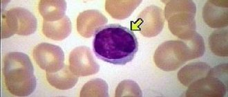
Symptoms of leukemia in cats
The disease is insidious in that it does not appear immediately, but when the number of leukocytes in the blood exceeds acceptable limits. In this case, helping your pet is very difficult, if possible at all.
Therefore, it is necessary to show your furry puppy to the veterinarian twice a year so as not to miss the first symptoms of the disease.
VLK in cats manifests itself with the following symptoms:
- drowsiness;
- lack of interest in games;
- frequent rises in temperature;
- loss of appetite;
- lethargy;
- salivation;
- frequent colds, cystitis;
- digestive problems;
- enlarged lymph nodes;
- discharge from the eyes and nose;
- bulging eyes;
- pallor of mucous membranes.
The symptoms are nonspecific, and the owner will not be able to independently figure out what is bothering his pet. By self-medicating, you can waste time and ultimately lose your pet.
Life expectancy of a cat after infection
How long a cat can live after contracting viral leukemia depends on its immunity. Typically, it takes 3 to 10 years from infection to death. The incubation period ranges from four days to eight months.
Unfortunately, 90% of infected animals die in the 4th year of the disease. This high mortality rate is due to the fact that obvious symptoms appear 3 years after the cat is infected.
With good care, the animal remains a carrier of the infection for a long time, but does not get sick, and clinical signs of the virus do not appear. However, carrying the virus poses a danger to other cats.
Leukemia: symptoms, causes of development and how to treat this terrible disease
Additional education:
"Hematology"
Russian Medical Academy of Postgraduate Education
Contacts
The appearance and development of blood cancer may include a list of syndromes and characteristic manifestations.
In most situations, leukemia symptoms include headaches, a tendency to increased bruising, bleeding and fever.
Symptoms for different types of lymphocytic leukemia are almost the same. They can appear completely suddenly and rapidly (acute form), or develop over years (chronic).
Modern medicine has not identified a clear reason why signs of leukemia may appear in adults, indicating a violation of a basic and vital process in the body. Risk factors include radiation exposure, exposure to carcinogenic chemicals, smoking, and hereditary factors. Children are extremely susceptible to this disease.
First indicators of leukemia in adults
The first signs of leukemia are those changes in well-being that cannot be ignored under any circumstances. In adults, blood cancer can manifest symptoms in the form of the following physical abnormalities:
- Excessive sweating is the most characteristic symptom that occurs even when at relative rest and was absent previously; it is at night that most often sweating is an indicator of anemia;
- Enlarged lymph nodes are an equally important sign, which should be detected immediately by a doctor. We are talking about nodes in the neck, armpits, groin, above the collarbones, the fact is that leukocytes live in them, and if they increase sharply in the blood, they will appear. abnormal formations in these areas;
- Bone and joint pain - leukemia often causes similar symptoms in adults, but this is not always an indicator of blood cancer, since many other diseases are accompanied by similar changes in well-being, the situation arises due to an excess of leukocytes that have lost the ability to function and develop, usually specialists during an X-ray examination can immediately determine the cause of the deviation.
Symptoms of a particularly dangerous nature in leukemia are weakness and fatigue, accompanied by general malaise and problems with sleep. You can notice the first signs of leukemia and problems with brain activity - memory and mental abilities begin to suffer greatly.
Symptoms of leukemia: what signs are detected in children
In children, the first signs of blood cancer that they themselves usually notice are bone and joint pain. Due to pain, the ability to walk is lost in some cases. In 97% of situations, childhood cancer occurs in an acute form, arising due to congenital pathology.
In a child, the first symptoms of blood cancer are nonspecific, such as fatigue, loss of appetite, and an unreasonable increase in body temperature.
Children who have leukemia often show symptoms of pale skin, sometimes with a yellow or sallow tint.
Against the background of infiltration due to leukemia, which affects the functionality of the mucous membranes, stomatitis or gingivitis can often occur. Just like in adults, there is an increase in lymph nodes.
Hepatosplenomegaly is a phenomenon related to leukemic hyperplasia of the spleen and liver. Sialadenopathy – lymph nodes.
Being acute, the disease causes in childhood symptoms of blood cancer of such a nature as the presence of hemorrhagic and anemic syndromes.
The level at which anemia occurs in a child will be relative to the state of the bone marrow and the severity of the proliferation of immature cells.
Dangerous disorders
Cardiovascular disorders in cancer that affects the blood, symptoms manifest themselves in the form of arrhythmia and tachycardia. The cardiac boundaries may expand (this can be seen on x-ray). Changes in the heart muscle of a diffuse nature occur. There is a decrease in ejection fraction.
Intoxication syndrome will cause fever, sweating, weakness, anorexia, and nausea are possible. Leukemia in particular situations has signs of immunodeficiency syndrome - an infectious-inflammatory process is layered. May become critical. Death often occurs in cases of sepsis or severe pneumonia.
The most dangerous complication (especially in childhood) is leukemic infiltration of the meninges and the brain itself, as well as the nerve trunks. Signs of neuroleukemia are: a feeling of nausea and headaches, a feeling of cloudiness in the head, stiffness of the neck muscles. If we are talking about the infiltration of the substance in the spinal cord, then paraparesis of the legs and other disorders may develop.
Disease syndromes
Syndromes of a pronounced nature in leukemia can occur in a very serious form. The most common symptoms of blood cancer are:
- Hemorrhagic syndrome as a sign of blood cancer - exhibits severe symptoms in leukemia that affects the blood. This is a tendency to such a phenomenon as skin hemorrhage. It manifests itself in leukemia in adults in the form of bleeding of the mucous membranes, bruising appears too much, it can rash, etc.
- Anemic syndrome - this occurrence of a symptom of leukemia affects the body in the form of pallor of the skin and mucous membranes, loss of strength, fatigue, shortness of breath, irritability, increased heart rate even under conditions of minor exertion, a burning sensation on the tongue, loss of sensitivity, and taste disorders are possible.
The leading role in the primary diagnosis of the pediatrician. Subsequently, the examination will be undertaken by a pediatric oncohematologist. Diagnostic methods are based on examination of bone marrow and peripheral blood.
In acute leukemia, changes in the general blood test will be characteristic, such as anemia, increased ESR, reticulocytopenia; a typical indicator can be when the so-called “leukemic failure” phenomenon appears.
“Leukemic failure” represents a condition in which the analysis reveals mature cells in small numbers, and the youngest ones are in advantage, while transitional forms of cells are absent. The decisive argument that allows us to make a decision that a child is developing leukemia is blast cells in a content of 30% or more.
However, it is important to remember that this type of disease can be cured and not show up in a blood test in about 10% of cases. Therefore, if there are any other clinical manifestations that indicate a problem, bone marrow puncture should be done, as well as cytochemical tests.
Leukemia rash
Manifestations of leukemia on the skin, such as a rash, may not always occur until other psychosomatic signs are present. Most often, this problem already occurs with a full clinical picture of the symptoms of blood cancer.
The largest group of people with leukemic rashes belongs specifically to the hemorrhagic syndrome. Psychosomatic changes in the skin can be in the following form:
- Urtico-pruriginous elements;
- Pemphigoid rash;
- Trophic skin changes;
- Eczematous changes.
This rash can appear mainly on the torso; it usually does not appear on the skin of the face or extremities. Depending on the type of leukemia, skin-related disorders can be observed both in the early stages of the disease and in the later stages.
Acute imphoblastic leukemia
The disease is typical mainly for children from 3 to 7 years old. Basic symptoms relevant for acute lymphoblastic leukemia:
- Intoxication - can manifest itself as fever (caused by infections), weakness, weight loss, how quickly - depends on the stage of the disease;
- Hyperplastic syndrome - lymph nodes of all groups are enlarged, abdominal pain can occur due to an increase above normal in the size of organs such as the liver and spleen, there is a susceptibility to an increase in bone marrow of a tumor nature, which can lead to pain and aching sensations in the joints;
- Anemic syndrome - due to a lack of red blood cells, pale skin, tachycardia, and bleeding gums occur;
- Increased testicular size – affects 30% of boys, unilateral or bilateral;
- Edema processes from which the optic nerve suffers and situations with bleeding into the retina - usually due to the presence of leukemic plaques in the fundus;
- Failures in the breathing process - occur due to enlargement of the lymph nodes in the mediastinum, and this is a risk of developing respiratory failure.
In very rare cases, kidney damage may develop. This occurs due to infiltration. At the same time, clinical symptoms may be absent in this situation.
Source: //CardioPlanet.ru/zabolevaniya/krov/lejkemiya-simptomy
Types and forms of leukemia
An animal may have one or more retrovirus strains: A, B, C or T.
Depending on the state of immunity, leukemia in cats can occur in several forms:
- Transient (temporary). The initial period of the disease, until leukemia affects the bone marrow. At this stage, in rare cases, a strong immune system can develop a powerful response and destroy the pathogen completely. Such an animal receives lifelong immunity against viral leukemia. Typically, no more than two months pass from the onset of the disease, and these cats do not become a source of danger to others.
- Latent . This variety can occur when the immune system is strong. Replications of the virus are already present in the tissue structure, but the infection cannot multiply and become active. The animal remains a carrier, it may feel great, however, it poses a danger to healthy fellow tribesmen.
- A persistent or replication form of a viral organism. The weakened immune system no longer resists the penetration of the virus into the bone marrow structure. Signs of leukemia increase, primarily anemia. Particular harm is caused to the gastrointestinal tract, skin, bladder, and respiratory system.
Leukemia can be differentiated by type of localization:
- Thoracic leukemia is characterized by the accumulation of fluid in the chest area, due to which the animal becomes increasingly suffocated. Differential analysis with the wet form of viral peritonitis is required due to the similarity of symptoms;
- abdominal is characterized by damage to the gastrointestinal tract and resembles an intestinal disorder or jaundice; constipation, dehydration, and cachexia also develop;
- multifocal affects several organs at once and has a very poor prognosis due to the difficulty of diagnosis.
Classifications[ | ]
Considering the nature of proliferating clonal cells
diseases can be divided into two large groups:
T-cell
and
B-cell
chronic lymphoproliferative diseases. Within these groups, based on clinical appearance and tumor origin, the following are distinguished:
- leukemia itself, which always occurs with the involvement of peripheral blood;
- leukemic stage (leukemia) of lymphoma, in which the source of the disease is peripheral lymphoid tissue, but the clinical manifestations, response to chemotherapy and prognosis correspond to leukemia;
- lymphomatous forms, in which blood is extremely rarely involved in the pathological process.
According to another classification, diseases are also combined into two groups:
- First group
: chronic lymphocytic leukemia, - Sézary's disease (lymphomatosis of the skin),
- T-cell lymphocytic leukemia,
- prolymphocytic leukemia (B-cell),
- hairy cell leukemia (B-cell).
– paraproteinemic leukemia:
- multiple myeloma,
Chronic lymphocytic leukemia is of greatest importance.
Diagnostics
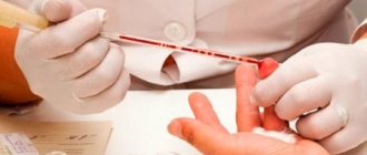
In order to confirm or exclude viral leukemia, and if confirmed, determine its type and form, the veterinarian performs a series of tests:
- clinical blood test - to determine the level of leukocytes in the blood, erythrocyte sedimentation rate, the presence of anemia;
- PCR - polymerase chain reaction - study of peripheral blood;
- immunofluorescence blood test , which allows you to detect antibodies to the virus and determine its type;
- ELISA is an enzyme-linked immunosorbent assay that detects the presence of viral waste products in the blood.
Attention! If, while symptoms persist, all tests for VLK are negative, it is too early to calm down. It is necessary to retake them after three months and conduct a differentiated study, since the same symptoms may indicate completely different diseases.
Today, veterinary clinics are increasingly doing rapid tests for leukemia ; they provide a high degree of reliability, but it is better to play it safe and conduct a comprehensive analysis.
It also doesn’t hurt to conduct ultrasound and x-ray studies, which can help identify the presence of malignant neoplasms and see abnormalities in the functioning of internal organs.
In some cases, the following are recommended for diagnosis:
These are invasive or minimally invasive tests and are done under general or local anesthesia.
Classifications
Considering the nature of proliferating clonal cells
diseases can be divided into two large groups:
T-cell
and
B-cell
chronic lymphoproliferative diseases. Within these groups, based on clinical appearance and tumor origin, the following are distinguished:
- leukemia itself, which always occurs with the involvement of peripheral blood;
- leukemic stage (leukemia) of lymphoma, in which the source of the disease is peripheral lymphoid tissue, but the clinical manifestations, response to chemotherapy and prognosis correspond to leukemia;
- lymphomatous forms, in which blood is extremely rarely involved in the pathological process.
According to another classification, diseases are also combined into two groups:
- First group
: chronic lymphocytic leukemia, - Sézary's disease (lymphomatosis of the skin),
- T-cell lymphocytic leukemia,
- prolymphocytic leukemia (B-cell),
- hairy cell leukemia (B-cell).
– paraproteinemic leukemia:
- multiple myeloma,
Chronic lymphocytic leukemia is of greatest importance.
Treatment of viral leukemia
It is not yet possible to completely cure a cat from leukemia. But timely symptomatic therapy will significantly alleviate the pet’s condition and prolong its life. The selection of drugs is carried out by a veterinarian strictly individually, depending on the condition, tests and symptoms of the patient.
Modern veterinary science suggests that in cases of deteriorating health of the purr, we should act in two directions:
- Stimulation and strengthening of the animal's immunity . The following proven drugs are used:
- Raltegravir.
- Feliferon.
- Interferon.
- AZT.
Antibiotics and vitamin complexes are also used.
- Symptomatic therapy:
- with the help of medications, symptoms of diseases developing due to decreased immunity and inhibition of hematopoiesis are relieved;
- a good but temporary effect is achieved by blood transfusion , this procedure will have to be repeated every 2 weeks; if oncology is diagnosed, chemotherapy (in particular, Vincristine), usually in this way it is possible to stop the pathological process.
In addition, owners will have to reconsider their approach to feeding their pet. Food must be complete and of high quality, heat-treated to prevent germs, bacteria and other pathogens from entering the weakened body of a pet.
A cat diagnosed with viral leukemia will have to be kept in quarantine for life , contact with other cats must be prevented, and its bed, dishes, toys, and litter tray must be kept clean. If there are other purrs in the house, then after contact with the carrier you need to thoroughly wash your hands and, if possible, change your clothes. It is useful to regularly quartz the room in which the carrier cat lives (during quartzing, the animal must be removed from the room).
Important! Remember that euthanizing a cat is a last resort. Never give up in the fight against the disease, even without resorting to treatment.
Treatment
If the treatment of leukemia is favorable and all prognoses are positive, then non-specific skin rashes can be treated using antihistamines, antipruritics, and tranquilizers.
In addition, creams with corticosteroids, menthol in alcohol (1-3%), ointments with anesthesin (3-5%) are often used.
If skin rashes become too widespread and cover large areas of the skin, and they may be accompanied by unbearable itching and even pain, it is recommended to use corticosteroids prednisolone 15-30 mg per day. Here you need to clearly distinguish between skin itching due to nervousness and itching due to leukemia; with the first, the material on our website, dedicated specifically to the mental problems of itching, will help.
Patients with specific skin lesions should use appropriate (depending on the form of leukemia) treatment methods.
Materials used in the article:
https://sblpb.ru/vidy-sypi/382-syp-pri-lejkoze
https://rodinkam.net/zabolevaniya-kozhi/vysypaniya-na-kozhe-pri-lejkoze/
https://www.neboleem.net/adelfan.php
Post Views: 336
Prevention
You should not allow your pet to come into contact with stray and unexamined fellow pets, place it in dubious pet hotels and in unverified foster care, or use other people’s toys, trays, treats, food, bowls, beds, or medicines.
After the death of the previous cat from leukemia, it is recommended to get a new purr no earlier than 2 days after the death of the sick cat and only after cleaning the apartment. It is better to purchase new bowls and trays.
But the best way to protect your pet from viral leukemia is timely vaccination , which must be preceded by a carrier test. Kittens are vaccinated at the age of 2 months and repeat this procedure annually. The vaccine does not kill the infection, but increases the body's resistance to the virus and has a cumulative effect. Therefore, it is important not to forget to vaccinate your pet.
Viral leukemia is one of the most dangerous pathologies in cats. The prognosis for the disease is unfavorable, but still, in our time, lymphosarcoma is not a death sentence, so it is important to recognize VLK at the initial stages of development using comprehensive diagnostics. A timely visit to a specialist will help improve the quality of life of your furry cat and extend his days. To prevent infection, it is necessary to follow preventive measures and pay attention to the general condition of the animal.
Acute lymphoblastic
This leukemia most often occurs in children aged 1 to 6 years.
In boys, the initial stage of the pathology is accompanied by enlargement of the testicles. In addition to the bone marrow, the acute lymphoblastic form is characterized by damage to the thymus gland and internal organs.
In this case, the following complications are observed:
- anemia. Causes pallor, tachycardia and increased fatigue;
- hyperplastic syndrome. Enlarged liver and spleen, which is accompanied by pain in the abdominal area;
- ailments;
- increased body temperature;
- weight loss.
Acute leukemia develops rapidly. With this form of the disease, it is almost impossible to achieve complete remission - the blasts remain in the human bone tissue forever.
The first symptoms of the disease make themselves felt 2–3 months after the onset of the disease. But due to the fact that the above symptoms are felt, this type of blood cancer is easier to detect. Timely diagnosis makes it possible to most effectively slow down the development of cancer.
VIRAL LEUKEMIA OF CATS
Author: Alexandra Fotchenkova Doctor-therapist at the Bely Klyk veterinary clinic
Chronic, or so-called “sluggish” viral infections of cats raise many questions among both animal owners and professional breeders. The disease is often asymptomatic, “carriage” can be hidden - an infected animal, without special examination, can remain “above suspicion” for a long time and come into contact with relatives, exposing them to the risk of infection.
However, such situations are a consequence of insufficient awareness of cat owners, because in practice, identifying and controlling such diseases is a completely feasible task.
In this article we will look at one of the common chronic infections – feline viral leukemia. How to diagnose and control the disease and how to protect cats from infection?
Myeloblastic
In this form of the disease, damage to the DNA structure in bone marrow cells occurs in blasts. Its cause is the influence of radioactive radiation or inhalation of benzene vapors. At the same time, there is a decrease in the concentration of healthy cells in the blood of all types. The pathology can be complicated by inflammatory processes in the joints.
Myeloblastic leukocytosis is diagnosed by analyzing a bone marrow sample. Chemotherapy provides the most effective results.
Her course lasts several weeks.
A timely course of treatment gives a great chance of remission, but it has the following disadvantages:
- reagents have a detrimental effect not only on sick cells, but also on healthy cells;
- severe side effects are observed: nausea, weakness and weakened immune system.
In addition to chemistry, radiation therapy is used to destroy blasts. In severe cases, a bone transplant from a genetically related donor may be necessary. The introduction of stem cells allows the loss of blood components to be restored.
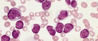
Photo: Myeloblastic leukemia
What is the prognosis for acute lymphoblastic leukemia is written here.
In this article you can find out what acute and chronic leukemia is.
The causative agent of feline leukemia virus is the FeLV virus.
The culprit of this disease is the RNA-containing oncogenic retrovirus FeLV. It was first described in 1964 by British scientist William Jarrett. In the process of large-scale scientific research on the leukemia virus, it was possible to establish that it provokes the development of lymphoma of malignant etiology in these animals.
This is one of the most common destructive viruses. FeLV is resistant to negative external factors and is highly contagious, i.e., it can be transmitted from infected individuals to healthy ones. The main feature of the feline leukemia virus is that it is secreted exclusively by cellular structures that are at the stage of division, therefore it is localized and subsequently spreads, as a rule, in those tissues that contain cells capable of rapid division. We are talking about bone marrow, epithelium of the respiratory and digestive tract.
Pathogenic effect of the felv virus
Viral leukemia in cats infects red bone marrow cells, which leads to damage to the pet's immune system. As a result of such exposure, the immune system is no longer able to perform its functions.
Because leukemia in cats has an increased number of immature white blood cells, the disease is called leukemia.
With leukemia, the body's resistance decreases, which leads to frequent illnesses. The pet begins to get sick more often, and with all sorts of complications, even death.
A dangerous disease often causes the formation of cancerous tumors in the body.
It is very difficult to determine the development of infection in a timely and correct manner, since the symptoms of leukemia can be asymptomatic for a long time. Leukemia in cats manifests itself in full by the time the disease is already advanced.
A pet can die from any serious infectious disease. The presence of leukemia in the animal’s body leads to weakening, the body simply cannot cope with the disease. Although, sometimes there were cases where the immune system was so strong that the animal recovered even without vaccination.
Routes of transmission of the virus
There are several ways to become infected with leukemia:
- contact with infected cats;
- through the milk of an infected mother;
- by airborne droplets;
- through common containers for water, drinking, toilet;
- sexually;
- intrauterine;
- through fleas.
Viral leukemia in cats is not transmitted to humans. In the entire history of medicine, there have been no cases of infection. This is explained very simply - by the different structure of organisms. The feline leukemia virus simply does not survive in the human body.
How does felv viral leukemia differ from fiv viral immunodeficiency in cats?
These diseases have a similar negative effect on the animal's immune system, but not in everything. Feline leukemia virus is different from immunodeficiency virus.
Description of feline viral leukemia
Not a single cat, no matter how well-groomed it is, is immune from viral leukemia, so every owner of such a pet is advised to know how this disease develops, because under unfavorable circumstances it can end in the death of the four-legged pet. The disease can occur in different forms, each of which has certain characteristics.
Mechanism of disease development
Infection occurs in the following ways:
- contact – during communication between a healthy animal and an infected cat or a latent virus carrier;
- aerogenic - through saliva, discharge from the eyes and nose;
- sexual – when mating with an infected partner;
- household - through food or things of an infected individual;
- transplacental – in utero (through the placenta);
- transmammary - the infection is transmitted to the kitten through milk from an infected mother.
Factors contributing to the manifestation of the disease:
- stress;
- weak immunity;
- latent infections;
- pathologies of viral, parasitic and bacterial etiology;
- acute inflammatory processes in the body.
Older cats, animals kept in nurseries and hotels, and those with an undeveloped or weakened immune system are at greater risk of contracting viral leukemia than others. Once in the cat's body, the virus rushes to organs containing cells with rapid division. It affects the red bone marrow, forcing it to synthesize mutated blood cells with an altered structure of leukocytes and lymphocytes. This is precisely why viral leukemia is often mistakenly mistaken for blood cancer, although the etiologies of these pathologies are different.
A virus that has entered a pet’s body can cause the following consequences:
- The cat will forever remain a virus carrier, but the infection will not develop due to the lack of conditions favorable for the reproduction of the retrovirus. Statistics show that invading FeLV is inactivated in 60% of cats.
- The retrovirus will exist in a latent form in the cat's body until it is activated by any pathology, severe emotional shock or taking medications that negatively affect the immune system.
- At the end of the incubation period, the animal will develop a complicated form of viral leukemia.
Forms of viral leukemia
When FeLV spreads in the tissues of the body, spreading through the lymphatic tract and accumulating in the lymph nodes, the pathology is still reversible. If it penetrates the bone marrow and its destruction begins, the cat’s body will no longer be able to overcome the infection, and the chance of recovery is almost zero. The pathological process can occur in different forms, which depend on the state of the animal’s immunity:
- Transitory, or temporary. Characteristic of the initial stages of the disease, until the retrovirus reaches the bone marrow. A strong immune system repels the attack of pathogenic agents, as a result of which the infection does not develop. This variant of the development of the disease is not diagnosed so often.
- Latent. A form of latent carriage is also possible if you have a strong immune system. FeLV is found in tissues, but there are no conditions for its activation and distribution throughout the body. An animal can be a latent virus carrier for a long time. It does not get sick, but it is a source of infection for others.
- Persistent. The virus multiplies because the weakened immune system is unable to fend off FeLV's attack on the bone marrow. The virus is carried throughout the body by leukocytes, its concentration in the bloodstream is high, so the manifestations of the disease increase over time.
- Atypical. It is a partially effective immune system response in which a retrovirus develops in a specific organ.
What is feline leukemia virus
The feline leukemia virus is a retrovirus and has a widespread but uneven distribution.
The virus attacks the cat's immune system, disrupting its function and making it susceptible to the development of secondary and opportunistic infections (those that a healthy immune system usually suppresses itself). In addition, the virus has an oncogenic effect, causing uncontrolled reproduction of cells affected by it with the development of lymphomas and leukemia. It is the defeat of the immune system that provides a wide variety of clinical manifestations in viral leukemia. The signs of the disease are especially pronounced in the organs and systems that perform the barrier function - the digestive and respiratory systems, as well as the skin.
The feline leukemia virus is unstable in the external environment and is easily destroyed by disinfectants, treatment with soap solutions, as well as drying or heating.
Feline leukemia virus is often confused with feline immunodeficiency virus because they are both retroviruses that attack the immune system while exhibiting similar symptoms. But there are differences between these pathogens:
- The feline immunodeficiency virus affects only mature cells of the immune system circulating in the blood, while the feline leukemia virus affects the hematopoietic organ - the bone marrow, causing the production of pathologically altered cells of the immune system, which are unable to perform protective functions and themselves participate in the course of the disease, forming tumors . Thus, immune disorders in viral leukemia are much more complex and severe than in viral immunodeficiency.
- With feline viral leukemia, lymphomas often develop due to the activity of immune system cells modified by the virus, while with viral immunodeficiency there is a decrease or loss of functions of immunocompetent cells. Malignant neoplasms are not typical for him.
- The feline immunodeficiency virus has a wide variety of subspecies, which makes it difficult to create a vaccine. Nevertheless, a vaccine has been created, but is used only in the United States. To protect against viral leukemia, vaccine prophylaxis has been developed and is available everywhere.
- In veterinary medicine, cases of complete recovery from viral leukemia are known. No similar data have been reported on viral immunodeficiency disease.
In some cases, the similarity of the symptoms of these two diseases is so pronounced that it is possible to accurately determine the pathogen only using laboratory diagnostic methods.
The feline leukemia virus infects cells that tend to divide rapidly
The source of infection is cats with viremia - a condition when the virus circulates in their blood and is actively excreted through saliva, excrement, sputum, and also through milk during lactation.
In addition to domestic cats, large wild cats also suffer from viral leukemia. This pathogen is not dangerous for people, dogs, and other domestic animals.
Clinical signs of feline viral leukemia vary widely from their complete absence with asymptomatic carriage, to the most severe forms of the disease with multiple organ damage, widespread tumor process and the active participation of secondary infection.
After infection, the virus develops as follows:
- Reproduction of the virus in the tonsils, retropharyngeal lymph nodes.
- Penetration into macrophages and B-lymphocytes of the blood, thereby creating conditions for the spread of viral infection.
- Colonization and the beginning of reproduction in the lymphoid tissue of the thymus, in the crypts of the epithelium and Peyer's patches of the intestine, as well as in the red bone marrow with infection of progenitor cells.
- Release of infected neutrophils and platelets from the bone marrow into the bloodstream.
- Penetration of the virus into epithelial cells and glands, where it multiplies and is released in large quantities with biological fluids and excrement.
At stages 2 and 3, as the disease develops, protective immune responses are suppressed, which corresponds to a period of 1–2 months after infection; stages 4 and 5 are characterized by the development of persistent (constant) viremia - this is a state of constant presence of viral particles in the blood; its development requires 1-3 months after infection.
With an adequate immune response, viremia is suppressed, and the disease does not develop further. A cat that has overcome viremia becomes latently (latently) infected, and reactivation of the infection in it is possible if an immunodeficiency state occurs. The cat's active immunity prevents the virus from multiplying, and the latter is detected only in the form of viral DNA integrated into the DNA of the host cell, the so-called provirus. This means that if favorable living conditions are maintained, this cat can live to a ripe old age.
If we turn to the statistical results obtained from examining infected shelter cats in the 80s of the last century, and use them as a forecast for the development of the disease, we find that:
- 30–40% of animals will develop persistent (constant) viremia and clinical manifestations of infectious leukemia;
- in 30–40% of cats, viremia will be inconsistent and periodic; this condition reflects the partial ability of the cat's immunity to restrain the aggression of the virus, while the virus may gradually colonize the bone marrow and lymphatic tissue, and symptoms of the disease may appear after several years, and a transition to latent infection is also possible if the cat's immune system wins;
- 20–30% of cats do not have viremia, but have antibodies - this is a latent infection when the immune system keeps the virus reproduction under control;
- in 5% of cats the course is atypical, when virus antigens are detected in the blood in the absence of viremia.
Therefore, the incubation period for this disease can last from several weeks to several years, and asymptomatic carriage is also possible.
Good care is important for a cat's strong immunity.
The most common clinical manifestations of feline viral leukemia are:
- immunosuppression (suppression of the function of the immune system) and predisposition to the development of various infections, manifested by: chronic inflammatory processes characteristic of weakening the barrier functions of the immune system, the treatment of which is difficult due to an immunodeficiency state: sinusitis - inflammation of the mucous membranes of the paranasal sinuses;
- rhinitis - inflammation of the nasal mucosa;
- gingivitis - inflammation of the gums;
- periodontitis;
- the appearance of abscesses (ulcers) on the skin;
- there is a predisposition to infection with feline viral peritonitis, as well as hemobartonellosis;
- alimentary form - manifested by lack of appetite, diarrhea, vomiting, tumor masses invade the small and large intestines, as well as the lymph nodes of the abdominal cavity;
We suggest you read: Wet tail colibacillosis
Less common:
- autoimmune manifestations: polyarthritis;
- hemolytic anemia;
- glomerulonephritis;
- fruit resorption;
- paresis;
Testing for possible carriage of the leukemia virus should be carried out in all cats, and especially:
- in animals at risk;
- if there are signs of disease, since viral leukemia is characterized by a variety of symptoms;
- in cats taken from the street or from a shelter, especially when there are already cats in the house;
- before prophylactic vaccination against viral leukemia.
Diagnostic methods:
- ELISA (enzyme-linked immunosorbent assay) - determines viral antigen in cases of damage to platelets, leukocytes, lymphocytes, diagnostic sensitivity 90%;
- PCR (polymerase chain reaction) - determines the DNA of the provirus (when the viral DNA is integrated into the genome of the host cell), the most reliable diagnostic method;
- PCR diagnostics aimed at detecting the RNA of the virus is a reliable evidence of viremia, which can even be used to study pooled samples in order to save money, for example, when examining nurseries;
- immunochromatography - also detects viral antigen in blood cells, diagnostic sensitivity is 90%;
- RIF (immunofluorescence reaction) - detects viral components in blood smears, diagnostic sensitivity is significantly lower than that of PCR diagnostics, a positive result usually indicates persistent viremia;
- Virus cultivation, the most sensitive method, especially in the early stages of the disease, is now used extremely rarely in routine practice.
Symptoms of VLK, diagnosis of the disease
In all cases, without exception, leukemia causes a significant weakening of the pet’s immunity. He begins to get sick often. The disease is severe, with complications and relapses. Other symptoms are associated with secondary infections:
- frequent and sudden changes in body temperature for no apparent reason;
- significant decrease in motor activity;
- excessive sleepiness;
- partial or complete loss of interest in food;
- frequent colds;
- disruption of the digestive system (vomiting, diarrhea);
- hypersalivation;
- enlarged lymph nodes;
- anemia of the mucous membranes.
Diagnosis of leukemia is possible only in a veterinary clinic. Diagnostic methods used to make a diagnosis:
Sources:
https://kotoholik.com/veterinaria/bolezni/lejkoz-u-koshek.html https://www.royal-canin.ru/college-pro/breeders/virusnaya-leykemiya-koshek/ https://murkoshka.ru /zdorovye/preparaty/vakcina-ot-lejkoza-koshek.html
Leukemides
Leukemids
is the general name for focal and diffuse lesions of the skin, subcutaneous tissue, genital mucosa and oral cavity, occurring in places of malignant proliferation of cells of the hematopoietic organs and reticular tissue.
They are extramarrow foci of hematopoiesis that develop in the dermis during the transition of leukemia to the terminal stage. The lesions have the appearance of erythema, erythroderma, raised papules, vesicles or bullae of light pink or dark red color, covered with smooth shiny skin, less often with crusts.
In the presence of a detailed clinical picture of leukemia, the diagnosis of leukemias does not cause difficulties. Radiation and PUVA therapy are used for treatment.
In the scientific literature, several synonyms are used to designate leukemias: specific hemoderma, hematoderma, hematodermatosis, reticulosis, reticulohemoblastosis. Depending on the type of leukemia, skin manifestations are found in 3-50% of patients.
In monocytic leukemia, damage to the dermis and hypodermis is observed in every second patient, in chronic lymphocytic leukemia - in every fourth. More often, leukemia is diagnosed in men. In patients of both sexes over 50 years of age, the likelihood of the formation of foci of extramedullary hematopoiesis is higher than in young people.
In children, and less often in adults, skin infiltrates can appear at the very beginning of the disease, forming one of the so-called “debut masks” of acute leukemia.
Leukemides
The development of blood cancer is based on malignant degeneration of hematopoietic cells of the bone marrow. Their active division leads to the formation of a clone of cancer cells, which within a short time displace normal elements from the bone marrow and begin to metastasize. Metastases of blasts and granulocytes into the dermis give rise to leukemias.
The appearance of metastatic foci in the lungs, heart, intestines, and brain leads to infiltration and disruption of the functioning of these organs. For some forms of leukemia, for example, for primary ulcers, the causative factors of appearance have been established: previous trauma, skin irritation with chemicals, rubbing parts of clothing.
But most forms of rash appear on unchanged skin.
Cancer cells enter the dermis and subcutaneous tissue through the blood and lymph. Having established themselves in the tissues, the cells of the metastatic focus begin to actively divide, forming large infiltrates, single or multiple elements of the rash.
Each of the foci of proliferation of the leukemic cell clone forms one element. The growth rate of elements is largely determined by the type of cancer cells and the rate of progression of the underlying disease.
As a rule, skin infiltrates are formed by blasts that are characteristic of this type of myeloid leukemia and have lost tissue specificity. Less common are formations consisting of mature granulocytes: promyelocytes and myelocytes, up to segmented ones.
Mature cell leukemias grow relatively slowly until they transform into blasts.
Accumulations of blasts in the dermis actively disseminate with the formation of new foci on the surface of the body and in the internal organs. In parallel with the dissemination of blasts, there is a process of disruption of blood circulation in the tissues. This is facilitated by thrombocytopenia and coagulopathy characteristic of blood diseases.
Thrombocytopenia leads to the appearance of subcutaneous hemorrhages, petechiae and ecchymoses. Coagulopathy is the cause of blood clots that block blood flow in vessels of different sizes.
As a result of a local circulatory disorder, leaf-shaped crusts form on the surface of the papules, and foci of leukemic infiltration become ulcerated.
The rash appears suddenly against the background of a previous illness. In one patient, the rash may be formed by elements of different sizes and types. New elements appear within a few days.
All this time, as a rule, there is a disturbance in the general condition and signs of intoxication.
In cases where leukemides appear at the onset of the disease, health can remain satisfactory for a long time.
Skin tumors do not cause any discomfort. Infiltrates on the mucous membranes are sharply painful; with their necrotic disintegration, the pain can become excruciating.
The shade of the skin over the neoplasms can vary from light green and pale pink to bluish-burgundy in blastic leukemias.
Mature cell formations are covered with unchanged skin of normal shade. The surface of leukemias is smooth and hairless.
Most often, small papules and plaques occur with leukemia. The elements are slightly raised above the surface of the skin, which is clearly visible in side lighting, have a rounded shape, and are located symmetrically on different parts of the body.
Consistency – from soft, doughy to dense. The attachment of the hemorrhagic component is characterized by the formation of blisters with a flaccid cap. For a long time, the elements remain unchanged or gradually increase in size.
Their spontaneous regression is possible.
Nodes can be single or multiple, isolated or drain. They often penetrate deeply into the hypodermis. Their size varies from millet grain to walnut. The shape is hemispherical or truncated.
The consistency is dense, in chronic myeloid leukemia and acute leukemia it is woody. The color is bright, rich red, less often burgundy. The clogged ducts of the sebaceous glands on the surface of the formations look like whitish grains or inclusions.
Individual nodes erode and undergo purulent-necrotic decay. Ulcers formed at the site of the nodes are resistant to local treatment.
Local leukemic infiltration, primary ulcers, and specific erythroderma are rare variants of hemodermatosis. Infiltrates are formed mainly on the torso and scalp, covered with longitudinal folds and deep grooves.
The usual localization of primary ulcers in myeloid leukemia is the lower extremities, groin area, and gums. Their diameter is 6-7 cm. The edges are uneven and undermined in places. The bottom is smooth, shiny, bright red, covered with blood crusts, purulent-necrotic plaque, and less often with granulations.
Specific erythroderma in leukemia is indistinguishable from nonspecific erythroderma in its clinical picture.
Damage to the skin in patients with leukemia occurs against the background of reduced immunity.
In this regard, the likelihood of a bacterial, fungal or viral infection increases many times over, which aggravates the patient’s general condition, complicates treatment, and worsens the prognosis regarding remission of the underlying disease and survival.
Severely painful leukemic infiltration of the oral cavity disrupts the process of chewing and eating. Weight loss can reach 10 kg per month, which quickly leads the patient to exhaustion. The spread of the purulent-necrotic process to the jaw bones increases the mobility of the teeth, provokes their loosening and loss.
Algorithms for carrying out diagnostic procedures in patients who initially consult a doctor about a skin disease and in patients with an already established diagnosis of myeloid leukemia are significantly different. In the first case, the examination is carried out by a dermatologist, in the second by a hematologist or oncologist. With damage to the mucous membranes, the patient can consult a dentist.
Primary diagnosis of leukemias is difficult. This is due to the numerous forms of skin rashes characteristic of hemoderma, the similarity of almost each of the forms with other dermatological diseases, and in some cases also to the insufficient sensitivity and specificity of diagnostic tests.
An accurate diagnosis can be made by:
- Careful history taking.
It involves identifying symptoms characteristic of the onset of leukemia, factors that increase the likelihood of developing oncology, conditions of infection with syphilis, HIV infection, leprosy, and other significant data. A detailed questioning of the patient allows us to reasonably prescribe diagnostic procedures and tests and interpret them correctly. - Blood tests
. A general blood test is prescribed, which can reveal abnormalities that allow one to suspect or completely exclude the diagnosis of myeloid leukemia, a serological test for syphilis, and HIV. If necessary, a blood test may be performed for Mycobacterium leprosy and other infections. - Histological examination
. Tissue samples are taken from the largest leukemias with the least pronounced inflammatory changes in the elements themselves and the surrounding skin. In punctates from leukemias, accumulations of blasts are detected, less often granulocytes at different stages of maturation. The method makes it possible to make a diagnosis in 50% of cases, since changes detected by microscopy are not always specific. - Molecular genetic research.
The polymerase chain reaction (PCR) method makes it possible to register clonal rearrangements of the T-cell receptor of lymphocytes and to establish the fact of monoclonal proliferation in lesions in the early stages of the disease. The probability of false positive results is 5%: monoclonal rearrangement is detected in a number of benign dermatoses, inflammatory processes, and systemic connective tissue diseases. - Immunohistochemical study.
The method is based on the use of fluorescent dyes, enzymes, or electron-dense particles labeled to identify tumor cells of a specific type. Antibodies selectively bind to blasts and make them visible under microscopy.
In dermatology, in patients without a history of cancer, leukemides are differentiated primarily from benign processes, such as diffuse neurodermatitis, lymphoplasia or primary reticulosis, mycosis fungoides, sarcoidosis.
In second place are infectious diseases with characteristic skin manifestations: lenticular papular syphilide, lepromatous type of leprosy. In patients with myeloid leukemia, leukemides must be differentiated from cutaneous manifestations of infections and complications of chemotherapy.
These are viral and bacterial rashes, side effects of medications, and other dermatological pathology that developed simultaneously with leukemia.
Therapeutic effects for hemoderma are aimed at treating the underlying disease. Putting myeloid leukemia into stable remission leads to a gradual regression of skin manifestations. If, as a result of chemotherapy, the manifestations of hematodermatosis persist, a local effect on the lesions is prescribed. For this purpose:
- Radiation therapy.
Cancer cells, unlike other cells in the body, are more sensitive to any damaging influences. The radiation therapy method is preferable for eliminating large single lesions. The power of X-ray radiation is selected in such a way as to affect the entire depth of leukemia. The number of procedures is determined individually, taking into account previously performed treatment, the patient’s condition, and the rate of reverse development of formations. - PUVA therapy.
The method involves exposing skin tumors to long-wave ultraviolet radiation, which is preceded by the administration or local application of drugs that increase the photosensitivity of tissues. PUVA therapy is used to treat multiple elements or large infiltrative lesions.
The prognosis is determined by the underlying disease. The use of modern treatment methods helps to increase the life expectancy of patients with leukemia. However, this increases the likelihood of developing serious complications of the disease, for example, damage to the central nervous system.
Leukemids, which appear in the early stages of leukemia development, with timely consultation with a doctor and proper diagnosis, allow you to quickly establish a diagnosis and begin treatment as early as possible.
The development of hemodermatosis with diagnosed blood cancer indicates the transition of myeloid leukemia to the final stage, which is an unfavorable prognostic sign.
Source: //www.KrasotaiMedicina.ru/diseases/zabolevanija_dermatologia/leukemid