Compaction of the aorta of the heart - what is it?
The aorta is the largest blood vessel in the human body.
Sometimes this vessel can be subject to deformation and the development of pathology—hardening of the aorta of the heart. Not everyone knows what it is, so when making a diagnosis they are at a loss. In fact, thickening of the aorta of the heart (we’ll look at what it is in detail below) is not a separate disease. Rather, this is the doctor’s general conclusion about the condition of the walls of the heart vessel.
During the compaction of this vessel, it loses its usual elasticity and density, which leads to the development of various serious diseases in the body. The thickening itself is caused by blood plaques that clog the aorta and lead to heart failure.
Compaction of the aorta of the heart (a cardiologist knows what it is and how to treat it) develops for the following reasons (the root of the disease):
- Hypertension is the most common cause of this condition. In this position, a person’s blood pressure constantly increases, which leads to thickening of the blood vessels and the formation of dense fibrous compounds in them. Moreover, the risk of aortic thickening also increases when the patient suffers from a number of infectious pathologies.
- Frequent consumption of foods high in fat, which, in turn, increases cholesterol levels in a person’s blood. This causes cholesterol plaques to accumulate in the arteries, causing them to become inflamed and thickened.
- Smoking and frequent drinking not only contribute to thickening of the artery, but also makes the vessels very brittle and susceptible to rupture.
- An elderly person, during which serious physiological changes are observed in the body (thickening of the walls of blood vessels).
- Vascular atherosclerosis is also a common cause of the development of this condition. Moreover, there is also the so-called atherosclerotic syndrome, in which the patient experiences serious circulatory disorders.
- Severe venereal diseases.
- Hereditary predisposition of a person.
- Severe infectious diseases, including damage to internal organs (lungs, liver, etc.).
If the aortic thickening is pronounced and treatment is not carried out for a long time, then this condition can lead to stenosis, disruption of the myocardial valve and its ventricles, as well as general heart failure.
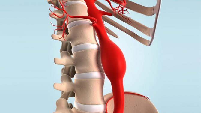
Moreover, if the disease progresses, the walls of the aorta in the area of plaque formation may begin to dissect, which threatens the development of an aortic aneurysm. This disease is extremely dangerous and requires long-term medical and surgical intervention, which is often unsuccessful.
Fortunately, aortic aneurysm develops quite rarely. No more than three cases are recorded per year.
Aortic thickening can be treated using both folk remedies and traditional medications.
Signs of the disease
UAS does not show any symptoms for a long time. As the lumen narrows, tissues and organs receive less and less nutrition, and alarming symptoms appear. But these signs are not specific, so they do not differentiate USA from other cardiovascular problems.
The symptoms of UAS depend on the location of the thickening:
- A seal located at the root of the aorta - at the exit from the heart or in the ascending part of the vessel - leads to disruption of the nutrition of the heart. The patient experiences symptoms characteristic of angina pectoris - chest pain, increased heart rate. The thickening located in this place leads to ischemia of the heart muscle, which, in turn, often leads to myocardial infarction.
- When the wall of the aorta in the descending part hardens, the brain is primarily affected. He experiences oxygen starvation, which is manifested by neurological symptoms - frequent dizziness, intense headaches, paresis and even fainting. This is an extremely dangerous condition that requires immediate medical attention.
- If the abdominal part of the vessel is thickened, symptoms occur that indicate problems in the gastrointestinal tract. The patient experiences abdominal pain and indigestion. Possible weight loss.
- The seal located in the abdominal part leads to poor circulation in the legs. The patient develops pain and cramps in the lower extremities, and lameness may appear. In severe cases, the person cannot walk.
Attention! Thickening of the abdominal aorta is a dangerous condition that requires urgent treatment. In difficult situations, peritonitis may even develop, which requires surgical intervention.
Aortic compaction on x-ray: symptoms of the disease
Compaction of the aorta (best seen on X-rays and CT scans) can occur for quite a long time without showing itself at all.
Over time, hardening of the aorta provokes a narrowing of the lumen of the vessels, loss of elasticity and the formation of fibrous growth in the artery, increased blood pressure, and the formation of aortic dissection - an aneurysm. In most cases, compaction of the aortic arch is detected during fluorography in people of different age categories - both young and elderly. In children, this pathology is detected somewhat less frequently.
The main signs of this condition are frequent attacks of angina pectoris, hypertensive crises, and acute heart failure.
Also, a sick person may suffer from chest pain, sleep disturbances, weakness and drowsiness.
In addition to thickening of the aortic arch during fluorography, this disease can be identified by frequent convulsions, changes in the density and structure of the aorta during examinations by a cardiologist, and pain in the heart area.
When the first signs of aortic thickening appear, a person is advised to consult a doctor and undergo a diagnosis.
It is possible to detect compaction of the aorta on an x-ray if the cause of the pathology is an infectious or atherosclerotic lesion.
Additional studies for aortic thickening and dysfunction of myocardial valves are:
- Ultrasonography.
- The compaction of the aortic arch is very clearly visible during fluorography.
- Contrast angiography.
- Radiography.
- MRI will detect all changes in the aorta and understand the extent of circulatory disorders.
In addition to drug therapy, it is important to follow a special diet when treating aortic thickening.
Treatment
Treatment of arterial compaction is complex, aimed at eliminating the root cause, and can be either medicinal or surgical.
For the first, drugs that lower blood pressure are prescribed: diuretics, adrenergic blockers, calcium channel blockers, anti-inflammatory drugs, for syphilis - penicillin, bismuth, arsenic, mercury.
The operation is performed in severe cases, for example, with inflammation of the peritoneum. The following methods are used:
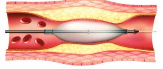
- Installation of a stent in a pathological area;
- Removal of a section of the vessel and replacing it with a prosthesis;
- Valve replacement.
Treatment of seals in the aortic arch and its other parts should, first of all, be aimed at eliminating the underlying disease that caused this pathology.
The risk group primarily includes patients with a history of hypertension, especially accompanied by frequent crises and sudden surges in pressure. Some chronic diseases, such as tuberculosis and tertiary syphilis, also cause the formation of plaques and growths on the walls of the aorta, as does smoking.
Such patients are required to be sent for further examination in order to have complete information about the prevalence of the disease and the risk of complications.
In the absence of pronounced progression of the disease, preventive measures are used aimed at eliminating provoking factors that cause the development of aortic thickening. Patients are recommended to undergo the necessary therapy if indicated.
The main method of combating the formation of atherosclerotic plaques is changing lifestyle, diet, moderate physical activity and walks in the fresh air. Patients are advised to avoid stressful situations.
Therapy is not aimed at the compaction in the aortic wall, but at the underlying disease.
If the symptom is detected against the background of hypertension and atherosclerosis, then an anti-cholesterol diet is used in therapy, a regimen with sufficient rest and stress prevention is recommended. The doctor prescribes statins (drugs that lower low-density lipoprotein levels). Treatment of hypertension is carried out according to the scheme with drugs of different groups.
Induration as a consequence of arteritis is treated with anti-inflammatory drugs when an active process is confirmed.
Antisyphilitic drugs are prescribed by venereologists at any stage.
At the compaction stage, it is impossible to achieve an effect, since part of the vessel is already completely “out of order.” All treatment is aimed at preventing possible complications:
- formation of dissecting aneurysm;
- myocardial ischemia;
- heart failure;
- damage to the kidney arteries.
Considering the availability of examination, it is necessary to pay attention not only to the underlying disease, but also to think about the consequences.
Hardening of the aortic walls: treatment methods
Thickening of the aortic walls is treated based on the patient’s symptoms, the degree of neglect of the pathology, as well as the cause that caused it.
Traditional drug therapy includes the following:
- Prescription of statin drugs (Lovastitin, Cholestyramine). This is practiced if the thickening of the aortic walls is caused by atherosclerotic syndrome or elevated cholesterol levels.
- Prescription of diuretics for the myocardium (Hypothiazide).
- If the thickening of the aortic root of the heart is caused by infectious diseases, the person is prescribed drugs based on penicillin - antibiotics.
In case of mitral or aortic stenosis, dissection of aortic fibers (impaired nutrition), acute heart failure, malnutrition and other dangerous conditions, the patient may be prescribed surgical treatment.
It involves special prosthetics of the affected area of the aorta or total organ transplantation. A CT scan and chest x-ray will help determine whether a person needs surgical treatment or not. Using these studies, you can see the thickness of the compaction of the aortic root of the heart, as well as the exact localization of the deposition of cholesterol plaques.
During the period of therapy, the patient needs to be under medical supervision, take prescribed medications, follow a diet and avoid stress.
As an additional treatment with folk remedies, the following can be used:
- Herbal treatment (herbal medicine).
- Acupuncture.
- Physiotherapeutic treatment.
Traditional medicine also offers the following recipes that will help eliminate compaction of the aortic root of the heart and general compaction of the aortic walls:
- Chop the garlic and pour boiling water over it. Add lemon juice and leave for three days. Take the finished product one teaspoon twice a day an hour before meals. The duration of treatment is at least four months.
- Make a tincture of water and rowan. Boil for an hour. Strain and take a teaspoon once a day.
- Mix rose hips, hops, mint and oregano in equal quantities. Pour boiling water and leave for two hours. Strain and take a spoon an hour before meals.
Prevention of hardening of the aortic root of the heart involves giving up bad habits, avoiding stress, moderate sports activities and a healthy lifestyle. It is also important to eat a balanced diet and promptly treat serious illnesses.
Compaction of the aorta is a pathology that can lead to a heart attack, aneurysm and many other dangerous pathologies, so do not underestimate this disease. It needs to be treated immediately after diagnosis.
Source: med88.ru
Prevention
Measures to prevent pathological changes in the aorta include following simple recommendations. Doctors advise:
- completely quit smoking and alcohol;
- balance physical activity with the body’s capabilities;
- adjust nutrition;
- sleep at least 8 hours a day;
- It is wise to alternate vigorous activity with rest.
Systematic planned examinations in combination with preventive measures guarantee timely detection of aortic compactions. This is a chance to prevent the progression of negative changes and the effectiveness of therapy that ensures vascular health.
Compaction of the aorta (valve leaflets, root, arch): symptoms, causes, how to treat
The aorta is the largest unpaired arterial vessel of the systemic circulation, coming from the heart and feeding all internal organs and systems, except the lungs. Throughout its entire length, the aorta has a uniform thickness and the same structure. Under the influence of unfavorable exogenous and endogenous factors, its structure is disrupted: atherosclerotic plaques or fibrous growths appear on the walls, disrupting normal blood flow.
Aortic compaction can be detected using various diagnostic methods: radiographic, fluorographic or ultrasound examination. Having discovered such a defect, specialists must find the main causes of the pathology and prescribe the correct treatment. Otherwise, the disease can lead to serious consequences: dissection of the aortic walls and rupture of the blood vessel. These complications are accompanied by large blood loss and often result in the death of the patient.
Depending on the location of the lesion, the following forms of pathology are distinguished:
- Compaction of the aortic root,
- Consolidation of the aortic arch,
- Consolidation of the ascending aorta,
- Consolidation of the descending aorta.
The main etiological factors of aortic compaction:
- Atherosclerosis is the main cause of the pathology. According to its structure, the aorta belongs to the vessels of the muscular-elastic type. They are the ones most exposed to LDL. Lipids are deposited on the inner lining of the aorta and lead to the development of rough fibrous tissue. Plaques form, protruding into the lumen of the vessel and growing with scar tissue. Compaction of the aortic root is always accompanied by spasm of the coronary arteries, which leads to the development of hypoxia and myocardial ischemia.
serious atherosclerotic changes in the aorta with the formation of a life-threatening aneurysm
Syphilitic infection occupies a special place among the causative factors of pathology. Thickening of the aorta is a late clinical sign of a long-term syphilitic process, developing 10-20 years after infection.
aortic valve seal
Factors contributing to thickening of the aorta: bad habits, foods high in cholesterol, hereditary predisposition, systematic overeating. Under the influence of these factors, the aorta automatically thickens and becomes highly sensitive to traumatic damage. The consequence of the pathology is sclerosis of the valve leaflets and the aortic ring. They lose their mobility, aortic stenosis and aortic regurgitation develop.
The compacted aorta does not manifest itself for a long time. Over time, the lumen of the blood vessels narrows, and the nutrition of the internal organs is disrupted. The clinical picture of the disease is determined by the location of the lesion.
- The narrowing of the coronary vessels, which occurs against the background of hardening of the aorta, is manifested by frequent attacks of angina or the development of myocardial infarction.
- The carotid artery arises from the thoracic aorta and supplies blood to the brain. When cerebral vessels are damaged, patients develop neurological symptoms, their general condition worsens, and the help of a specialist is required.
- Consolidation of the abdominal aorta is manifested by nagging pain in the abdomen, weight loss and symptoms of impaired digestion. In severe cases, signs of peritonitis appear: sharp pain, “acute abdomen,” fever and severe dyspepsia.
- Damage to the vessels supplying blood to the lower extremities leads to characteristic lameness. When walking, patients experience pain and cramps, forcing them to stop.
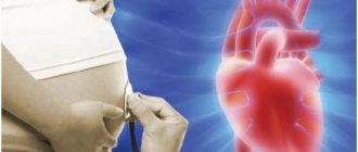
Aortic thickening is detected by chance, since this pathology is often asymptomatic. Pathological foci occur in certain areas of the blood vessel or its entire length. There are no specific symptoms of aortic thickening. The doctor can make a preliminary diagnosis based on the patient’s complaints, clinical picture, auscultatory examination data - emphasis of the second tone on the aorta, characteristic murmur. A large difference between blood pressure numbers also indicates involvement of the aorta in the pathological process.
The most reliable diagnostic methods are:
- X-ray examination in direct and lateral projections allows us to examine the shadow of the heart and large blood vessels. In persons with aortic compaction, its shadow is elongated, there is a bend along the vessel and a pathological turn, the aorta is dilated, and the intensity of the shadow is increased.
- Contrast angiography is the most effective diagnostic method.
- Consolidation of the aortic arch is often discovered accidentally during fluorography.
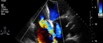
- Ultrasound examination and Dopplerography.
- Magnetic resonance imaging allows you to identify changes in the aorta and determine the degree of circulatory disturbance.
Treatment of the pathology is complex and serious, aimed at eliminating the cause . Patients are advised to lead a healthy lifestyle, eat right, fight bad habits, walk in the fresh air, avoid stress, and be under the supervision of a doctor.
- If the cause of the pathology is atherosclerosis, patients are prescribed an anti-cholesterol diet and medications that reduce the level of LDL in the blood - statins Fluvastatin, Lovastatin, fibrates Clofibrate, Fenofibrate, drugs that enhance the excretion of bile acids - Cholestyramine, Cholestipol "
- To treat hypertension, drugs of different groups are used: diuretics - Hypothiazide, Veroshpiron, adrenergic blockers - Atenolol, Bisoprolol, slow calcium channel blockers - Nifedipine, Amlodipine.
- Peritonitis requires emergency surgery.
- If thickening of the aorta is a consequence of arteritis, anti-inflammatory therapy is carried out.
- Venereologists prescribe antisyphilitic drugs - drugs of penicillin, bismuth, arsenic, mercury.
Surgical treatment for thickening of the aortic valve leaflets involves repair or replacement of the valve. In the case of aortic dissection along its length, or the formation of an aneurysm, the issue of vessel prosthetics or installation of a stent in the thickening zone is resolved.
Traditional medicine to strengthen the structure of the aorta:
- The garlic is peeled, crushed, and poured with boiling water. The infusion is stirred several times during the day, lemon juice is added and infused for a week in a dark and cool place. Take the resulting remedy one teaspoon three times a day half an hour before meals. The duration of treatment is three months.
- An infusion is prepared from rowan bark and water. The resulting product is boiled for two hours, cooled and filtered. Take a tablespoon three times a day.
- A collection of clover grass, rose hips and hawthorn berries, hops, mint, oregano, motherwort, and sweet clover is poured with boiling water, left for two hours, filtered and taken three times a day before meals for a month.
Currently, there are many treatment methods aimed at restoring the structure of the aorta. But before starting such therapy, you need to consult with your doctor.
Diagnostics
As mentioned above, aortic thickening is detected by chance. A specialist can make a diagnosis based on the patient’s complaints and pressure deviations from the norm.
If aortic thickening is suspected, the patient is sent for a more accurate diagnosis using the following methods:
- Radiography. On X-ray, the VA looks like an elongated shadow, a bend along the vessel and a pathological turn, dilatation of the vessel, the shadow is more intense.
- The most effective method is contrast X-ray examination of blood vessels.
- The presence of a blood vessel seal is randomly determined by fluorography.
- Ultrasound and Doppler method.
- MRI makes it possible to detect pathology in the aorta and determine the degree of blood flow disturbance.
The most effective diagnostic methods are ultrasound of the aorta and contrast angiography. This way you can identify compaction not only locally, but throughout the entire length of the vessel.
Often signs of the disease are discovered when the patient is being examined for other diseases.
As for treatment, the method is determined by the nature of the manifestation. For example, a dissected aneurysm cannot be treated without emergency surgery. Moreover, the operation may not always be successful. Surgery will be required for peritonitis and hardening of the aortic root.
Aortic valve insufficiency, caused by thickening of the leaflets and deterioration of their mobility, includes medication, special or surgical treatment. Plastic surgery and prosthetics are common. Significant damage to the structure of the heart may require transplantation of a donor organ.
Moderate physical activity is an excellent prevention of vascular diseases
In cases where the disorder does not progress, preventive measures are prescribed, the task of which is to eliminate factors that cause changes in the structure of the walls of the aorta of the heart. This means that you will need to undergo comprehensive treatment of the underlying disease and prevention of aortic compaction.
Preventive measures include:
- giving up bad and destructive habits;
- proper diet;
- moderate physical activity;
- daily exposure to fresh air;
- elimination of stressful situations;
- further, regular monitoring.
As mentioned above, most often aortic thickening is detected accidentally. Nevertheless, there are a number of signs that may lead a doctor to think about such a pathology:
- When measuring blood pressure, there is a large difference between the upper and lower readings;
- When listening to the heart, a specific murmur is heard, and the second sound in the aorta intensifies.
To confirm the thickening of the aortic walls, the following diagnostic studies are performed:
- X-ray of the chest cavity in frontal and lateral projections. The radiologist will be able to see characteristic changes in the image that confirm the pathology. If a change in the abdominal aorta is suspected, then an image of the abdominal cavity is taken.
- Ultrasound examination, Dopplerography. These methods allow a more accurate and localized study of the damaged area of the vessel, as well as identify circulatory disorders and changes in the internal organs, if any.
- Magnetic resonance imaging.
- Contrast angiography.
To obtain information about the state of the circulatory system, patients need to undergo an X-ray or ultrasound examination.
Such diagnostic techniques will allow us to study the structures of the aorta and identify areas of damage. If thickening of the aortic root is detected, it is important to take measures to determine the causes of this phenomenon. Only after this can you decide on the tactics of further treatment.
When age-related changes are a provoking factor, patients are prescribed a course of therapy aimed at inhibiting the further development of the pathology. Also mandatory components of complex treatment are moderate physical activity, nutritional correction, stabilization of the emotional state, and strengthening of the body.
In order to prevent complications and serious consequences, it is important to give up bad habits, lead a healthy and active lifestyle, and follow all the instructions of your doctor.
If there are diseases that cause thickening of the walls of the aorta, measures must be taken to eliminate them. In rare cases, patients are prescribed surgical treatment.
Home » Diseases » Cardiology » Cardiology (general) » Compaction of the aortic root of the heart
The long absence of clinical manifestations is the reason that the detection of the disease is sometimes a complete surprise to the patient himself.
The fact that the aortic arch is compacted can be shown by a fluorogram, so annual fluorography is of important preventive value.
A fluorographic image demonstrating an altered structure of a blood vessel with calcified areas may confirm the onset of atherosclerosis.
The appearance of alarming symptoms is an indication for a more thorough medical examination.
Considering the fact that pathological changes can affect both isolated sections of the artery (most often these are the walls of its descending part, the compacted aortic root, the aortic valve leaflets) and the entire vessel, during the diagnostic examination the following is performed:
- Ultrasound of blood vessels;
- contrast angiography.
Video: the main cause of aortic thickening is atherosclerosis
Source: sosudinfo.ru
Aortic thickening is not a diagnosis, but a conclusion about the condition of the wall of the largest vessel in the body, obtained through various methods of diagnostic examination of the chest and heart organs. The most accessible method is radiography or fluorography. The methods allow us to examine the aorta, starting from the point of origin from the left ventricle (root) and then in the ascending section, arcuate bend, descending section up to the abdominal section.
We will consider what changes the wall compaction causes and how this threatens the functioning of the body, depending on the most common causes.
How to Diagnose the Problem
Aortic thickening is a change that can only be detected by instrumental diagnostic methods:
- Ultrasound of the problem area;
- X-ray of the chest (fluorography) and abdominal cavity;
- computer and magnetic resonance imaging (CT and MRI of the chest and abdominal cavities);
- contrast aorto-arteriography.
Methods for diagnosing aortic compaction
Atherosclerosis as the main cause of compaction
The aorta is a vessel of the muscular-elastic type, so it is one of the first to be exposed to low-density lipoproteins.
The ejection force of the heart is greatest at the level of the ascending aorta. This promotes the pressing of cholesterol derivatives into the shell. The process is especially intense in hypertension.
The accumulation of lipids in the inner membrane contributes first to the development of tender, and then rough fibrous, hyalinized tissue. Plaques are formed from several layers of fatty compounds, calcium salts and fibrin. They protrude into the lumen of the vessel and grow into scar tissue. This causes uneven compaction of the aortic walls, calcification, loss of elasticity with subsequent expansion.
It is very important that compaction of the aortic root leads to a narrowing of the mouths of smaller vessels extending from this area, including the coronary arteries. This means the subsequent formation of myocardial ischemia.
On fluorography, the aorta is dilated
Before we begin to understand this disease in detail, it is worth remembering the principle of operation of the human heart.
Even people far from medicine imagine the main function of the heart muscle - pumping blood. The walls of the aorta along their entire length should ideally have the same thickness and not thicken anywhere. But in a number of diseases, as well as in the aging process, this condition is violated.
Causes of aortic thickening
But the same processes occur with age. True, aging, as a rule, primarily affects the inner surface of the walls of the aorta, making them less elastic and thickening them.
What is atherosclerosis
And although the processes occurring in the aorta during atherosclerosis differ from those that can be observed during aging, there is still no clear answer to the question of why atherosclerosis begins to develop.
With this pathology, the walls of the aorta are compacted, that is, empty formations protruding into the lumen, containing only fats (lipids) - the so-called atherosclerotic plaques - gradually form inside it.
Who may have atherosclerosis
If the aorta of the heart is thickened and dilated, this disease is called an aneurysm. It is dangerous due to the possibility of rupture, which in 75% leads to death. By the way, this pathology is observed more often in men than in women.
Compaction of the aorta is dangerous due to the threat of dissection
It is not the fact that your aorta is sealed that is life-threatening. What does it mean?
This, fortunately, does not happen very often, but sometimes blood flows between the formed layers in the wall of the vessel, which ultimately leads to its rupture and enormous blood loss.
This pathology can only be treated surgically, but even timely intervention does not save 90% of those suffering from death.
How is thickening of the aortic walls treated: general medical criteria
In the case when other diseases are not detected, the patient is recommended to adhere to a diet, avoid stress and take frequent walks in the air.
Treatment of thickening of the aortic walls with folk remedies
If the walls of the aorta are thickened, this problem can be treated with folk remedies. The most common of these is the use of garlic oil.
The described remedy is taken a teaspoon three times a day 30 minutes before meals. One course of such treatment lasts three months, after which you need to stop for a month and then repeat the treatment again.
Forewarned is forearmed
//.ru/article/171363/aorta-uplotnena—chto-eto-znachit-lechenie-uplotneniya-aortyi
What does it mean if the aorta is thickened or dilated?
Modern developments make it possible to thoroughly examine the aorta, starting from its root and ending with examination of the abdominal area.
What is aortic thickening?
- Due to the largest blood vessel , that is, the aorta, all internal organs are nourished. Only human lungs are excluded from this chain.
- For the normal functioning of the circulatory system, not only the correct structure of the vessel is required, but also the absence of defects in its components.
- With age or due to the presence of pathologies , it can be observed that the aorta is deployed or has other defects. Existing growths or atherosclerotic plaques also interfere with the blood circulation process.
- Unfortunately, there is a health hazard due to the existing pathology. Often, complications can even lead to death. For this reason, careful patient monitoring and medical health support are required.
Symptoms and signs
It is noted that in the descending section there is a division into the abdominal and thoracic regions; these signs will have fundamental differences:
It is worth noting that today the presence of such changes is noted not only in older people. The relatively young population may have a history of significant defects. Fortunately, childhood diseases of this type almost never occur, with the possible exception of hereditary factors.
An interesting fact, but medicine has noted cases when, at a younger age, doctors wrote that the aorta was moderately thickened. However, the further development of the pathology was in a frozen state and serious changes occurred already in old age.
Diagnostics
- In most patients, aortic thickening is discovered incidentally. The patient has a wide range of symptoms, but they are not particularly specific to this disease.
- Sometimes doctors make preliminary diagnoses , but everything happens based on data after an auscultatory examination.
Then the presence of strange noises is noted, which can be attributed to the specific symptoms of the defect. - Patients should worry about their health when there is a big difference in blood pressure readings. There should not be a huge gap between two indicator numbers.
Today, there are quite a few diagnostic techniques that convey accurate data:
Causes
Due to the fact that the disease manifests itself in old age, the main prerequisite for changes in the aorta is considered a natural process. That is, along with a person’s aging, he also acquires some diseases.
However, there are more obvious reasons for the thickening of the aorta and its other changes:
Doctors note that syphilitic infection plays a big role in the formation of the defect. Thickening of the aorta due to this infection is not found immediately, but 10 years after the person is infected.
REVIEW FROM OUR READER!
I noticed changes within a week: the constant pain and tingling in my heart that had tormented me before receded, and after 2 weeks disappeared completely. Try it too, and if anyone is interested, below is the link to the article. Read more "
In order to cure the pathology, serious drug therapy is required. The selection of drugs is approached carefully, as a whole range of measures is required.
Complications and consequences
The risk to health and life is not the fact that the aorta is lengthened or thickened, but the disease resulting from the defect. Therefore, in order to avoid dire consequences, you need to undergo a full examination.
- Of course, atherosclerosis , which usually occurs in combination with hardening of the aorta, is considered a dangerous disease. But it is worth remembering that unforeseen situations will not arise if you take the right medications.
- Refusal of adequate medical care can result in heart attack, stroke, peritonitis, and additional diseases of the circulatory system. In some cases there is a danger to human life.
- As practice shows , you can live with an altered aorta, since for some the pathology was undetected for a long period. However, this is not a reason to let the further course of events take its course.
- Only a physician of a narrow specialty can provide assistance to such patients. And so that the problem does not come as a surprise, it is worth undergoing regular examinations after 40 years.
- Clinics have mandatory medical examination programs that are offered to the public free of charge. To do this, it is enough to have a compulsory insurance policy.
For questions regarding cooperation, please contact us by e-mail: [email protected]
The information on the site is provided for informational purposes only. Consult your doctor before making any decisions. The management of KardioHelp.com is not responsible for the use of information posted on the site.
© – All rights reserved. Online magazine about cardiovascular diseases KardioHelp.com
Copying material is possible only with an active link to the site.
//kardiohelp.com/drugoe/aorta-uplotnena.html No comments yet!
Source: //slovovracha.ru/rekomencacii-specialistov/pri-fljuorografii-aorta-rashirna.html
Involvement of hypertension
Hypertension not only contributes to the accumulation of cholesterol deposits in the wall, but complicates the course and complicates the treatment of the disease. Increased blood pressure is considered one of the factors of maximum damage to the vascular bed in the area of the aortic arch.
The compacted wall of the vessel causes a rapid rise in the wave of cardiac contraction with an increase in blood pressure during each systole of the heart. An increase in systolic pressure with a decrease in diastolic pressure is typical. The higher the speed of the pulse wave, the more pronounced the atherosclerotic damage to the main vessel of the body.
Causes
There are many reasons for the formation of aortic compaction on fluorography, the main ones being atherosclerosis and hypertension.
Atherosclerosis is a slow process of cholesterol deposition on the vessel wall, leading to the formation of plaque.
There are many reasons for the occurrence of pathology:
- Metabolic disease;
- Diabetes;
- Arterial hypertension;
- Smoking;
- Insufficient physical activity;
- Stress at work and at home;
- Poor nutrition;
- Genetics.
The pathology is difficult to determine in the early stages without undergoing any special diagnostic methods, since the formation of cholesterol plaque occurs asymptomatically. The aorta is large enough to withstand a small growth. When the latter becomes so large that the main unpaired vessel begins to be unable to cope with the large amount of pumped blood, the patient begins to feel fatigue, shortness of breath after physical work, and a strong heartbeat. When the upper part is affected, pain occurs in the back of the chest.
As the disease progresses, cholesterol deposits move to the outgoing vessels. Thus, with pathology of the vascular system of the arms and head, the patient will feel a frequent loss of body position in space, pain in the neck and head, stiffness, the body becomes cold, and blood pressure surges. With deposits in the lower part, the kidneys may fail and difficulties in digesting food may arise. Coldness of the lower extremities, gait changes, the person begins to limp, and ulcers may appear that do not heal.
Aortic aneurysm
Aortic hypertension. Constant high pressure leads to thickening of the walls of the vessel, their tone is disrupted, elasticity decreases, and they become thicker.
Causes of aortic hypertension:
- Bad habits, alcohol and smoking lead to wear and tear of the entire cardiovascular system;
- Dietary disorder. An extra ten kilograms leads to an increase in pressure by two or three millimeters of mercury.
- Lack or excess of physical activity. Violation of the vessel walls increases by fifty percent during sedentary work. Too much daily exercise increases blood pressure.
- High salt intake;
- Stress at work and at home;
- Lack of potassium and vitamin D;
- Lack of sleep;
- Hereditary predisposition.
There are several degrees of development:
- The increase in pressure does not spread to vital organs;
- One organ is affected;
- The pathology has spread to several organs; a stroke, heart attack, kidney or eye disorders may occur.
Other reasons leading to compaction may include infectious diseases of a chronic nature, for example, sepsis, rickettsiosis, tuberculosis, brucellosis and some others. Some autoimmune diseases associated with the aorta and even long-term drug treatment. As a person ages, the heart expands to the left side, and the cardiac aorta becomes denser.
Syphilis compaction is distinguished separately, since it develops twenty years after the diagnosis of the underlying disease.
Factors for the occurrence of two main and common causes were described above, highlighting the main ones that cause consolidation of the aorta on x-ray:
- Drinking alcohol, smoking, foods containing high amounts of cholesterol, bad genetics, overeating. Under their influence, the vessel involuntarily begins to thicken and becomes more sensitive to damage. Later, thickening of the valves may occur, making it difficult for them to pass blood.
Possible negative consequences
Compaction of the aortic valve leaflets and the aorta itself can lead to a wide range of dangerous consequences. Increased pressure in the arteries, which is accompanied by a high load on their walls, leads to a gradual dissection of the aortic walls. By the way, there is another reason that provokes this phenomenon - too intense physical activity.
Exposure to these factors can lead to aortic vessel aneurysm, which is characterized by bulging and stretching of the walls. If the necessary treatment measures are not taken in time, further development of the aneurysm may lead to sudden rupture of the artery. Most people with this diagnosis die.
There is only one method of treating these complications - surgical intervention by cardiac surgeons. If peritonitis may develop due to painful compaction of the aortic root, in this case surgical methods of therapy will also be required.