What etiological factors are at work?
Mitral endocardiosis in dogs (MEC) is characterized by thickening of the mitral valve and loss of its elasticity, as a result of which the valves do not close completely.
Valvular endocardiosis develops as a result of the action of two main factors on the body: genetics and age. In veterinary practice, this is considered the most adequate cause of the development of the disease and is considered first when the animal is admitted. But this does not mean that all dogs without exception that have a “crooked” gene will necessarily have heart problems from birth.
The “triggering” of endocardiosis can provoke:
- inflammatory processes in the body, systematic hypothermia, chronicity of the process with any localization;
- constant stress, improper maintenance, emotional overload, worries;
- physical activity, long-term forced training, long-distance running.
Age is considered a factor in the progression of the disease, this is explained by long-term observation of dogs susceptible to EMC. It was found that endocardiosis is rare in young and young individuals, even if they are at risk. The onset of the disease is noted in middle-aged dogs, the peak of pathology occurs at 8-11 years.
Important! Endocardial disease in dogs is more common in small and medium-sized breeds. Predisposed: Schnauzers, Pekingese, Toy and Small Poodles, Chihuahuas, Spitz, Spaniels. Their large dogs the mitral valve is the weakest point in Dalmatians, Germans.
Types of disease
In addition to distinguishing by the nature of occurrence, veterinarians also distinguish varieties by what transformations occur in the cardiac tissue. These should include:
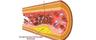
- Hypertrophic cardiomyopathy. It is characterized by a proportional increase in both the overall size of the heart and the thickness of the walls of the atria and ventricles. If the animal is elderly, then it begins to lack the strength to maintain the normal functioning of the grown organ. The reduction in the volumes of the atria and ventricles results in the dog’s blood receiving half as much oxygen and nutrients. In its advanced form, it can cause myocardial infarction or sudden cardiac arrest in the dog.
- Dilated cardiomyopathy. Experts are convinced that this type of pathology occurs most often in dogs. Sometimes dilation smoothly follows from the type of cardiomyopathy described above. In this case, the heart wall becomes amorphous, its texture loses its primary elasticity. This results in the organ being unable to contract normally. The dog develops severe shortness of breath and increased fatigue. The prognosis for cure is extremely unfavorable.
- Restrictive. In cardiac tissue, uncontrolled formation of fibrous fibers occurs, which are made from a vital organ - an unsuccessful analogue of cartilage. As a result, low myocardial contractility, leading to problems with oxygen entering the pet’s body. The symptoms are aggravated by the fact that the shaggy friend develops distinct painful sensations in the chest area.
- Mixed. It is characterized by a combination of signs of fibrosis of cardiac tissue with its hypertrophy. Variations in pathological processes may include concomitant expansion of the ventricles or atria of the organ.

Endocardiosis: what should the owner do?
When a breeder knows that his pet is at risk for endocardiosis, there is no need to panic. EMC is not a death sentence; the prognosis for the disease is favorable, provided that gentle management measures are applied to the animal. But this does not mean depriving the dog of its usual joys - playing, running, actively participating in the life of its owners.
To minimize the risks of disease progression, cardiologists at the Ros-Vet Center recommend:
- examine the animal 1-2 times a year;
- know the basic parameters of ill health, in particular - an increase in respiratory rate at rest (from 27 times/min.). If it is higher, this is a reason to consult a doctor;
- establish a balanced diet; fat deposition under the skin and on internal organs is especially dangerous for dogs with heart problems. This means that in no case should the pet be fed prohibited food from the table or encouraged to lie for a long time;
- There should always be moderate exercise; it’s not difficult to take your dog for a walk 2-3 times a day in good weather, at a measured pace and periodic elements of the game (“fetch a stick”).
That is, in fact, there are no special “privileges” for dogs at risk for endocardiosis; they should not be overly pampered or overfed. But it is also impossible to trigger mechanisms that can increase the rate of development of the disease.
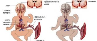
Causes of hydropericardium in the fetus, children and adults
The appearance of fluid in the pericardial cavity is an unfavorable sign, since most often it is an indicator of decompensation of the process. This condition can be detected even during fetal development. Moreover, the causes of pathology in children and adults are different.
Small hydropericardium in a child
You can see fluid in the pericardial sac as early as 20 weeks of pregnancy. Normally, the distance between the pericardial layers exceeds 2 mm. If there is more fluid than needed for lubrication, this may be a sign of:
- heart defect,
- hydrops fetalis,
- Rh incompatibility,
- protein starvation,
- anemia,
- immunity disorders,
- infectious process,
- tumors.
In children after 3 years of age, the criterion for small hydropericardium is the divergence of the leaves up to 10 mm. It occurs in rheumatic and autoimmune diseases, heart defects, and myocarditis.
Etiology in adulthood
Most often, this condition complicates the course of heart failure and is a sign of its decompensation. In addition, factors that provoke the appearance of excess fluid in the pericardial cavity may be:
- bruises, chest injuries;
- mediastinal tumors;
- cardiac surgery;
- nephrotic syndrome;
- exhaustion, protein starvation;
- tuberculosis;
- myxedema;
- autoimmune processes;
- radiation and chemotherapy treatment.
Hydropericardium
Reactive hydropericardium during infarction
It occurs in the first days of the disease and is characteristic of transmural necrosis, that is, the damage covers all layers of the heart wall. It also happens with small-focal infarction, which is located under the outer lining of the heart. Most often it does not last long and does not require special treatment.
Endocardiosis: what kind of disease is it?
EMC cannot be considered solely a “disease of old age,” although it develops in the 2nd half of a dog’s life. The heart is a four-chamber biopump that ensures blood movement through vessels of different sizes. Between its sections there are “plug” valves that allow blood to move only in a certain direction. With endocardiosis, the mitral valve ceases to close tightly, a “leak” occurs, and blood flows back, which creates increased pressure in one of the sections.
A failure in one mechanism always entails an imbalance in other anatomical structures. As a result of a critical accumulation of blood volume, it stagnates and the pressure in the vessels increases.
Important! Deformation of the left valve entails the development of pulmonary edema; if the right valve malfunctions, effusion (liquid fraction) begins to accumulate in the abdominal cavity.
According to statistics from cardiologists at the Ros-Vet EC, pathology is most often detected:
- in the left side of the heart (70%);
- in the right (5%);
- bilateral involvement (25%).
It is precisely this pathogenesis of the development of the disease that is responsible for the typical symptoms of endocardiosis in dogs - fainting, shortness of breath, high fatigue. If they appear, the owner must immediately take the pet to a veterinary clinic for a full examination.
What is dilated cardiomyopathy?
Cardiomyopathy is a pathology of the heart muscle, which is accompanied by disruption of the myocardium, the pumping function of the organ decreases, and blood circulation slows down. As a result, the pet is diagnosed with arrhythmia and heart failure.
The disease is classified into primary and secondary types. The first occurs against the background of congenital developmental defects, but is rare. The second develops as a complication against the background of a viral, bacterial or fungal disease.
The disease is also divided according to the type of changes in cardiac tissue. The most common type is dilated cardiomyopathy. The walls of the heart lose their elasticity, which prevents them from contracting. All chambers expand, the rhythm is disrupted.
What are the symptoms of endocardiosis?
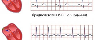
CHF – chronic heart failure does not always develop in the early stages; this is a typical sign of EMC in dogs and doctors pay special attention to it. CHF is considered an irreversible phenomenon; the prognosis is not always favorable and depends on the stage of the disease:
- latent (without symptoms), occasionally the pet may breathe heavily, cough, get tired, but the owner attributes this to a one-time physical activity;
- obvious symptoms appear as an increase in symptoms at the beginning of the pathological process and a noticeable deterioration in the condition with the critical development of CHF.
In the latter case, even an inattentive owner will notice that the dog is often depressed and does not want to walk, play, or simply show active emotions. He has difficulty breathing, and is often plagued by a debilitating cough and swelling of the paws (with severe damage to the right ventricle).
But all these signs do not always indicate endocardiosis. Cardiac insufficiency may be a consequence of other causes that need to be differentiated as a result of a thorough examination of the dog.
Obvious signs of endocardiosis:
- pulse weak and incomplete;
- cyanotic tongue, hoarse breathing;
- pale, anemic mucous membranes;
- thirst with lack of appetite;
- open mouth breathing;
- pinkish foam in the nostrils (pulmonary edema);
- ascites (fluid in the abdominal cavity);
- jugular veins are distended.
The dog's sleep is very restless, as it often has to change position; when lying down, the animal begins to choke. During bouts of coughing or physical activity, the dog may faint, this is a typical sign of problems with the mitral valve.
Treatment of a sick pet
The treatment strategy depends on the phase of the disease and the condition of the pet. Unfortunately, there is no hope for a complete recovery, especially in the case of the primary type of pathology. Therapy will be aimed at increasing life expectancy, improving its quality, and alleviating unpleasant symptoms.
Emergency treatment
Ambulance is required due to the development of acute heart failure, which threatens the life of the animal. This treatment is carried out exclusively within the clinic and includes:
- Intravenous injections of Furosemide 5%. The drug quickly reduces pressure in the left ventricle of the heart and pulmonary artery, causing veins to dilate.
- Nitroglycerin spray or ointment (applied under the arms), the dosage is prescribed by the doctor depending on the condition. The remedy is designed to stop the attack.
- Medicines for inotropic support (Dopamine, Digoxin). Their goal is to maximally saturate the tissues with oxygen, without increasing cardiac output.
- Oxygen therapy (the animal is in an oxygen chamber). Gas with an increased O2 content is supplied to it. This is one of the first resuscitation measures, since in critical conditions all cells of the body suffer from hypoxia.
- In case of severe anxiety, the pet is given a sedative.
- If fluid has accumulated in the pleural cavity, thoracentesis is performed as part of first aid. A puncture is made in the pleura to pump out the contents and normalize respiratory function.
After an attack, the animal will need constant monitoring. The dog will stay in the clinic for 2-3 days, after which it will be prescribed maintenance treatment.
Video: What is DCM in dogs, its symptoms, treatment and prognosis
Diagnosis of pathology
At the Ros-Vet Center, a full examination of a dog with suspected endocardiosis is carried out. This is necessary to confirm the diagnosis or identify problems with a similar course.
What they do:
- carry out a visual examination (auscultation, palpation);
- radiography (degree of enlargement of the heart chambers);
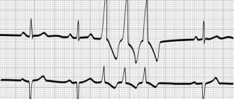
- electrocardiography (ECG);
- echocardiography (EchoCG);
- Ultrasound of the heart.
Laboratory tests of urine and blood for endocardiosis are not very informative, but this does not mean that they do not need to be done. Based on them, the general health of the pet is determined, what accompanying path processes occur in the body. Also differentiated from: pneumonia, pharyngitis, dirofilariasis, tracheal collapse, chronic bronchitis, etc.
Treatment of endocardiosis in dogs
Drugs are prescribed only by a veterinarian based on the stage of the disease. The goals of conservative therapy are to relieve symptoms, maintain the animal’s heart in working condition as much as possible, and correct neurohormonal activity, which often causes deterioration of the condition.
With obvious signs of EMC:
- diuretics (prevention of edema);
- ACE (cardiac stimulation);
- beta blockers (arrhythmia);
- diuretics and vasoconstrictors (cough).
The latter prescription is associated with the accumulation of effusion in the bronchi, which causes the appearance of a characteristic “suffocating” cough. Corticosteroids are needed in the later stages with the development of chronic cough and the formation of bronchitis.
Refractory congestive failure is difficult to treat and requires an increase in drug dosage. So, to relieve recurring pulmonary edema, the animal is limited in movement and given diuretics. ACE in the maximum dose to stabilize cardiac activity. Be sure to check kidney function, electrolyte levels, food without salt, etc.
Surgical intervention
Modern equipment in the surgical room allows the doctor to treat endocardiosis surgically. The mitral valve is being replaced. The indication for intervention is stagnation of blood in the heart, which cannot be eliminated with medication. But secondary cardiomyopathy is a contraindication to surgery; it develops in advanced cases. That is why the dog owner needs not to let the disease progress, but to consult a doctor in time.
Symptoms of dropsy of the pericardial sac
In the case of accumulation of transudate in small volumes, symptoms of the disease may be absent for a long time until the volume of fluid begins to increase. As its volume increases from 80 ml or more, clinical signs begin to appear.
The initial symptoms of hydropericardium in patients are:
- General weakness, fatigue.
- Constant shortness of breath, which does not depend on physical activity.
- The appearance of pressing pain behind the sternum, which intensifies when bending forward. Such pain is prolonged, as compression of the coronary arteries occurs and myocardial ischemia develops.
- Choking of a paroxysmal nature.
- Swelling of the face, upper limbs, feet and legs.
- Protrusion of the neck veins.
- Tachycardia, which is constantly recorded.
- Decreased blood pressure.
With an increase in transudate content, the patients’ condition worsens and the following symptoms develop:
- Hiccups appear, compression of the esophagus leads to disruption of the swallowing process.
- Weakness is growing.
- The heaviness and pain in the chest intensifies.
- The heart increases in size.
- Shortness of breath does not decrease at rest, the respiratory rate reaches 30 per minute.
- There is stagnation of blood in the internal organs.
- Cyanosis of mucous membranes and skin.
- The skin becomes covered with sticky, cold sweat.
- Patients become agitated due to the fear of death.
- A decrease in blood pressure can lead to loss of consciousness.
Important! To avoid the development of cardiac tamponade, even at the slightest sign of the disease, medical attention is necessary. Lack of treatment, even with the slightest deterioration in the patient’s condition, leads to death.
How to care for a dog with endocardiosis
An animal with “heart problems” requires special attention. So the owner must:
- ensure complete peace, this means that each family member is obliged not to annoy the dog, not to play loud music, not to shout;
- ensure good air ventilation;
- give cool, clean water, food that does not provoke thirst (except dry food, salt).
The dog requires help with external care; you need to clear your nose and mouth so that it can breathe easier.
Forecast
Possible complications of EHR include persistent cardiac arrhythmia, decompensation, chord separation, left atrium rupture (instant death). With a slight development of endocardiosis and a gentle lifestyle, the pet can live a long life. Severe EMC with treatment gives the dog a chance to live for at least a year.
Special treatment and prognosis
Treatment and prognosis of pericardial disease depend on the underlying cause. Unfortunately, most dogs with pericardial effusions have neoplasia and most cats have systemic disease and cardiomyopathy, so the long-term prognosis is poor. However, some diseases, such as idiopathic pericardial effusions, have a better prognosis.
Cardiac neoplasms At the time of diagnosis, hemangiosarcoma as the cause of pericardial effusion is already a highly malignant and metastatic tumor. Intermittent pericardiocentesis, pericardial window, or percutaneous balloon pericardiotomy (Cobbetal., 1996) may provide temporary relief to the animal, but the overall prognosis is poor. Some veterinarians do not advise pericardiectomy in patients with right atrial hemangiosarcoma due to the potential for bleeding. Surgical removal of the tumor is usually unsuccessful, and postoperative complications are possible. The authors have used intrapericardial cisplatin injections in some cases, but there is insufficient information to draw definitive conclusions. One report noted a decrease in the size of right atrial hemangiosarcoma after combination chemotherapy (deMadronet et al., 1987).
For dogs with chemodectoma, pericardiectomy can be used to reduce symptoms of cardiac tamponade. These tumors grow slowly and metastasize slowly, therefore, reducing the symptoms of pericardial effusions can improve the patient's condition and increase life expectancy. These tumors are inoperable because they are attached to large blood vessels. When these tumors grow large, they begin to compress cardiac structures, but surgery is unlikely to provide long-term symptomatic improvement. After pericardiectomy, animals can live up to 3 years. Mesothelioma is difficult to diagnose without surgery, which is most often a subdiaphragmatic pericardiectomy. After surgery, mesothelioma often spreads into the pleural space. At this stage, periodic thoracentesis and intrathoracic cisplatin are necessary to slow the rate of fluid accumulation. Cats with pericardial effusions secondary to lymphosarcoma respond well to combination chemotherapy.
Idiopathic Pericardial Effusions in Dogs In approximately 50% of cases, idiopathic pericardial effusions are cured after one or two pericardiocentesis. In other cases, pericardial effusions can recur, either 10 days or 5 years after pericardiocentesis. For idiopathic pericardial effusions, systemic or intrapericardial corticosteroids are sometimes given, but their effectiveness has not been well documented and we therefore cannot recommend them. Also, one preliminary report suggested that azatiprine (Imuran, GlaxoWellcome) reduces recurrence of idiopathic pericardial effusions (Bussadori, 1995). For recurrent pericardial effusions after pericardiocentesis, corticosteroids (prednisone, 1 mg/kg every 24 hours for 2 to 3 weeks) should be considered, although this treatment has not yet been evaluated in a clinical setting. If a preliminary diagnosis of idiopathic pericardial effusion is made, echocardiography is recommended every 10-14 days for 2 months. After the third recurrence of pericardial effusion, pericardiectomy is recommended. After surgery, most dogs become symptom free, with only a minority of patients experiencing relapses.
Infectious pericarditis Animals with fungal or bacterial pericarditis require aggressive therapy. Appropriate drugs are prescribed based on microbiological identification of the type of microorganism and/or the results of cultivation and antibiotic sensitivity tests. An indwelling pericardial catheter can be placed for drainage and lavage, but surgery and pericardiectomy are best. The prognosis for infective pericarditis is generally guarded to poor; The main complication is constrictive pericarditis.
Left atrial rupture Pericardiocentesis is performed only in patients with unstable tamponade, removing a small amount of fluid each time until clinical symptoms improve. The prognosis for these animals will be poor. The rupture may heal, but these animals remain prone to recurrent ruptures and generally have progressive cardiac damage at the time pericardial effusion develops. In cases of traumatic atrial ruptures and recurrent effusions, surgery is indicated.
Mixed Causes Vitamin K and fresh plasma transfusion are indicated in animals with pericardial hemorrhage following vitamin K antagonist poisoning. Pericardial effusions as a consequence of heart disease are treated with heart medications appropriate for the disease. No specific treatment has been developed for cats with IPC, so the prognosis in this case is unfavorable.