Myxomatous mitral valve degeneration - symptoms and treatment
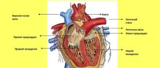
The heart is not only, as many people think, the organ of love, but also the engine of our body. It has a large number of functions, but perhaps the most important is pumping blood through the vessels of our body, which supplies organs and tissues with oxygen and allows us to exist.
Myxomatous degeneration of the uterus
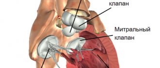
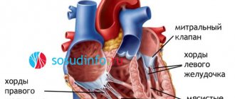
MD MV is a disease characterized by compaction of the mitral valve leaflets, which prevents their complete closure and contributes to the occurrence of regurgitation (backflow) of blood into the cavity of the left atrium.
General information about the defect and causes
When considering myxomatous degeneration of the mitral valve leaflets, the question arises: what is it? So, this is a pathological condition, which is not the most dangerous for the body: if the defect is detected in a timely manner, there are measures to take and preventive programs are recommended.
Myxomatous degeneration of the valve leaflets is a stretching or increase in their thickness, which, as the disease progresses, begins to interfere with the complete closure of the valve at the time of systole and cannot resist reverse blood flow. Most often, this defect is diagnosed in older and middle-aged people.
In total, there are three degrees of development of the pathological process:
- the first degree is characterized by an increase in the thickness of the valves ranging from 3 mm to 5 mm, which does not interfere with closure;
- on the second, the thickening reaches 8 mm, which leads to valve deformation, single ruptures of the chords and impaired closure;
- at the third stage, when the thickness of the leaflets increases above 8 mm, the valve does not close and regurgitation of blood occurs (reverse flow), in which part of it returns to the atrium.
The causes of pathology can be many factors
The initial stage is not life-threatening, but progression of myxomatous degeneration and transition to later stages can lead to mitral valve insufficiency, stroke, infective endocarditis, and death.
Read it! Supraventricular extrasystole: what is it?
To date, no specific causes that can lead to this defect have been identified. In some cases, heredity is a dangerous factor. A pattern has been identified in which patients with this pathology have problems with growth. Doctors do not rule out the influence of hormonal imbalances, but this factor is still being studied.
Myxomatous mitral valve degeneration - symptoms and treatment
The heart is not only, as many people think, the organ of love, but also the engine of our body. It has a large number of functions, but perhaps the most important is pumping blood through the vessels of our body, which supplies organs and tissues with oxygen and allows us to exist.
Myxomatous degeneration of the uterus
MD MV is a disease characterized by compaction of the mitral valve leaflets, which prevents their complete closure and contributes to the occurrence of regurgitation (backflow) of blood into the cavity of the left atrium.
Stress echocardiography for mitral valve prolapse
Stress echocardiography in the assessment of MR in patients with severe mitral valve prolapse and a “dangling” leaflet.
Indications: severe asymptomatic/low-symptomatic mitral regurgitation of an organic nature with preserved left ventricular function at rest.
Methodology: bicycle ergometry in a horizontal/semi-horizontal position according to the protocol 25 (50) W x 2 minutes
Indicators:
- exercise tolerance,
- clinical symptoms,
- heart rate dynamics
- blood pressure dynamics
The following questions need to be answered:
- Dynamics of mitral insufficiency (increasing?)
- MPAP (exceeds 60 mm Hg during physical activity?)
- The response of the left ventricle to physical activity is contractile reserve (does the ejection fraction increase and does the ejection fraction decrease?). If the ejection fraction increases by no more than 4-5% or decreases, these are candidates for surgical treatment.
Video 2. Echocardiography. Parasternal longitudinal section. Mitral regurgitation of the 1st degree is visualized against the background of mitral valve prolapse. Start in Video 1.
Myxomatous mitral valve degeneration - symptoms and treatment
The heart is not only, as many people think, the organ of love, but also the engine of our body. It has a large number of functions, but perhaps the most important is pumping blood through the vessels of our body, which supplies organs and tissues with oxygen and allows us to exist.
Myxomatous degeneration of the uterus
MD MV is a disease characterized by compaction of the mitral valve leaflets, which prevents their complete closure and contributes to the occurrence of regurgitation (backflow) of blood into the cavity of the left atrium.
Features of myxomatous degeneration
Myxomatous mitral valve degeneration is a common cardiac pathology. This disease has many names (degeneration, endocardiosis or valve prolapse). This disease is associated with the mitral valve, which separates the left atrium and left ventricle. All names are descriptions of age-related degeneration of the structural parts of the heart valves, which is manifested by stretching and thickening of the valve leaflets. In this case, the closure of the valves is disrupted and regurgitation (backflow of blood) appears through one or a pair of valves with an audible murmur in the heart. Subsequently, degenerative changes and an increase in reverse blood flow intensify, and the parts of the heart expand. Other complications may also occur (cardiac arrhythmia, failure and other dangerous conditions).
Diagnosis and treatment methods
The presence of pathology is indicated by what the doctor can hear by auscultation (listening). To confirm the diagnosis, prescribe:
- electrocardiogram;
- echocardiography (a type of ultrasound of the heart);
- X-ray of the chest area.
Attention! Genetic tests and blood tests are currently not required to identify this defect.
At the initial stage, when myxomatous degeneration of the mitral valve leaflets does not interfere with the functioning of the heart and does not affect the general condition of the body, active treatment, much less surgical intervention, is not required. However, the patient must be registered with a cardiologist and undergo regular examinations.
There are currently no effective drugs that could completely stop and eliminate this pathological disease. Therefore, as the pathology progresses, medications are prescribed that help eliminate symptoms and significantly slow down the dangerous process. Such medications include those that remove excess accumulated fluid from the body, are aimed at maintaining the performance of the heart muscle and improving blood circulation, and regulating the heart rate.
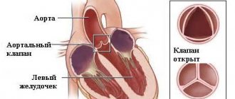
In cases where the pathology has led to mitral regurgitation and blood regurgitation, surgical intervention may be indicated (you can watch the video on the Internet resource), in which it is possible:
- preservation of the valve with plastic surgery of the valves or their replacement;
- prosthetics (the affected mitral valve is removed and a biological or artificial prosthesis is placed in its place).
Despite the fact that the causes of the development of myxomatous mitral valve degeneration have not been fully established, and it is difficult to talk about specific prevention, there are some important recommendations.
- You should definitely be under the supervision of a doctor and undergo regular preventive examinations.
- Lead a completely healthy lifestyle (excluding all bad habits).
- Create and adhere to a work and rest schedule.
- Review your diet, include only healthy foods (more vegetables and fruits, quail eggs). Focus on foods that contain heart-healthy components (for example, potassium-rich foods - dried apricots, prunes, cabbage, rose hips). Avoid strong black tea and coffee.
18.09.2014
Mitral valve prolapse
General information about mitral valve prolapse, its clinical picture and diagnosis
Mitral valve prolapse
- sagging (bending) of the mitral valve leaflets into the left atrium.
Mitral valve prolapse syndrome, or Barlow syndrome
, described by J. Barlow in 1963 and occurs extremely often - in 5-10% of people in the population.
It is necessary to distinguish between true prolapse of the valves and their wave-like sagging
(billowing).
In many cases, mitral valve prolapse is asymptomatic
(has no symptoms), in some cases,
arrhythmias
(heart rhythm disturbances),
the presence of a characteristic noise when listening to tones
,
chest pain
, and
shortness of breath
.
Emotional lability, fatigue and other nonspecific clinical signs
are also recognized as associated with mitral valve prolapse .
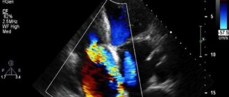
Prolapse, or sagging, of one or both cusps of the mitral valve during systole into the cavity of the left atrium is considered true only if it is recorded in two echocardiographic positions: apical four-chamber and parasternal along the long axis of the left ventricle.
Diagnosis of mitral valve prolapse is carried out during an echocardiographic study carried out in B-mode, M-mode, color and spectral Doppler modes.
In the expert practice of cardiac ultrasound, only a combination of all modes allows one to obtain holistic views.
about the nature of the process, the presence and severity of hemodynamic disorders.
In a number of countries, there is a rule to perform any surgical interventions in patients with prolapse syndrome under the guise of antibiotics in order to prevent complications.
Currently in our country there is overdiagnosis of mitral valve prolapse in children and adolescents.
This may be due to incorrect execution of the research procedure (technique) - incorrect identification of the apical position of the heart. In addition, a slight sagging of the base of the anterior leaflet of the mitral valve in children and adolescents up to 3–5 mm is normal if it is not accompanied by pathological regurgitation. In addition, the valve leaflets and chords develop faster than the fibrous rings, therefore, sagging of the leaflets is more often recorded in childhood and adolescence.
It is necessary to distinguish between physiological mitral valve prolapse
– without disrupting its function, and
pathological mitral valve prolapse
– with pathological mitral regurgitation.
Mitral valve prolapse syndrome is characterized by
young age of patients – usually 20–50 years; predominance of women; the presence of noise - a “click” during auscultation, thickening of the valves and their systolic displacement during echocardiography, hypotension, as well as mitral regurgitation during Doppler examination, the degree of which exceeds physiological.
Myxomatous degeneration is not uncommon.
(proliferation of the middle layer of the mitral valve leaflet - spongiosis and changes in the structure of the valve leaflets and chords) of the mitral valve leaflets, signs of which are found in older patients - 40-70 years old, among whom males predominate. In these cases, pathological mitral regurgitation is detected by echocardiography, and there are pronounced changes in the leaflets that require cardiac surgical correction.
Myxomatous degeneration of the mitral valve leaflets, as one of the most common causes of mitral valve prolapse, can affect the leaflets of all heart valves, but the most common lesion is the mitral valve.
Over the past few years, the number of people suffering from this pathology has increased significantly throughout the world. Just 10 years ago, the majority of patients with myxomatous degeneration were patients with Marfan syndrome. Currently, the connection between unfavorable environmental factors and the use of a number of weight loss drugs in the occurrence of this pathology has been proven. The number of patients over 70 years of age suffering from myxomatous degeneration has increased significantly.
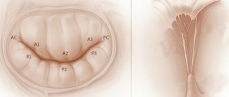
An echocardiographic examination clearly shows scalloped, “serpentine”, uneven, thickened cusps of the mitral valve. They prolapse into systole into the cavity of the left atrium. Rounded protrusions are formed on the valves, resembling small tumors - myxomas. This is where the name of this pathology comes from. You can often observe separation of the valve chords.
Most patients with myxomatous mitral valve degeneration demonstrate cardiac arrhythmias—atrial fibrillation or frequent ventricular extrasystole. The causes of arrhythmia are pathological mitral regurgitation against the background of myxomatous changes in the valve leaflets and, as a result, increased pressure in the cavity of the left atrium.
Patients with myxomatous degeneration require dynamic monitoring; those with significant mitral regurgitation require consultation with a cardiac surgeon.
The nature of changes in the structure of the mitral valve leaflets and the occurrence of pathological mitral regurgitation can contribute to infection of the valve. Differential echocardiographic diagnosis in this case may be difficult.
Differential diagnosis of myxomatous degeneration
valve cusps should be performed with infective endocarditis and Lamblet's growths. Clinical and laboratory diagnostics play an important role in this. Thus, with myxomatous degeneration there is no inflammatory reaction recorded during laboratory testing.
Secondary mitral valve prolapse occurs in the following situations:
- Marfan syndrome is a mesenchymal dysplasia. It is accompanied by a characteristic appearance of the patient (“Marfan-like type”) - increased joint flexibility, aortoanular ectasia, frequent development of aortic aneurysm and intimal detachment of the aorta in the thoracic ascending region and myxomatous degeneration of the valves and subvalvular structures. In this case, all heart valves prolapse. The degree of prolapse is usually significant. Pathological valvular regurgitation is recorded.
Hypertrophic cardiomyopathy. In this case, mitral valve prolapse is associated with increased pressure in the cavity of the left ventricle during systole. Prolapse is especially pronounced in patients with obstructive hypertrophic cardiomyopathy.
Ehlers-Danlos syndrome - connective tissue dysplasia syndrome - is a hereditary defect of hemostasis with damage to collagen structures. Accompanied by increased joint flexibility, increased skin stretching, bleeding and prolapse of heart valves
Dysfunction of the papillary muscle due to myocardial infarction or cardiac injury is accompanied by prolapse of the valve leaflet and significant mitral regurgitation.
Severance of the chords of the valve leaflet against the background of infective endocarditis, myxomatous degeneration, myocardial infarction, rheumatic lesions, etc. leads to prolapse of the leaflet and pathological valve regurgitation.
Assessment of the degree of mitral valve prolapse is carried out by assessing the severity of sagging of the leaflets:
- Minor mitral valve prolapse – 3–6 mm (1st degree mitral valve prolapse).
Moderate mitral valve prolapse – 6–9 mm (2nd degree mitral valve prolapse)
Significant mitral valve prolapse – more than 9 mm (grade 3 mitral valve prolapse).
It must be remembered that the degree of prolapse and the degree of mitral regurgitation may not correlate with each other
. For example, when the chords are torn off at the end of the mitral valve leaflet, prolapse of up to 3 mm can be seen and grade 3–4 mitral regurgitation can be recorded.
Depending on the cause, primary mitral valve prolapse is distinguished (idiopathic, hereditary, congenital), which is an independent pathology not associated with any disease and caused by genetic or congenital failure of connective tissue. Mitral valve prolapse with differentiated STD (Marfan syndrome, Ehlers-Danlos syndrome (types I-III), osteogenesis imperfecta (types I and III), elastic pseudoxanthoma, increased skin extensibility (cutis laxa)) is currently classified as a variant of primary mitral valve prolapse .
Secondary mitral valve prolapse develops as a result of any disease and accounts for 5% of all cases of valve prolapse.
Forecasting
Prognosis for myxomatous mitral valve degeneration is related to the stage of the disease at the time of diagnosis, including the speed of development of the disease. This pathology manifests itself in some patients at an early age, and in some patients the disease progresses rapidly. Mostly, the development of myxomatous valve degeneration occurs very slowly and quietly, over several years, which is why some patients may be asymptomatic for a long period after the detection of systolic murmurs. If heart failure develops, the average life expectancy with optimal treatment is 6-18 months.
What symptoms does it manifest?
At the beginning of its appearance, the pathological process may not be accompanied by certain symptoms due to the fact that no disturbances in the functioning of the organ occur.
With the development of the defect and the transition to the second and third degrees, myxomatous degeneration of the mitral valve is accompanied by quite characteristic signs:
- periodic pain in the left side of the chest, stabbing in nature and short-term manifestation;
- deterioration of general condition (increased fatigue, decreased physical activity, weakness, decreased appetite);
- the appearance of shortness of breath even with little physical activity;
- feeling of lack of air;
- dizziness, lightheadedness and fainting.
In some cases, an additional symptom may be a cough. At first it is dry, and then with phlegm and splashes of blood.
Diagnostics
Mitral valve myxomatosis is determined by the results of several studies:
- assessment of patient complaints;
- anamnesis data;
- objective examination;
- additional examination methods.
During the examination, characteristic auscultatory signs of pathology are:
- systolic click;
- midsystolic murmur;
- holosystolic murmur.
A distinctive feature of the auscultatory picture in myxomatous degeneration is its variability (the ability to change from visit to visit).
From an additional examination, the doctor prescribes:
Such extensive diagnostics are necessary to determine further tactics for patient management and monitor therapy.