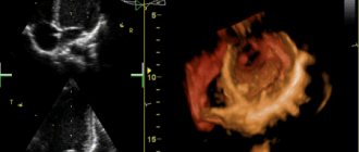
Ultrasound of the heart
Hello.
Please help me figure it out. My son is 17 years old. The doctor heard a murmur in his heart and sent him for an ultrasound. We got the following results. EchoCG. Conclusion. Bicuspid aortic valve. The valve flaps are sealed. Cardiac cavities: dimensions within the age norm. Contractility is preserved. Mitral valve prolapse, art. 1, with slight regurgitation. Regurgitation on the tricuspid valve, stage 1. How serious is this? What do we have to do? Thank you. Good afternoon. Please comment on the child's echocardiography. Aorta 0.1 cm. Opening amplitude 8 mm. Left atrium - 14mm. The mitral valve is normal. There is an antiphase. There is no prolapse. Interventricular septum: diastole – 4 mm, systole – 3 mm. Posterior wall of the left ventricle: diastole – 3 mm, systole 4 mm. Overall contractility: EF 67%. UO 18ml. FU 38%. Right ventricle - 8mm. Pulmonary artery 10mm. Interatrial septum: defect with a diameter of 8 mm with left-right shunting. The interventricular septum is normal. There is no free fluid in the pericardial cavity. An additional chord is visualized in the cavity of the left ventricle. Conclusion: congenital heart defect: ASD. Dilatation of the pancreas. Moderate valvular stenosis of the pulmonary artery.
Hello. Please look at the results of the heart ultrasound. Aorta: the lumen of the aortic root is 40 mm, the walls are thickened and dense. The aortic valve leaflets are thickened, dense, calcified (++). In systole, the divergence of the leaflets is 18 mm. The anteroposterior size of the left atrium is 40 mm. Right atrium 31x45. EDD of the right ventricle 35 mm. EF 63%. ESD of the left ventricle 50. ESD of the left ventricle 32. IVS 10 mm. The movement of the IVS is without any peculiarities. ZSLZh 10 mm. The movement of the LV is without any peculiarities. Mitral valve: the movement of the leaflets in diastole is multidirectional. The mitral valve leaflets are compacted, changed, calcified (+). Peak E = peak A. The movement of the mitral valve is M-shaped. The blood flow velocity at the aorta is 110, at the level of the MV - 80, the tricuspid valve - 50, and the pulmonary artery -100. The cavities of the heart are not dilated. Regurgitation: aortic 1-2 degrees, mitral 1-2 degrees. Calcification of the aortic valve - grade 2, mitral valve - grade 1. Diastolic function is impaired. No violations of local and global contractility were detected. I am 69 years old. I have atherosclerosis of the cervical arteries; with prolonged physical activity, pain appears in the heart area. What is this connected with?
Good afternoon. Please explain the results of the heart ultrasound. Left atrium 31 mm. The aorta at the level of the sinuses of Valsalva is 41 mm, the ascending aorta is 39 mm. The divergence of the aortic valve leaflets is 17 mm. Right ventricle 33 mm. The outflow tract of the right ventricle is 27 mm. The movement of the mitral valve leaflets is multidirectional. MZhP 8mm. The amplitude of the IVS is 3 mm. Posterior wall of the left ventricle 11mm; the amplitude of the left ventricle is 5 mm. ESD 65mm ESD 48mm EDV 225 ml ESD 112 ml Ejection fraction 50% Dopplerography Regurgitation on the LA 2nd stage. at a speed of 1.1 m/sec. Gradient - 4.6 mm Hg. Art. Left ventricular diastolic dysfunction. Conclusion: the left and right parts of the heart are enlarged. The aorta and valves are sealed. PA valve insufficiency, stage 1. Hypokinesia of the LV walls. Myocardial contractility is reduced. Type 1 LV diastolic dysfunction. I have diabetes and sometimes have tachycardia. How is your heart condition?
Hello. Irina, 55 years old. For several months I have been worried about severe shortness of breath, and with minimal exercise I feel very bad. I did an ultrasound of the heart and got the following result: Irregular rhythm 78 beats/min. Congenital heart defect. ASD 7-8 mm with blood discharge from left to right with signs of hemodynamic overload of the right heart. Dilation of the right heart. Pulmonary hypertension stage 1. Aortic insufficiency of the 1st degree. Consolidation of the aorta and aortic valve. Tricuspid regurgitation grade 2-3. Compaction of the mitral ring. Mitral regurgitation grade 2-3. Do these changes require surgical treatment? Is surgery performed at this age? Thank you.
Hello. Please decipher the results of the ultrasound of the heart. Left atrium - 37 mm, right atrium - 48 mm, right ventricle - 28 mm, anterior wall of the right ventricle - 5 mm. Aorta: aortic diameter at the level of the cusps - 27 mm, aortic valve cusps - somewhat compacted, cusp divergence - 18 mm, Pg max - 9 mm, V max - 1.5 m/sec, regurgitation degree 1, T 1/2 regurgitation flow - 51 ms. Mitral valve: leaflets are sealed, peak A: Vmax - 0.5 m/sec, peak E: Vmax - 1.1 m/sec, Pg max - 5 mm. RT, regurgitation - 1-2 degrees, mitral orifice area - 4.8 cm2, T 1/2 pressure gradient - 42 ms, E/a - 2.2, IVRT - 62 ms, DT - 132 ms. Left ventricle: ESD - 65 mm, EDV - 207 ml, ESR - 51 mm, ESV - 115 ml, stroke volume - 94 ml, heart rate - 94, ejection fraction - 45%, shortening index - 22%, local contractility - not impaired . MZhP: MZhP syst. — 11 mm, IVS dia. — 10 mm, syst. excursion of the IVS - 2 mm, TZSLZh diast. — 12 mm, TZSLZh syst. - 14 mm, systolic excursion of the posterior wall of the LV - 4 mm. Tricuspid valve: leaflets – no changes, regurgitation – 1st degree, Pg max – 2.2 mm. Pulmonary valve: leaflets - without features, PA trunk - 24 mm - at the upper limit of normal, Vmax - 0.98 m/sec, Pg max - 3.6 mm, 1st degree regurgitation, collapse of the IVC on inspiration - less than 50%, signs of pulmonary hypertension, systolic pressure in the LA - 30 mm Hg. Art.
Good afternoon. Please look at the ultrasound of the heart. Are the results normal? There is no dilatation of the heart cavities. EDD of the left ventricle is 3.5 cm, the anterior-posterior size of the left atrium is 2.3 cm. There is no myocardial hypertrophy. TZSLZH - 0.7 cm. The aorta is not dilated and is mobile. Pulmonary artery: no changes. Valvular apparatus: slight prolapse of the anterior valvular leaflet into the cavity of the left atrium in systole up to 0.2 cm - within the physiological norm. Left ventricular ejection fraction 65%. There is no myocardial asynergia. Doppler analysis: without features.
What is the mitral valve of the heart?
They talk and sing about this organ, and paint pictures about it. The human heart is an amazing organ about which we can talk a lot. Have we ever thought that the sound of the heart is the result of the work of its valves? How they work is discussed in our article.
Of course, if you talk about everything at once, you will need a lot of space. Therefore, let's talk about the valvular apparatus of the heart, or more precisely about the mitral valve in humans. Every interested person knows that the heart has valves and how many there are. But this is not all the information about the work of the heart. One day, someone managed to determine that the human heart pumps blood through its chambers and vessels at a speed of 40 km per hour. Agree, this is not a small number. To achieve this, you need not only valves, but also other structures that this organ has. Therefore, there is the concept of the valvular apparatus of the heart.
During pregnancy
Half of the female population has a pathology, but not everyone is aware of it. When undergoing a mandatory medical examination, mitral valve prolapse is most often discovered in pregnant women. Many women immediately wonder about the compatibility of the disease and future motherhood.
What is valvular heart disease?
The following step-by-step illustration explains the normal function of the left ventricular valves. When the left ventricle relaxes, the aortic valve closes and the mitral valve opens. When the left ventricle contracts again, the mitral valve closes and the aortic valve opens to allow blood to flow into the aorta.
- This allows blood to flow from the left atrium into the left ventricle.
- When the left atrium contracts, more blood flows into the left ventricle.
Heart valves can have a variety of dysfunctions, including the following.
The occurrence of valve deflection is associated with increased load on the heart and increased blood ejection into the aorta. Fetal hypoxia and growth retardation are a consequence of hypoxia caused by pathology.
If there was no regurgitation in the first months of pregnancy, despite the diagnosed prolapse, then the risk of developing pathology is almost zero.
The valve does not close completely and this causes blood to flow back through the valve. The blood must find an alternative route, usually through another birth defect such as interatrial or interventricular communication. When the heart valves fail to open or close properly, the consequences for the heart can be serious as it becomes increasingly difficult to pump blood properly throughout the body.
All rights reserved. This information is not intended to be a substitute for professional medical care. Only your doctor can diagnose and treat your health problem. The heart has four cavities separated by a septum and four valves: the aorta; mitral; tricuspid and pulmonary.
Most often, the consequence of the disease in pregnant women is premature birth or weakened labor. In this case, a caesarean section becomes necessary. If arrhythmia may occur, drug treatment is also required.
Moderate or severe course of the disease is determined by the following symptoms:
- Autonomic-vascular dysfunction, expressed by heart pain, lack of air, cardiac arrhythmia, chills;
- Vascular disorders, including frequent headaches, swelling;
- Hemorrhagic, which occurs in the form of bruises with the slightest pressure and bleeding from the nose;
- Psychopathological: sudden feeling of anxiety and fear, mood swings.
When these signs appear, a pregnant woman should be constantly monitored by a cardiologist and receive a referral for childbirth at a specialized perinatal center.
- Avoid sudden changes in temperature, heat and cold;
- Do not sit for a long time;
- Rest in a reclining position.
- Planning pregnancy is a necessary measure to reduce the risk of pathology in a woman.
consultation needed: cardiological terms
A question for everyone, especially cardiologists.
Translated regular papers to people for consultations abroad. Among other things, there are the results of Echo-CG (a person has coronary artery disease and a previous myocardial infarction). I stumbled on this: The mitral valve has sealed leaflets, antiphase is present.
1. What does antiphase mean in this context? (I know what a mitral valve is and what it is needed for. ;-)) From the Internet I was able to glean only that it should be present.
2. What do “compacted flaps” mean – are they thickened or have reduced elasticity?
Operation
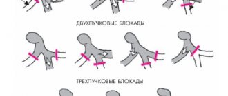
The conduction system of the heart constantly sends impulses, which causes it to pump blood into the ventricles from the atria, into the vessels from the ventricles, and to all internal organs and tissues of the human body from the vessels.
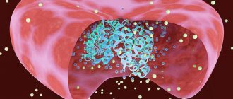
At the exit of the left ventricle is the mitral valve, the halves of which are tightly closed. After the cavity of the left ventricle is completely filled with blood, the valves of the MV close.
At this moment, the chords are stretched to their maximum. Thanks to this, the likelihood of the snort halves sagging into the left atrium is minimized.
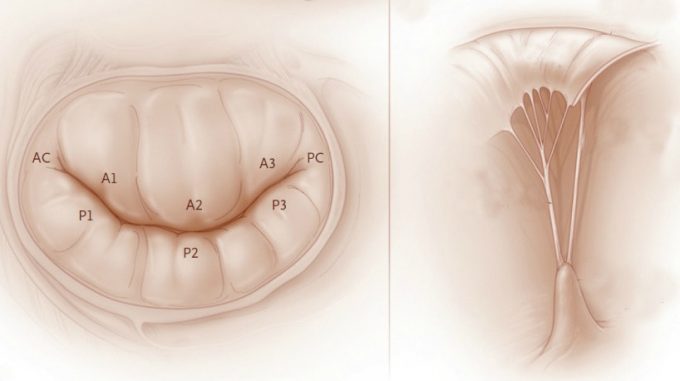
The mitral valve performs the following main functions:
- In the relaxation phase, the snort allows free blood flow by lowering its flaps. Thanks to this, blood can freely pass into the ventricle from the atrium.
- At the moment of systole, the halves of the valve close, occupying the upper position. As a result, the process of blocking the reverse flow of blood occurs - regurgitation.
The operation of the entire valve mechanism of the human cardiovascular system, including the mitral valve, has a strictly streamlined system. Thanks to this, the heart contracts with a certain periodicity and frequency. If the structure or operation of one of the elements of this system fails, the functioning of the SSS as a whole occurs.
Answered: 13
“Dense” on EchoCG means that they have increased echogenicity. In practice, this means that the valves contain more connective tissue compared to ordinary ones. In reality, this is a thickening and a decrease in elasticity and mobility.
Thank you. Do you know how it is usually written in English - indurated, hardened, or simply of increased echogenicity?
Did you translate the last term verbatim or take it from the dictionary? In Russian medical language, increased echogenicity is also called hyperechogenicity. Maybe you can find an analogue. You are not translating scientific work, but the results of analyses. Therefore, the closer to the original, the better.
hyperechoic, of course, is often written.
I am more concerned about the notorious “antiphase”.
Treatment
Vegetative-vascular dystonia and valve prolapse are similar in symptoms, so their treatment is almost the same. As in the case of VSD, medications cannot affect the prolapse itself, only its symptoms. Sometimes there are cases that prolapse is accompanied by an additional chord in the left ventricle.
Surgical intervention is only necessary when regurgitation is severe and circulation is impaired. Valvuloplasty is performed - the mitral valve is sutured. Only in cases where the operation turned out to be ineffective or could not be carried out according to any indicators, is it possible to replace the valve with an artificial analogue.
In all other cases, when the disease is asymptomatic or its manifestations do not affect life activities, exercise therapy and taking sedatives are indicated. Massage, acupuncture, and vitamins may be prescribed.
No special treatment is required for mitral valve prolapse.
English translation: leaflet opposition
*Mitral valve: the leaflets are sealed. There is antiphase.*
Explanation:
At Anna’s request, I am publishing here my letter sent to her a couple of days ago.
> Unfortunately, I understand little about echocardiography, and I have never encountered the term “antiphase” at all. I scoured the Internet; The only thing that came to my mind was leaflet opposition, which is normal and is disturbed in case of pathology of the mitral valve (poor leaflet opposition, faulty leaflet > opposition). > > > > A couple of quotes: > > Book Current Emergency Diagnosis > > . Echocardiography is the most valuable technique for assessing mitral > stenosis. The valve is thickened, opens poorly, and closes slowly. The > anterior and posterior ¶leaflets are fixed and move together, rather than in > ¶opposite directions. > > > > Internet, https://www.echoincontext.com/int1/skillI1_02.asp
> > . The pathological changes in the valve apparatus produce dramatic > alterations to the echocardiographic images (Fig. 2). On the M-mode > recording, thickening of the leaflets and reduction in their mobility can be > appreciated. Commissural fusion alters the motion of the ¶posterior leaflet; > instead of moving in the ¶opposite direction to that of the anterior, it is > pulled forward when the valve opens at the beginning of diastole. In the > normal valve, rapid early diastolic filling of the left ventricle means that > the valve can partially close in mid-diastole, and it then reopens during > atrial systole. > > > > But this is a matter of guesswork. If you want, I'll try to ask our > director tomorrow, he is the author of the book “Clinical Echocardiography”. > It would be nice if you provided more context.
consultation needed: cardiological terms
A question for everyone, especially cardiologists.
Translated regular papers to people for consultations abroad. Among other things, there are the results of Echo-CG (a person has coronary artery disease and a previous myocardial infarction). I stumbled on this: The mitral valve has sealed leaflets, antiphase is present.
1. What does antiphase mean in this context? (I know what a mitral valve is and what it is needed for. ;-)) From the Internet I was able to glean only that it should be present.
2. What do “compacted flaps” mean – are they thickened or have reduced elasticity?