Pathological hyperemia
Here the cause is pathological factors, both external and internal:
- toxins – in case of poisoning or certain diseases, as well as medications;
- chemicals - meaning toxins that act externally;
- burns and frostbite;
- mechanical effects, for example, venous hyperemia, which occurs when veins are compressed by a tumor or tissue edema;
- allergies - arterial hyperemia often appears as increased sensitivity of blood vessels to an irritant;
- infectious diseases - in this case, an increase in temperature is the cause of hyperemia, and not a consequence;
- inflammation of various kinds.
The pathological form can be quite short, for example, in acute respiratory disease, but no less often it becomes protracted. Treatment should provide not only a way to get rid of the underlying disease, but also from the unpleasant consequences.
Pathological forms are divided into two types
- Acute – accompanies inflammation, infectious diseases, and poisoning.
- Chronic – associated with metabolic disorders and inflammation of internal organs. May be accompanied by redness of the skin, although this is not necessary. Often, flushing of the facial skin is associated with a disruption in the functioning of the digestive organs, and the characteristic location of the reddened areas can tell an attentive observer the true cause of the disease.
Types of disease
Blood flow, or rather, blood stagnation, occurs for three reasons: either because the arteries are too active, or because the veins are insufficiently functional, or for both reasons. According to the mechanism of formation, the disease is divided into active, passive and mixed hyperemia.
Active form
Active or arterial occurs as a result of excessive blood flow to organs or tissues. In this case, there is an expansion of arterial vessels in the area, acceleration of blood flow and an increase in the number of active vessels.
Arterial hyperemia is characterized by the following symptoms:
- high blood flow speed leads to increased blood pressure in the area. Obviously, if prolonged, this phenomenon will have serious consequences;
- the difference between the amount of oxygen in venous and arterial blood decreases. In this case, the tissues or organ receive a normal or increased amount of oxygen, but the regime itself requires increased work of the heart and lungs;
- increase in temperature - an influx of a large volume of hot blood, of course, causes a local increase in temperature and an increase in tissue volume;
- arterial hyperemia causes increased lymph formation, resulting in swelling of the hyperemic tissue;
- if arterial hyperemia appears on the skin, the color of the spots will be bright red.
The narrowing or expansion of arterial vessels does not occur spontaneously, but due to the work of vasodilator nerves. Arterial hyperemia is formed according to two different patterns.
- Neurotonic – increased tone of the vasodilator nerves. This mechanism is realized in all physiological factors - redness of the face under the influence of anger or joy, a rush of blood to the working organ, and in some pathological ones. The latter include, for example, a neuroviral infection that acts directly on nerve fibers.
- Neuroparalytic arterial hyperemia is the result of a decrease in the tone of the vasodilator nerves. A typical example is an increase in blood flow to an organ after temporary anemia. The reasons are both physiological in nature - compression, and pathological - accumulation of ascitic fluid, which impedes the flow of arterial blood.
In medicine, arterial hyperemia is often used as a therapeutic technique: after all, increased blood flow helps to quickly saturate tissues with oxygen and remove waste products. The same mechanism is used in such a familiar procedure as venipuncture. A tourniquet is applied to the patient's shoulder and loosened after 2 minutes.
As a result, the blood flow in the arteries sharply increases, and accordingly, the blood supply to the veins also increases. This means that it becomes much easier to take blood from a vein.
Anemia (ischemia)
Anemia is the reduced blood supply to a tissue, organ, or part of the body as a result of insufficient blood flow. In tissue during ischemia, not only hypoxia develops, but also a deficiency of metabolites used by the cell in the process of glycolysis, which activates under conditions of decreased oxygen delivery, which explains the accelerated development of damage.
By prevalence, anemia can be divided into:
-general (anemia)
A disease of the hematopoietic system and is characterized by insufficient levels of red blood cells and hemoglobin.
-local
Tissue changes that occur during anemia are associated with hypoxia or anoxia (oxygen starvation). Depending on the cause of ischemia, the moment of suddenness of its occurrence, the duration of hypoxia and the degree of sensitivity of the tissue to it during ischemia, either subtle changes occur at the level of ultrastructures, or gross destructive changes, up to a heart attack.
Cell damage is also observed when blood flow is restored - reperfusion syndrome
, which includes three components:
1) calcium overload
Reperfusion of ischemic cells that have lost the ability to synthesize sufficient levels of ATP leads to loss of control over ion exchange. An intracellular increase in calcium triggers apoptosis or activates enzymes that disrupt cell membrane structures.
2) formation of reactive oxygen species
Ischemia induces the generation of reactive oxygen species such as superoxide, peroxide, and hydroxyl radical. Free radicals cause a cascade of damage to cell membranes, proteins and chromosomes.
3) development of inflammation
Also, reactive oxygen species activate the inflammatory cascade.
For acute anemia
dystrophic and necrobiotic changes occur.
In this case, glycogen disappears from the tissue, a decrease in the activity of redox enzymes and destruction of mitochondria. Acute ischemia should be considered as a pre-infarction condition. With prolonged anemia,
atrophy of parenchymal elements and sclerosis develop.
Coloring
: for diagnosis, various tetrazolium salts and potassium tellurite are used, which are reduced outside the ischemic areas and color the tissue gray or black, while the ischemic areas are not colored.
Depending on the reasons and conditions
occurrences are distinguished:
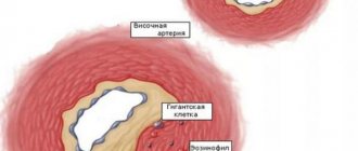
a) angiospastic anemia
Occurs as a result of artery spasm due to the action of any irritants. For example, painful stimulation can cause spasm of the arteries and anemia in certain areas of the body. Angiospastic ischemia appears both with negative emotional affects (“angiospasm of unreacted emotions”) and with exposure to low temperatures.
Angiospasm underlies the veno-arterial effect: with an increase in venous pressure, arteriolar spasm develops.
b) obstructive anemia
It develops as a result of the closure of the lumen of the artery by a thrombus (often resulting in vasospasm) or embolus, as a result of the proliferation of connective tissue in the lumen of the artery during inflammation of its wall (obliterating endarteritis), narrowing of the lumen of the artery by an atherosclerotic plaque.
c) compression anemia
Appears when an artery is compressed by a tumor, effusion, tourniquet, or ligature.
d) ischemia as a result of blood redistribution
Observed in cases of hyperemia after anemia. For example, cerebral ischemia when fluid is removed from the abdominal cavity, where a large mass of blood rushes.
Anemia due to arterial spasm is usually short-lived and does not cause any particular distress. However, with prolonged spasms, dystrophy and heart attack may develop. Acute obstructive anemia is especially dangerous, as it often leads to a heart attack. Long-term anemia sooner or later leads to atrophy and sclerosis.
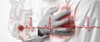
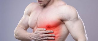
Venous hyperemia: differences from the arterial form, danger in pathology
Venous hyperemia is more understandably called “stagnant” or “passive”. For it you need:
- mechanical obstruction, compression of blood outflow pathways through the main veins by a tumor, scar tissue degeneration, pregnant uterus, strangulated hernia;
- decreased strength of heart contractions;
- reducing the suction role of the chest and diaphragm in case of wounds and injuries, enlarged abdomen;
- impaired valve mechanism of the veins for pumping blood and maintaining an upright position (varicose veins);
- increased viscosity and coagulability of blood, significantly complicating circulation;
- tendency to low blood pressure or acute shock;
- thrombosis or embolism of the venous bed.
The following signs are typical for venous hyperemia:
- bluish color of the skin and mucous membranes in visible areas (limbs, face);
- decrease in temperature in the affected organ and tissues;
- swelling of surrounding tissues.
The pathological mechanism causes a sharp drop in blood flow speed. Fluid enters the interstitial space. Swelling is usually well expressed. The result is tissue hypoxia - oxygen starvation.
Low-mobility blood with platelet aggregations creates a threat of thrombosis and embolization of internal organs. Oxygen deficiency stops metabolism and helps stop the elimination of toxins. Against this background, the addition of infection causes gangrene. And blood platelets form conglomerates of cells. Along with fibrin, the veins begin to block with thrombotic masses, which further increase stagnation.
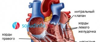
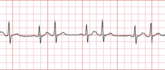
Examination of the fundus using an ophthalmoscope is of diagnostic importance.
In clinical conditions, we can talk about the predominant role of some type of hyperemia, since they are associated and cause a general disturbance of microcirculation.
One example of hyperemia in an inflammatory disease is the manifestation of conjunctivitis, you can learn about this in this article.
To clarify, ultrasound and Doppler ultrasound methods are used. They make it possible to identify congestion of internal organs and record its cause.
Treatment
Arterial and venous hyperemia are extremely unpleasant diseases, which, with a very pronounced symptomatic picture, cause the development of embarrassment, complexes and self-doubt. Skin hyperemia is a disease that is easily treatable. There are also preventive measures, the implementation of which will help prevent the recurrence of the disease in the future.
Treatment of the pathology is complex, including taking medications and traditional medicine methods that help eliminate unpleasant symptoms. In addition, physiotherapeutic methods can be used in the treatment of hyperemia. Physiotherapy helps to quickly eliminate unpleasant signs of the disease.
Despite the fact that hyperemia responds well to drug treatment and traditional methods, therapy must be started in a timely manner. If the disease has been advanced and the cause of its development is internal pathology, medications may not provide the desired therapeutic response. In such a situation, the only effective treatment is surgery.
The operation is selected depending on what disturbances in the functioning of the heart muscle and vascular system caused the hyperemia. Surgical interventions such as eliminating congenital and acquired heart defects, removing blockages - blood clots, and restoring blood circulation in internal organs are often performed.
Treatment of hyperemia includes mandatory diet. The following products should be on the menu daily:
- the basis of the diet is fresh fruits and vegetables (root vegetables);
- cereals;
- sprouted grains;
- cereals;
- dairy products with a minimum percentage of fat content.
Important information: How to treat periarteritis nodosa in women and its symptoms
In case of venous or arterial hyperemia, the following products are prohibited:
- Fatty meats and fish. Preference in creating a menu should be given to lean meat - chicken and turkey, beef and veal, lamb.
- Fatty dairy and fermented milk products - cheese and cottage cheese, cream and sour cream.
- Chicken eggs are allowed to be consumed, but rarely and in limited quantities, since the high protein content in this product prevents proper blood circulation.
- Sweets and confectionery, chocolate, ice cream.
- Spices and seasonings, it is recommended to avoid table salt; sea salt will bring more benefits to the body.
- Alcoholic drinks are strictly forbidden to consume, they negatively affect the condition of the liver, impair blood circulation, leading to aggravation of the symptomatic picture of the disease.
It is also necessary to give up such a bad habit as smoking. Tobacco has an extremely negative effect on the functioning of the heart muscle, leading to blockages of blood vessels with a total circulatory disorder.
Drug therapy
Venous and arterial congestion should begin to be treated by eliminating the cause of its occurrence and selecting appropriate medications. If hyperemia is caused by infectious diseases, antibacterial drugs are prescribed; in case of hormonal imbalance, hormone therapy is carried out. In addition to eliminating internal causes, facial hyperemia is treated with drugs aimed at normalizing blood circulation - Rhodamine, Vazonite and Trental.
To protect the skin from the negative effects of external irritating factors and relieve the symptomatic picture, local spectrum drugs are prescribed - Nezulin (used for allergic hyperemia, has a pronounced antihistamine effect), Sinaflan, Iricar.
Folk remedies
Arterial hyperemia and venous congestion in the early stages of the development of pathology are effectively treated using traditional medicine recipes. If the disease is advanced and has a pronounced clinical picture, alternative treatment is used to eliminate external unpleasant symptoms. Recommended folk recipes:
- Mix 2% boric acid and Hoffmann drops in equal proportions, apply the resulting composition to the face 1-2 times a day.
- Facial ointment - mix 20 g of Vaseline and 3 g of salol thoroughly, add 10 g of zinc ointment to the resulting mixture, mix. After the first 2-3 procedures of applying the prepared ointment, you need to see a doctor so that he can assess the condition and reaction of the skin to the drug. During the entire period of use of the ointment, you must refrain from washing your face, using cosmetics and shaving cream.
- Plantain - crush the leaves of the plant, after washing them under running water. You need to crush the leaf until it releases juice. Apply the leaves to those places of the skin where there is redness and cover with a band-aid for reliable fixation. You need to keep this bandage for 10 hours.
- Beetroot juice - chop the vegetable on a grater, wrap the pulp in a bandage, press down a little so that the juice appears, and apply to the red skin, keeping this mask on for 10 minutes. After the mask is removed, the skin must be wiped with chamomile decoction. It is important that after rubbing with chamomile, the skin dries naturally.
- Aloe and cucumber mask. Mix 1 tsp. aloe juice with juice obtained from one cucumber (only seasonal vegetables are suitable, without pesticides). Apply the mixture to the skin and keep it as a mask for 15 minutes.
Important information: How to treat moderate myocardial hypoxia (oxygen starvation of the heart) and what are its symptoms
Types of arterial hyperemia
There are several most characteristic forms, provoked by different reasons.
- Inflammatory – usually focal. Accompanies inflammation of an organ or tissue in a specific area of the body. It is secondary in nature and disappears after the main cause is eliminated.
- Blood flow after anemia, that is, temporary restriction of blood flow. After eliminating the cause - compression, swelling, blood temporarily fills the vessels, trying to saturate the tissues with oxygen.
- Redistributive hyperemia - for example, decompression sickness in divers. A consequence of the small difference between the volume of oxygen in arterial and venous blood.
- Congestion based on arteriovenous grafting, for example, when, during injury, part of the venous blood enters the artery.
- Collateral - occurs as compensation for the difficult movement of blood through the main vessels. The blood flow is partially redistributed to secondary vessels until the obstacle in the main line is eliminated - a tumor, a blood clot.
Passive form
Venous hyperemia, or stasis, is the result of insufficient work of the veins. The blood that comes through the arteries in normal or excessive quantities does not return in full and stagnates in the tissues or organ.
- expansion of the vein due to varicose veins or, for example, overheating disrupts the normal functioning of the venous valves. As a result, the blood is not pushed out completely, but returns back;
- increased blood viscosity, which makes it difficult to circulate;
- decrease in pressure;
- venous hyperemia is a mandatory sign of a disturbance in the pumping heart muscle;
- blockage of a vein - thrombosis or embolism;
- compression of the vein - mechanical external, or internal - due to tumor, edema, and so on.
Venous hyperemia does not find useful use, except in special cases when it is necessary to stop the development of infection for a short period of time.
Its direct consequence is hypoxia of an organ or tissue, since normal blood circulation is disrupted and the organ does not receive the proper amount of oxygen.
The signs of the passive form are:
- local decrease in temperature in the affected area;
- cyanosis of the skin and mucous membranes;
- an increase in the cross-section of veins and venules - swelling of the veins becomes noticeable;
- edema is also dangerous because it exerts additional compressive pressure, interfering with already poor blood flow;
- slowing down and stopping blood circulation in the capillaries;
- with a prolonged course of the disease, external and internal hemorrhages are possible;
- stasis provokes the development of organ hypoxia and metabolic disorders.
If passive hyperemia manifests itself externally - cyanosis, swelling of veins close to the skin, it is easy to detect and diagnose. But when it comes to internal organs, special methods are used for examination - Doppler scanning, ultrasound, and so on.