Mitral valve prolapse of the 1st degree is the initial phase of congenital or acquired heart disease. The essence of the deviation is a violation of the elasticity of the anatomical structure.
The MV acts as a septum between the left atrium and the same ventricle. With the development of prolapse, a reverse outflow of blood (regurgitation) into the previous chamber is observed. Hence the drop in hemodynamics and the impossibility of normal functioning of the organ.
Treatment is strictly surgical. Prospects depend on the duration of the pathological process, the presence or absence of concomitant conditions. Prosthetics are usually indicated. Plastic surgery has a small list of indications.
The diagnosis is made by a cardiologist. At stage 1, dynamic observation may be prescribed. Surgery is resorted to in difficult cases and when the defect progresses.
Mechanism of cardiac activity
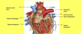
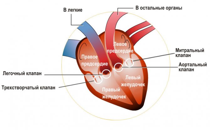
The heart is the most important muscle in the physiological structure of a person, providing, like a pump, the correct blood circulation cycle in the body. The heart cavity consists of 4 chambers, on the right and left sides there are the ventricles and atria. They work crosswise, forming two circles of blood circulation.
The small circle carries out gas exchange, that is, thanks to this function, a person inhales oxygen entering the right ventricle, and exhales carbon dioxide at the exit from the left atrium. The large circle, starting its movement from the left ventricle, supplies the entire human body with blood and passes into the atrium on the right side.
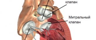
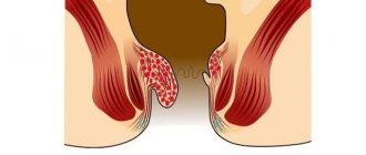
Of course, the heart itself is not capable of functioning, so there are small helpers in the form of valves, they direct the blood in the right direction. The mitral valve (MV) is located between the atrium and the ventricle, on the left side of the heart cavity, and consists of two valves that form a “ring”. The valves direct blood flow from the atrium to the ventricle; thanks to the timely opening of the valves, they do not allow it back, thereby creating a powerful push for the release of blood into the main artery.
Features of the diagnostic search for this disorder of the heart valve apparatus
It is not possible to recognize the presence of this disorder in the functioning of the heart valve apparatus using all diagnostic methods. Among the simplest methods, it is not always possible to recognize the presence of prolapse by auscultation, although this condition is characterized by its own sound phenomenon in the form of a click during late systole. Percussion can determine the characteristic vertical “hanging” position of the heart, but this is observed in a relatively small number of patients and is not considered an accurate criterion.
No specific changes were detected during electrocardiographic examination. Some patients may exhibit sinus tachycardia, as well as supraventricular extrasystoles. For a more detailed assessment of the heart rhythm during the day, Holter monitoring is used (with this diagnosis there is no urgent need for such a study).
The radiographic method allows us to identify only a few features that indicate this pathology - the configuration of the heart shadow in the form of a “hanging drop”; in some patients, the pulmonary artery is distinguished against the background of the cardiac shadow. Often there are no changes at all on the x-ray. This method is not informative.
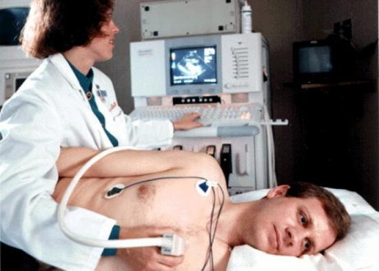
Echocardioscopy is the most accurate and reliable way to diagnose this dysfunction of the valve apparatus of the body's main pump.
The ultrasound diagnostic method gives an idea of the structure and functioning of the leaflets, and also allows you to assess the strength of the reverse flow of blood, if any (in the criteria for the severity of this pathology there is a point regarding how powerful the reverse flow is and how deep into the atrium it extends).
What is it - stage 1 mitral valve prolapse?
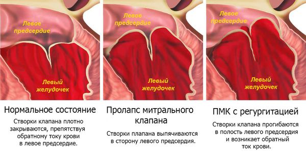
The health of the heart largely depends on the functioning of its “helpers”. In a standard situation, the valve ring closes tightly and does not allow blood to pass through.
Grade 1 mitral valve prolapse is a cardiac abnormality where one or both valves are deformed, preventing the valve from forming a tight closure, thereby creating the risk of blood flow leaking back into the atrium.
According to its origin, prolapse is represented by 2 types:
Primary MVP is a congenital defect that occurs frequently and occurs for the following reasons:
- hereditary predisposition;
- a genetic disease associated with connective tissue pathology (for example, Marfan syndrome);
- congenital anomaly of the body.
Secondary, also known as acquired, is quite rare and appears during life, when another disease affects the structure of connective tissues:
- rheumatism is the most common autoimmune disease that provokes valve deformation;
- lupus;
- myocardial infarction;
- vegetative dystonia.
Along with the above, the disease can occur in two different forms:
- ascultative - characterized by changes in cardiac activity and. accompanied by clicks or clicks in breathing, detected by simple listening using a stethoscope and an ECG.
- silent - painless, detected only on echocardiography. Symptoms of this form of grade 1 mitral valve prolapse usually do not appear.
Mitral valve prolapse
Character of valve deformation
Valve prolapse is caused by sagging or deflection of the valve flaps, the size of which ranges from 3 to 5 mm; there are no symptoms and the disease does not cause any discomfort.
Medical practice notes that prolapse syndrome most often proceeds favorably and does not involve any difficulties. But the likelihood of complications still exists and depends largely on individual symptoms. These include the following diseases: heart disease, MV insufficiency, abnormal changes in other valves in the cardiac cavity.
Degrees of regurgitation
Regurgitation is a disorder that can occur when the valve is deformed, characterized by the formation of a gap between the leaflets, resulting in blood flow back into the atrium. There are 3 stages of severity of regurgitation, depending on its volume:
- 1st degree provokes the occurrence of blood turbulence - the gap between the valves is insignificant, the blood rises and affects the walls of the valve;
- Stage 2 is characterized by blood flow reaching ½ of the atrium;
- 3rd degree - blood fills more than half of the atrium;
- Stage 4 is considered the most severe, due to the fact that the blood flow completely fills the atrium.
The essence of the concept of regurgitation
Regurgitation is a medical term. Translated from Latin, it means “to flood again.” Cardiologists call this term the pathological process in which blood is thrown back into the chamber of the heart.
The tricuspid valve is one of the four heart valves. It is tricuspid in structure. Normally, the valves periodically open, allowing blood to flow further from the heart chamber. Its task is to prevent blood flow from returning, to regulate blood flow. Pathological changes can occur in any of the heart valves. Depending on which valve is affected, a different clinical picture will be observed.
The most common types of regurgitation are mitral and aortic. Tricuspid is less common. Also, one patient may experience pathology of several valves at once.
Classification by localization of prolapse
The defect may deform the anterior, posterior, or both leaflets of the valve. Grade 1 anterior leaflet mitral valve prolapse is much more common.
Front flap
The disease caused by sagging of the anterior flap is most often caused by congenital pathology, and is usually diagnosed at an early age. Pathology prevents the normal formation of connective tissue, and as a result contributes to its deformation.
Rear flap
Such deformation occurs less frequently than in the first option. More typical is acquired mitral valve prolapse of the 1st degree with regurgitation of the 1st degree in an ascultative form.
Double-sided
In fact, each of the listed options, as well as a bilateral anomaly, are found in medical practice. At present, the importance of the location of the prolapse of a particular valve has not been established by science. The form of deformation itself and the presence of progressiveness are much more important.
Causes
There are primary and secondary factors that provoke different forms of pathology.
Primary factors:
- Heredity. May cause triscupid valve insufficiency. The problem begins during fetal development. Its mechanism has not yet been established. If one of the parents has such a pathology, the risk that the child will also acquire it is reduced to 12-15%. Spontaneous defects may develop in the perinatal period.
- Cardiac adhesions. Appear during inflammatory processes, especially if they are infectious in nature. Such fibrin small strands disrupt the normal anatomy of the heart.
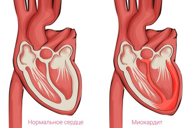
- Inflammatory cardiac pathologies. It could be myocarditis, etc. The tissues of cardiac structures are quickly destroyed. Urgent hospital treatment is required. Antibiotics, diuretics, steroids and NSAIDs are required.
- Heart attack. After it, healthy tissues are replaced by scar tissues, which cannot conduct the signal and contract normally. The tricuspid valve can become completely overgrown, and its functional insufficiency and stenosis may develop. Severe regurgitation is immediately observed. Urgent surgery is required.
- Rheumatism. This is a chronic inflammation in which a short remission is often interrupted by another relapse.
Secondary factors:
- pulmonary hypertension and pathologies of the development of heart structures. It is necessary to treat urgently, at an early stage. At risk are smokers, asthmatics, and those with a long course;
- cardiomyopathy;
- pathologies of the endocrine system (excessive production of adrenal hormones, hyperthyroidism, diabetes, etc.).
Symptoms in adults
Symptoms of grade 1 mitral valve prolapse in adults are not recorded in most cases. Often, such an anomaly in patients is discovered by chance, for example, during a diagnosis or routine examination.
It is worth paying attention if you are bothered by short pain or discomfort. Symptoms may also be accompanied by non-cardiac signs:
- increased sweating;
- causeless attacks of fear or aggression (panic attacks);
- shortness of breath, especially during physical exertion - this symptom is typical in half of the cases;
- a decrease in pressure up to the point of fainting is rare, occurring in approximately 10–15% of patients.
Prevention and prognosis
A healthy lifestyle is recommended to prevent any disease. A normal work and rest schedule, reasonable physical activity, lack of stress and harmful addictions makes the likelihood of getting sick minimal. A very important aspect is the timely and correct treatment of colds, the obligatory clarification of the causes of systematic ailments. If the patient has undergone surgery for a heart defect or other pathology, it is very important to follow all clinical recommendations of cardiologists and carry out rehabilitation measures.
The prognosis for tricuspid regurgitation is positive; in childhood it can level out with age. For an adult, grade 1 pathology detected by chance is considered a functional norm that does not require treatment. If tricuspid insufficiency occurs in combination with other complications, the patient is assigned a disability group.
Be attentive to yourself and your children, do not ignore periodic ailments. Dangerous conditions may be masked behind them.
Features in children
Mitral valve prolapse in 1st degree in adults and children are very similar. In children, this syndrome is detected much more often. In the age group from 7 to 15 years, girls are more susceptible to anomalies of ascultative expression. In general, the incidence of silent infantile prolapse is equally common in both boys and girls. The progression of prolapse largely depends not only on the direct characteristics of the disease, but also on associated factors.
Much attention is paid to the state of the child’s autonomic nervous system, which affects metabolism and the presence and outcome of prolapse in adult family members.
MVP can be congenital or acquired. The size of the deformed valve ranges from 3 to 5 mm and can occur with or without regurgitation.
The symptoms of prolapse in children are ambiguous, but in most cases no abnormal changes are felt.
Prolapse of the anterior leaflet of the mitral valve 1st degree with regurgitation may be accompanied by the following symptoms:
- tachycardia;
- heaviness or short-term pain in the chest area;
- aggression, tearfulness, depressive mood.
Due to the possible progression of the disease with age, it is extremely important to visit a specialist at least once a year. For children with the ascultative type of MVP, it is recommended to visit a cardiologist at least 2 times a year and undergo an ultrasound examination of the heart.
Drug treatment
Moderate tricuspid valve insufficiency is not considered an indication for surgery. In this case, treatment is carried out with the help of medications. The standard treatment regimen involves the use of the following medications:
- Diuretics (Britomar, Hydrochlorothiazide). Eliminate congestion in the body, accelerate the process of fluid removal.
- Potassium preparations (Panangin, Asparkam). Help the body not accumulate excess fluid.
- Venous dilators (Corvaton, Nitrosorbide). Reduce the load on the heart through blood deposition.
- Anticoagulants (Warfarex, Warfarin).
- Cardiac glycosides (Digoxin, Korglykon). Helps in the fight against arrhythmia.
- Beta-blockers (Diltiazem, Carvedilol). Reduce the frequency of contractions of the left ventricle.
The regimen and dosage of drugs are determined individually, taking into account the severity of the disease.

Management of pregnancy with this diagnosis
If a diagnosis of grade 1 mitral valve prolapse is made, pregnancy, as a rule, proceeds favorably and without symptoms.
This is the most common heart pathology, occurring in 17% of young women. The state of health and general well-being for 9 months largely depends on the form, degree and nature of the pathology.
In general, there are no special contraindications for women in labor with MVP, since minor deformation does not affect the state of health. But if the disease is accompanied by weakened heart activity, it is likely that a cesarean section will be used. In addition, changes in heart rhythm sometimes occur.
General recommendations for managing pregnancy with grade 1 mitral valve prolapse syndrome:
- do not overcool or overheat;
- you cannot sit for a long time;
- exclude prolonged stay in baths and saunas;
- get more rest;
- eat foods high in potassium and magnesium;
- take vitamin and mineral complexes;
- walks in the open air.
There is a possibility that pregnancy will be accompanied by gestosis, which is characterized by the following symptoms:
- swelling;
- increased blood pressure;
- convulsions;
- decrease in protein content in the body.
Such complications can lead to insufficient oxygen supply to the fetus. In medical practice, the possible occurrence of premature birth is noted. Treatment of MVP with medications is used only in special episodes caused by severe prolapse.
In most cases, MVP is transmitted to the child, so it is very important to manage the pregnancy under the supervision of a cardiologist.
Risk factors
The pathological process can be started if:
- Smoking a lot for a long time.
- Don't know how to drink alcohol.
- Drug addiction.
- Prolonged immobilization, when a person moves very little. This happens with injuries and illnesses. Six months of such inactivity is enough for the pathological process to begin.
- Long-term treatment with potentially dangerous drugs, for example, glycosides, progestins, antiarrhythmics, hormonals, antibiotics.
- Work in hot, chemical production, in a mine.
If several negative factors are exposed at once, the likelihood of developing pathology increases significantly.
ATTENTION! Most often, this pathology is provoked by heart failure, pulmonary hypertension, and obstruction of the pulmonary arteries. Less commonly, it is caused by rheumatism, infectious diseases, especially endocarditis, taking certain medications, etc.
Is it possible to play sports?
Physical activity for mitral valve prolapse syndrome, with stage 1 established, is not contraindicated, and is sometimes mandatory.
It is important to understand that each diagnosis has individual limitations and recommendations, so you must always consult with your doctor. For patients with a sedentary lifestyle, special techniques with complexes of therapeutic physical exercises have been developed.
Children with this diagnosis are allowed physical activity if there are no signs contributing to the progression of the disease. In other cases, there are a number of restrictions that are set by the cardiologist.
It is possible and necessary to play sports with grade 1 prolapse, with deformation of the mitral “slamming” valve, but with moderate amounts of exercise.
In cases where the pathology is accompanied by one of the following symptoms, high-intensity training is strictly prohibited, otherwise there is a risk of worsening the pathology of MVP or causing an exacerbation:
- fainting;
- tachycardia;
- heart failure MK;
- thromboembolism - blockage of a blood vessel with a thrombus;
- low contractility of the myocardium (heart);
If there are no such signs, you can engage in high-intensity training (for example, CrossFit).
It is worth noting that for professional athletes with the characteristic above-mentioned signs it is possible to engage in the following activities:
- billiards;
- bowling;
- curling;
- shooting;
- golf.
In addition, for athletes with a “sailing” valve who want to take part in any competitions, there are a number of certain restrictions established by special international organizations.
What sports should you not do:
- athletics;
- intense jumping;
- wrestling, boxing;
- lifting heavy weights.
Do they take you into the army?
In the generally accepted sense, when diagnosed with mitral valve prolapse of the 1st degree, they are taken into the army in cases where the disease is not accompanied by regurgitation.
Regulatory acts of the Russian Federation establish the classification of conscripts according to their state of health. During a medical examination, doctors as part of a military commission determine the degree of suitability of a young man for service and make a corresponding note in his personal file. For example, group “B” means suitability with minor restrictions.
For conscripts diagnosed with stage 1 mitral floating valve prolapse with grade 1 cusp regurgitation, the army is also mandatory, with possible restrictions.
According to the current legislation of the Russian Federation, the release of a conscript is possible on the following grounds:
- stage 3 MVP syndrome;
- the presence of heart failure of functional classes 3 and 4.
Such conscripts may be exempt from service altogether or receive Group “B” status, which means eligibility with significant restrictions. According to the classification, young people from this category may be drafted in the event of a state of war being declared in the country.
Since each patient's clinical presentation is different, a medical opinion must be obtained to determine fitness for duty.
Diagnostics
Conducted by a cardiologist. The survey presents certain difficulties at this stage. There are still few symptoms, the clinical picture may be completely absent. Careful management of the person will be required.
What methods are used:
- Oral questioning of the patient regarding complaints.
- Taking anamnesis. Lifestyle, family history, other issues, including bad habits.
- Measurement of blood pressure, heart rate. Both indicators are normal or slightly changed.
- Daily monitoring. Both blood pressure and heart rate are recorded for 24 hours. In dynamics, much more data can be obtained, especially if the patient continues normal daily activities.
- Electrocardiography. Used to assess the functional activity of cardiac structures. There are not many deviations, sometimes none at all.
Echocardiography. Ultrasound method for assessing the cardiovascular system. The main way to diagnose mitral valve prolapse.
- Auscultation. Listening to heart sounds. The diagnosis of MVP is made, among other things, on the basis of sinus noise - this is how the reverse flow of blood manifests itself.
- MRI as needed.
The pathological process is determined relatively simply. Echocardiography is sufficient. Other methods are aimed at establishing the degree of severity and complications.
How to treat?
Drug treatment of hereditary or congenital prolapse caused by the presence of a 1st degree mitral valve without regurgitation is not required, since such a diagnosis is not considered a disease. The cardiologist simply registers the patient for an annual preventive examination and also prescribes physical therapy.
To improve your overall health, you need to take vitamin and mineral complexes. In addition, it is worth eating foods that help strengthen and improve immunity (dried apricots, bananas, walnuts, raisins, grapes, figs).
The cardiologist determines how to treat grade 1 mitral valve prolapse with regurgitation, taking into account the state of health and individual contraindications. Typically, sedatives, anticoagulants that prevent the formation of blood clots in blood vessels, and cardiotrophic drugs are prescribed. In addition to medications, a set of physical exercises is also selected to strengthen the cardiovascular system.
Additional recommendations for promoting health:
- take a contrast shower or harden yourself;
- take long walks in the fresh air more often;
- proper nutrition;
- sleep at least 8 hours.
Set of diagnostic measures
Diagnosis of PTC is an important step in the process of treating the disease. It must be comprehensive and timely, which will increase the patient’s chances of recovery. A doctor can assume the development of a disease in a patient based on complaints, as well as objective examination data: palpation, percussion, auscultation of the heart.
The following instrumental research methods can confirm the diagnosis:
- electrocardiography, which allows you to determine dilatation of the right ventricle;
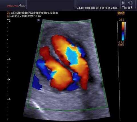
- echocardiography, which makes it possible to diagnose the presence of flexion of the tricuspid valve leaflets and regurgitation;
- catheterization of the right heart, which can be used to determine the increase in pressure in the heart chambers and lungs;
- X-ray of the chest cavity can confirm the diagnosis of pulmonary hypertension;
- computed tomography and MRI to detect enlargement of the right heart.
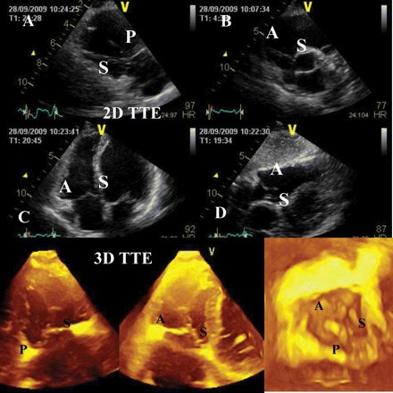
Echocardiogram of the heart with tricuspid valve prolapse