The pathological structure of the tricuspid valve, its attachment downwards (inside the right ventricle) is called Ebstein's anomaly. This defect can be congenital. Children are born weakened, with difficulty breathing and cyanosis of the skin. Paroxysmal tachycardia and circulatory failure are often associated. Treatment requires surgery to replace the valve and reconstruct the opening between the chambers of the right half of the heart.
Causes of development in the fetus
The following factors for the development of fetal anomalies have been proven:
- taking medications containing lithium salts by a pregnant woman;
- rubella virus, measles;
- scarlet fever;
- diabetes;
- thyrotoxicosis;
- alcoholism;
- use of psychotropic medications;
- toxicosis of the gestational period;
- threat of miscarriage;
- hereditary predisposition.
The effects of infections and intoxications in the first trimester of pregnancy are especially dangerous.
We recommend reading the article about tricuspid valve disease. From it you will learn about the causes of the pathology and its symptoms, diagnostics, possible complications and prognosis for patients.
And here is more information about combined heart defects.
Reasons and essence
Unfortunately, it is impossible to say reliably about the reasons for the development of Ebstein’s anomaly today. There are the following causes of pathology:
- Genetic factor is the ability of an organism to inherit genetic information from its parents. We are talking about mutations, hereditary predisposition.
- Physical factors are the harmful influence of the external environment. The most dangerous of them are electromagnetic and radioactive radiation.
- Chemical factors - the presence of bad habits. These include smoking, drinking alcohol, drug addiction and taking dangerous medications by pregnant women. A separate role in the development of Ebstein's anomaly is given to lithium preparations.
- A biological factor is the presence of an infectious disease in a pregnant woman that negatively affects the fetus.
Features of hemodynamics
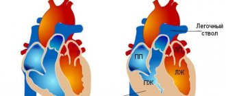
In this pathology, the valve leaflets are deformed and displaced into the lumen of the right ventricle. They are located below the physiological opening between the atrium and the ventricle.
Therefore, the right atrium is enlarged due to the “attachment” of part of the ventricle, and the latter becomes disproportionately small.
At the moment of systole of the atrium, its upper part contracts, and the lower part only during the systolic period of the ventricle. At this point, part of the blood from the right ventricle returns to the atrium. There is little blood left in the right ventricle, which reduces its flow to the lungs. And the atrium fills with blood, expands, its muscle layer hypertrophies.
High pressure in the atrium causes a window in the atrial septum to open. A reset is formed from right to left. It helps relieve the right atrium, but at the same time reduces the oxygen content in arterial blood due to its mixing with venous blood.
Etiology
The peculiarity of the disease is that Ebstein's anomaly is formed long before the child is born. The pathology leads to intrauterine fetal death in 85% of cases.
According to the timing of detection, the disease in most cases is detected from the first to the nineteenth week of the baby’s life. 67% of children survive their first year, but only 59% of them will celebrate their tenth birthday.
Main risk factors:
- excess lithium enters the body of a pregnant woman - occurs with uncontrolled use of medications;
- severe infections suffered during pregnancy - measles, scarlet fever or rubella;
- exacerbation of chronic diseases during gestation, for example, diabetes, anemia or thyrotoxicosis;
- addiction to bad habits;
- severe gestation;
- irradiation of the body;
- risk of spontaneous miscarriage.
It is worth noting that the formation of the child’s cardiovascular system occurs in the first trimester, in particular from 1 to 9 weeks. During this period, the influence of negative factors often leads to the development of such a defect.
An important role in the formation of congenital Ebstein anomaly is played by burdened heredity - in 8% of patients the influence of genetic predisposition is noted.
Symptoms in a newborn
Asymptomatic defects with Ebstein's anomaly practically do not occur. The most severe developmental disorders lead to fetal death during gestation. Most often, children are already weakened in the first months of birth; physical activity (feeding, crying) causes severe shortness of breath and bluish skin.
At an older age, they complain of heart pain and palpitations. The fingers look like drumsticks, and the nails look like convex watch glasses. Due to the enlargement of the right atrium, a protrusion of the chest is formed, similar to the cardiac hump.
Heart with Ebstein's anomaly
Symptoms
In total, modern medicine distinguishes three forms of the disease, on which the observed symptoms of Ebstein’s anomaly in children and adults depend:
- The first of them is the most gentle, but at the same time the least common. She has virtually no symptoms.
- The second is accompanied by severe circulatory disorders of the body, depending on heart rhythm disturbances, which may or may not occur with it.
- The third is the hardest. Its distinguishing feature is persistent decompensation.
A large number of patients are found to have: cardiac tachycardia, WPW, abnormal development of the nail plate and fingertips.
In the future, insufficient functionality of the right ventricle, pathological processes in the hepatic gland, and enlarged neck veins may appear.
A congenital pathology such as Ebstein's anomaly, in its severe forms, can terminate the life of a child even before his birth. In an alternative perspective, its development will be consistent with age, and symptoms will either simply not be present or will go unnoticed.
But most often, Ebstein's anomaly in newborns is visible already in the first months of life. He will have noticeable bluish limbs and a triangular zone of the nasolabial fold - manifestations of acrocyanosis, increased fatigue, pain attacks in the heart and its accelerated beating.
Blueness of the nasolabial fold is a hallmark symptom of Ebstein's anomaly
Complications caused by the disease
With this anomaly, the right atrium experiences increased load, and the ventricle ejects less blood than necessary. One of the early complications is circulatory failure of the right ventricular type. It is characterized by:
- difficulty breathing,
- enlarged liver
- increased pulsation of dilated veins of the neck.
Without treatment, Ebstein's anomaly tends to progress. With cardiac decompensation and severe arrhythmia, death is possible.
What is this disease?
Ebstein's anomaly can appear in both men and women with equal probability. Unlike other types of heart defects, patients with this pathology have a high chance of avoiding death and surviving safely to adulthood. In patients with this pathology, the septum of the tricuspid valve is displaced compared to normal. Because of this feature, the right ventricle increases in size. The valve itself, depending on the degree of the disease, may have deformed walls. In some patients, portions of the valve walls are missing. Such changes lead to poor circulation and cardiac rhythm disturbances.
Diagnosis in children and adults
During examination, attention is paid to the cyanotic color of the skin, deformation of the fingers and chest. The borders of the heart are shifted to the right, a 3- or 4-beat rhythm is heard, a murmur to the right of the sternum, the 2nd tone is bifurcated.
If a tricuspid valve defect is suspected, the following examination is performed:
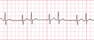
- ECG - the axis is deviated to the right, the right atrium is hypertrophied and dilated, rhythm disturbances in the form of paroxysmal tachycardia, atrial fibrillation and flutter, Wolff-Parkinson-White syndrome, blockade of the right branch of the Hiss pathway.
- FCG – systolic murmur over the projection of the right ventricle, 1 tone is weakened, 2 is bifurcated, 3 and 4 are high-amplitude.
- X-ray – the right atrium gives the contour of the heart the shape of a ball, the pulmonary fields are transparent.
- Ultrasound of the heart - the valve leaflets are displaced downwards, close slowly, the right ventricle is poorly filled, blood is discharged through the atrial septum to the left. In the fetus, these abnormalities can be detected in approximately 65 - 70% of cases.
- CT and MRI make it possible to detail the degree of valve non-closure, the presence and intensity of blood shunting, and the size of the heart chambers.
- Ventriculography and probing of the cardiac cavities are used only when it is difficult to make a diagnosis.
Pathophysiology
Embryological development of the cusps and chords of the tricuspid valve involves undermining the free wall of the right ventricle. This process continues to the level of the atrioventricular (AV) junction. In Ebstein's anomaly, this undermining process is incomplete and does not reach the level of the AV junction. In addition, the apical parts of the valve tissue, which are usually subject to resorption, are not able to completely resorb. This leads to distortion and displacement of the leaflets of the tricuspid valve, and part of the right ventricle becomes atrial. In one study of 50 hearts with the anomaly, the entire right ventricle was found to be morphologically abnormal.
Ebstein's anomaly is usually associated with other congenital, structural, or conduction system diseases, including intracardiac shunts, valvular lesions, and accessory conduction pathways (eg, Wolff-Parkinson-White syndrome).
The hemodynamic consequences of Ebstein's anomaly arise from displaced and malformed tricuspid leaves and the atrium of the right ventricle. An anomaly of the leaflet leads to tricuspid regurgitation. The degree of regurgitation depends on the degree of leaflet displacement, ranging from mild regurgitation with minimally displaced tricuspid leaflets to severe regurgitation with extreme displacement.
The atrial portion of the right ventricle, although anatomically part of the right atrium, contracts and relaxes with the right ventricle. This discordant contraction leads to stagnation of blood in the right atrium. During ventricular systole, the atrial portion of the right ventricle contracts with the rest of the right ventricle, which causes blood to flow back into the right atrium, increasing the effects of tricuspid regurgitation.
Signs and symptoms
Patients may experience a variety of symptoms that are associated with the anatomical pathologies of Ebstein's anomaly, their hemodynamic effects, or concomitant disease of the structural and conduction systems, including the following:
- Cyanosis
- Fatigue and shortness of breath
- Heart palpitations, sudden cardiac death
- Symptoms of right-sided heart failure, particularly edema and ascites
Other less common symptoms include the following:
- Brain abscess as a result of shunting the blood circulation from right to left
- Bacterial endocarditis
- Paradoxical embolism, stroke and transient ischemic attacks
Cyanosis is quite common and often results from right-to-left shunting at the atrial level or severe heart failure. It may be transient during neonatal life, with recurrence in adult life; may also develop for the first time in adult life. Temporary appearance/worsening of cyanosis in adult life occurs due to paroxysmal arrhythmias; Once established, cyanosis gradually regresses.
Fatigue and shortness of breath are associated with poor cardiac output, which is secondary to right-sided ventricular failure, and decreased left ventricular ejection fraction.
Palpitations and sudden cardiac death may occur as a result of paroxysmal supraventricular tachycardia or Wolff-Parkinson-White syndrome in 1/3 of patients. Fatal ventricular arrhythmias may also occur due to the presence of accessory pathways.
Ebstein's anomaly is a congenital disorder of often unknown cause.
Environmental factors relevant to the etiology of the disease include the following maternal factors:
- Maternal use of lithium in the first trimester of pregnancy
- Maternal use of benzodiazepines during pregnancy
- Exposure to varnishing substances
- History of previous maternal fetal loss
Ebstein's anomaly probably accounts for 0.5% of congenital heart defects. Its true prevalence is unknown because mild forms are often undiagnosed. With the widespread use of echocardiography, more cases are being diagnosed.
Operation as a radical method of treatment
Conservative therapy is used only for temporary relief of the condition or in preparation for surgery. Prescribe cardiac glycosides, antiarrhythmic drugs that stimulate metabolic processes in the myocardium.
The only treatment is surgery. It is performed for heart failure and heart rhythm disturbances. In severe cases, a newborn child can be operated on, but it is best performed at 15 years of age.
Surgical intervention involves:
- plastic or prosthetic tricuspid valve,
- restoration of the integrity of the septum between the atria,
- cauterization of additional pathways,
- installation of a pacemaker (if necessary, a cardioverter).
Watch the video about Ebstein's anomaly and its correction:
Treatment of Ebstein's anomaly
Drug therapy for AE is necessary to correct the heart rhythm when signs of heart failure appear. Antiarrhythmic drugs include beta blockers (atenolol, metoprolol), calcium antagonists (verapamil, diltiazem). For heart failure, diuretics, ACE inhibitors, and cardiac glycosides are indicated. The choice of drug is determined by the patient’s age and the course of the pathology.
AE is one of those developmental defects whose manifestations cannot be corrected only by conservative methods, therefore the vast majority of patients require surgical treatment. The age of the operation and its type depend on the structural abnormalities in the heart itself, the severity of the defect and the nature of the hemodynamic disorders.
The most common types of operations for AE:
- Tricuspid valve repair;
- Valve replacement.
During plastic surgery, the excess part of the right atrium is eliminated, a single-leaf valve is created and the diameter of the valve fibrous ring is reduced. If there is a defect in the interatrial septum, the surgeon will also suture it. Both during plastic surgery and prosthetics, “extra” impulse pathways are crossed, which contribute to arrhythmias.
Newborns with severe overflow of the right atrium and an insufficient size of the oval window may require surgery in the first weeks of life. Its essence is to expand the oval window or a defect in the septum with a special device (balloon) to ensure the movement of “excess” blood to the left half of the heart. This measure is not radical, but it eliminates the risk to the baby’s life, and in the future the valve will still require plastic surgery or its replacement.
If it is impossible to perform plastic surgery on the valve, the only way to cure the defect is prosthetics. Its disadvantage is the presence of foreign material in the organ, but, on the other hand, it is a very reliable method of correcting congenital heart disease. It is advisable to carry it out in adolescence, when the volume of the heart is already as close as possible to that of adults, in order to avoid a discrepancy between the size of the valve and the area of the heart and, as a result, stenosis.
Prosthetics means removing the structures of the damaged valve and replacing it with an artificial analogue. Modern medicine proposes to supplement such operations with the introduction of stem cells, which provide additional material for restoring the missing mass of the myocardium of the right ventricle of the heart.
When it comes to the need to implant a prosthesis to replace a damaged valve, any parent will want to know what exactly will be installed in their child’s heart. Today, cardiologists can offer either a mechanical valve, consisting entirely of synthetic materials and metal, or a biological one, which is made from elements of human pericardium, or simply transplanting a pig valve corresponding in size to a human one.
These options have both advantages and disadvantages. A mechanical prosthesis requires lifelong use of anticoagulants, but it is durable and reliable. The biological valve does not require anticoagulant therapy, but also has a slightly shorter service life. The choice of the type of prosthesis remains with the cardiac surgeon, who evaluates the real clinical situation.
Forecast
Heart disease with Ebstein's anomaly is classified as severe. Without timely treatment, about 35% of patients die by the age of 10; most do not live to be 30 years old. Causes of death include right ventricular failure and ventricular fibrillation.
If the operation was successful, then the prognosis for life is favorable. Long-term results depend on the severity of myocardial hypertrophy and postoperative rhythm disturbances.
Ventricular fibrillation as a consequence of Ebstein's anomaly
Diagnostics
At the first visit or scheduled visit to the doctor, he may suspect the presence of heart pathology based on a number of data and signs:
| Method | What does it evaluate? |
| Survey | Possibility of harmful effects during early pregnancy Ultrasonography (ultrasound) data of the fetus: a heart defect is detected later than 21 weeks of intrauterine development in 87% of cases Complaints about any health problems |
| Inspection | Compliance of growth and development with norms for a given age category Breathing pattern Skin color of the face and neck at rest and during moderate physical activity Saphenous veins of the neck and upper extremities (their bulging is a sign of high pressure in the right atrium) |
To clarify the diagnosis, the following methods are used:
| Method | Features of changes in Ebstein's anomaly |
| Auscultation (listening) of the heart | Additional heart sound (gallop rhythm) Systolic and/or diastolic heart murmur |
| Electrocardiography | Signs of enlargement of the right ventricle and (or) atrium Partial or complete block of the His bundle Heart rhythm disturbances |
| X-ray of the lungs and heart muscle | Right heart enlargement or atrial isolation Signs of decreased blood flow to the lungs |
| Echocardiography (ultrasound of the heart) with blood flow assessment | Displacement of the tricuspid valve leaflets into the cavity of the right ventricle Valve failure Backflow of blood into the atrium during ventricular contraction (regurgitation) Defect of the wall between the atria Enlargement of the right ventricle and/or atrium |
| Electrophysiological study of the heart (assessment of the sources and pathways of the excitation wave) - carried out to identify disturbances in the rhythm of the heart muscle | Impairment of impulse conduction (blockade) along the main pathways The presence of additional pathways (usually multiple) Foci of formation of additional excitation impulses |
Echocardiogram of a patient with severe Ebstein's anomaly showing a severely displaced septal leaflet (arrow).
RA – right atrium, RV – atrial right ventricle, RV – right ventricle, LA – left atrium, LV – left ventricle The diagnosis of “Ebstein’s anomaly” is established only according to instrumental additional examination. Manifestations of the disease and patient examination data are nonspecific for the disease.
Treatment
Modern cardiology involves eliminating a disease called Ebstein's anomaly only through surgery. Sometimes, with a minor defect that does not cause serious problems, and if surgery is impossible, they turn to conservative methods of treatment.
Conservative (drug) therapy involves taking the following medications:
- antibacterial agents;
- cardiac glycosides;
- diuretics;
- disaggregants;
- beta blockers;
- calcium blockers.
One and a half ventricular correction is more often practiced as an operation in the surgical treatment of Ebstein’s anomaly, but other methods are worth highlighting:
- radiofrequency ablation;
- expansion of the natural window in the partition;
- formation of an artificial window - to relieve the load on the right atrium;
- complete closure of the communication between the atrium and the ventricle with the subsequent creation of a bypass connection between the vena cava and the arteries of the lungs;
- placing a shunt between the inferior vena cava and the pulmonary artery;
- tricuspid valve repair;
- valve transplantation - performed on patients over 15 years of age;
- implantation of a pacemaker or cardioverter-defibrillator.
Conservative therapy may be used in preparation for surgery.


