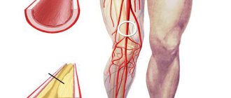
Occlusive thrombosis of deep vessels is a type of thrombus formation that involves complete blockage of the vascular lumen.
The development of occlusive thrombosis in most cases begins in the vessels of the leg (sural thrombosis), and in the case of late diagnosis and untimely treatment, the occlusion spreads along the vascular bed up to the great vena cava.
The danger of the disease lies in the fact that in its initial stages the venous blood flow is still functioning and the patient practically does not feel any discomfort.
Due to this feature, there are often cases of delayed diagnosis of occlusive thrombosis, serious trophic pathologies and patient disability.
In most cases, occlusive deep vein thrombosis is diagnosed; complete blockage of the superficial vessels of the lower extremities is less common.
There are also more frequent cases of diagnosing occlusive thrombosis on the left limb than on the right, due to the anatomical structure of the vascular system.
According to the ICD - the international classification of diseases - this disease is assigned code 180.
Causes of the disease
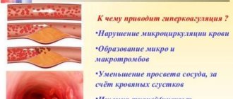
The general prerequisites for the development of thrombosis, including those of an occlusive nature, are three interrelated factors:
- Violation of the speed of blood flow, its slowdown, the formation of venous stagnation.
- Accelerated blood clotting, predisposition to thrombosis.
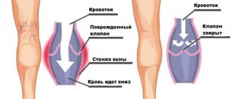
- Destruction of the healthy structure of the inner layer of the venous wall due to mechanical factors: injections, blows, operations, or due to varicose deformation.
Social factors in the development of leg vascular occlusion include:
- Reaching the age of 40 years and older.
- Surgical interventions.
- Infectious and oncological diseases.
- Hormone therapy, including for family planning.
- Prolonged physical inactivity, adynamia.
- Varicose veins.
- Frequent injections in the leg area.
- Habit of using tobacco and alcohol.
Prevention
To prevent thrombosis (including thromboendocarditis), a person must constantly take care of the condition of his blood vessels. To do this, it is advisable not to forget about a healthy drinking regime. You should give up alcohol and smoking. It is extremely important to lead an active lifestyle, and if your work involves sitting, then at least take regular breaks to do basic leg exercises. A very good form of physical activity to benefit blood vessels is walking. Among other things, women should not get carried away with wearing uncomfortable narrow shoes with high heels. And contrast foot baths or a contrast shower will significantly strengthen the walls of blood vessels.
It is always important to remember that thrombosis of the leg veins is a serious pathology that cannot be neglected. Therefore, at the slightest sign of illness, you should contact a specialist as soon as possible for competent help. If you follow all the recommendations of the attending physician and with adequate treatment, the prognosis for the patient is quite favorable.
Diagnostic methods
There are several ways to diagnose the disease.
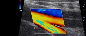
Doppler ultrasound scanning
Occlusive deep vein thrombosis of the lower extremities is diagnosed using duplex ultrasound scanning. This method is non-invasive, ensures accurate results, allows you to establish:
- Blood clot size.
- The degree of blockage of the venous lumen.
- Condition of the walls of blood vessels.
- The degree of disturbance of blood flow.
X-ray contrast venography
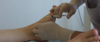
If an ultrasound scan does not allow the apex of the thrombus to be seen, an invasive method is used: radiopaque venography, when a contrast agent is injected into the vessel cavity, allowing an X-ray scan to be performed.
This analysis helps to establish the location, shape, size of the clot, the degree of destruction of the vascular wall, and the condition of the venous valves.
Blood tests
- OAC, to determine the presence of an inflammatory process in the body.
- A coagulogram is a blood test method that allows you to determine the rate of blood clotting.
- Study of D-dimer on the nature of blood clotting.
Diagnostics
In order to prevent pulmonary embolism or other serious pathologies, it is necessary to make an accurate diagnosis of the patient as soon as possible. Diagnostics are carried out using the following hardware methods:
- Phlebography using a contrast agent. Identifies areas affected by vein thrombosis.
- Duplex ultrasound of leg vessels. It also allows you to identify the location of a blood clot.
- Magnetic resonance phlebography. Determines the processes of defective filling of leg veins.
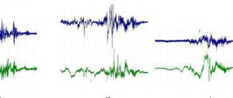
- Impedance plethysmography. Makes it possible to track the process of blood filling of blood vessels using the electrical resistance of tissues.
Treatment
If occlusive deep vein thrombosis is suspected, the patient requires immediate hospitalization.
The set of therapeutic measures is as follows:
- Effective treatment of a patient with occlusion of the veins of the lower extremities is possible only with bed rest, which should last at least 5-7 days. When in bed, the leg should be fixed at an angle of 50-60 degrees relative to the body.
- Taking or administering anticoagulants - drugs that reduce excessive blood clotting, such as Warfarin, Heparin, Clexane.
- To avoid the inflammatory process at the site of occlusion, patients need therapy with non-steroidal anti-inflammatory drugs - Trental, Diclofenac.
- To dissolve blood clots, patients are given thrombolytics by drip: Purolase, Fibrinolysin, Streptokinase.
- To normalize the condition of the venous walls, patients are prescribed phlebotonics: Detralex, Antistax, Phlebodia 600.
Forms of thrombophlebitis of the lower extremities
Thrombosis can take on various forms, each with its own characteristics and duration.
Highlight:
- Acute thrombophlebitis. It is characterized by a sudden appearance and a duration of thirty days.
- Subacute. It is recognizable by its long-term clinical symptoms, which last up to six months.
- Chronic thrombophlebitis is expressed by the appearance of painful sensations in the veins of the legs, which can be caused by various factors and causes.
- Migratory. This form is characterized by the appearance of periodic signs of thrombophlebitis of the lower extremities.
Based on the location of vessels and veins susceptible to thrombophlebitis, the following are distinguished:
- outer;
- interior.
According to the level of closure of the venous lumen, thrombosis is divided into:
- occlusal;
- non-occlusive.
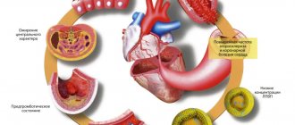
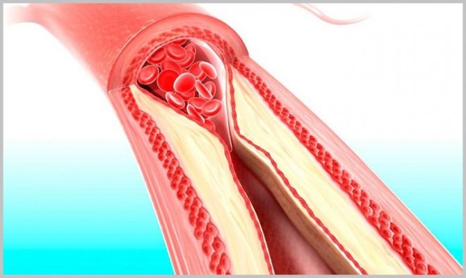
The formation of blood clots in occlusive thrombophlebitis of the deep veins of the legs is an absolute blockage of the venous lumen. In this condition, there is a risk of absolute cessation of blood circulation. Professional medical procedures are urgently required to avoid the progression of further spread of the disease.
The form of non-occlusive thrombosis is caused by the formation of floating thrombi. In such cases, the clots are concentrated at the base of the vessel, as they are a parietal type of thrombus. With this form, blood flow is not impaired, because the free venous area is washed with blood and has no significant obstacles.
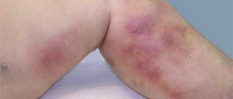
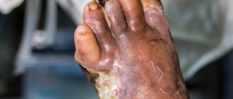
Since most often thrombosis of the sural veins of the lower extremities is asymptomatic, it is rarely possible to establish a diagnosis at the beginning of the disease. Only after a long period of time does thrombophlebitis show its signs. This period is characterized by a change in the color of the skin of the legs in places where blood clots form in the vessels.
Surgical intervention
If conservative measures are not enough, a decision is made on surgical intervention.
- Phlebectomy. The operation involves complete or partial removal of the occluded vessel. This operation is performed under general anesthesia and requires prolonged bed rest and extensive rehabilitation.
- Thrombectomy is the removal of a clot from a vessel by excision of the venous wall. After the thrombus is removed, the vessel cavity is cleaned, treated with an antibacterial solution and sutured.
- Endovascular thrombectomy is a minimally invasive method. It involves removing clots with a catheter while maintaining the integrity of the vein. A balloon catheter is inserted into the incision at the site of thrombus formation, which is filled with saline solution when it comes into contact with the clot, after which the clot is pulled out. The procedure is repeated several times until the vessel is completely cleaned.
Occlusive thrombosis of the veins of the lower extremities: symptoms and treatment
Occlusive thrombosis is a type of blockage of blood vessels when the lumen of the deep veins is completely blocked. This is a fairly serious disease that is asymptomatic in the early stages. It is important to be able to determine the initial stage of the disease in order to begin its treatment in time. If adequate measures are not taken, the consequences of thrombosis can be very serious.
Mixed reasons
High levels of blood characteristics can cause thrombosis in these situations. These include homocysteine; fibrinogen; VIII, IX, XI blood clotting factors.
Important! The appearance of blood clots is very often observed in people who lead a sedentary, “sofa” lifestyle. As a rule, they prefer to travel exclusively by transport and spend a lot of time at the computer.
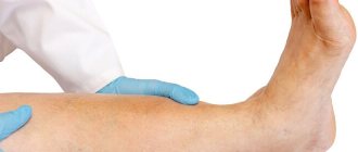
Symptoms
The clinical picture of the disease during the first two days is often vague. The characteristic symptoms of the disease depend on many reasons: the degree of spread through the vessels, stages, type and size of the affected area.
At the initial stage of the disease, the patient's general condition remains satisfactory. Then slight pain in the calf muscles begins to be noted. The next stage is swelling of one or both legs, which continues for several days.
There are also complaints of pain in the lower leg muscles. The pain itself is mild, and patients focus on swelling of the foot.
The initial period of the disease is manifested by the occurrence of severe pain when touching the affected area. The intensity of sensation may vary depending on any particular case.
The clinical picture of occlusive thrombosis turns out to be quite pronounced when all three pairs of deep veins of the leg are filled with blood clots. This is accompanied by very sharp pain, an unpleasant feeling of fullness and tension in the lower extremities, often combined with bluish skin and increased temperature (the onset of thrombophlebitis).
Important! It happens that along with such symptoms, chest pain begins. This may be a clear sign of a serious complication of occlusive thrombosis - pulmonary embolism.
Other symptoms that are quite often present in the patient:
- cramps of the calf muscles at night;
- skin redness;
- swelling and severe heaviness in the legs;
- burning sensation in the affected area;
- high sensitivity in areas where blood clots form, pain when touching the legs.
Occlusive thrombosis most often forms in the veins of the leg. If the necessary measures are not taken in time, the disease begins to gradually spread to other vessels. There are thrombosis of deep and superficial veins of the legs, rarely of the pelvis.
Complex therapy methods
Treatment of occlusive thrombosis must be carried out using conservative methods. The first 3-5 days require bed rest. Anti-inflammatory drugs (Melbek, Olfen) and anticoagulants that combat high blood clotting (Heparin, Clexane, Warfarin) are prescribed without fail.
The treatment process also includes:
- rheological drugs (Tivortin, Trental);
- prostaglandin drugs, for example, Vazaprostan (if necessary);
- glucocorticoids (Methylprednisolone).
Along with the use of medications, it is mandatory to wear compression garments, which, due to their special properties, improve blood flow.
Thrombolysis is performed in the hospital. This procedure is performed using a catheter, when a special substance is injected into the vein cavity to dissolve the formed blood clots.
Severe types of occlusive thrombosis most often require surgical intervention to eliminate thrombosis. During subsequent rehabilitation measures, physiotherapy is prescribed.
With timely therapy, the necessary patency of the veins is restored within six months. If you do not pay due attention to the treatment of the disease, this can lead to amputation of the leg due to the onset of gangrene.
Important! With occlusive thrombosis, approximately 70% of patients become disabled due to chronic venous insufficiency.
Regardless of the reasons for which deep vein thrombosis developed, remember that it must be detected and treated promptly. You need to contact an appropriate qualified specialist for help as soon as possible and unquestioningly follow all his recommendations.
Source: https://vseonogah.ru/zabolevaniya-sosudov/tromboz/okklyuzivnyj.html
Preventive measures
If you are at risk of developing the disease, as well as if you have a history of venous occlusions, patients should follow a number of preventive recommendations:
- Wear special high-compression jersey for a long time.
- Take anticoagulants, antiplatelet agents, phlebotonics in courses during the postoperative period, and sometimes throughout life.
- Regulate your diet, avoid foods that increase blood viscosity or provoke the deposition of cholesterol on the walls of blood vessels.
- Give up harmful addictions: tobacco, alcohol.
- Perform permitted physical exercise regularly.
Symptoms of sural vein thrombosis
Thrombosis of the sural veins is expressed by the following symptoms:
- Pain localized in the limb whose veins are affected by a blood clot. The pain is bursting in nature and occurs in the calf area.
- If you try to press on your leg along the vein, the pain will intensify.
- The pain increases during movement.
- The calf muscle may become hot to the touch. Sometimes a person suffers not only from local hyperthermia, but also from a general increase in body temperature.
- Swelling in the calf muscle area. Moreover, the leg can begin to increase in size literally before our eyes if we are talking about acute thrombosis of the sural veins.
- Increase in size of superficial veins.
It should be taken into account that thrombosis of the sural veins is not always accompanied by all of the above symptoms. Moreover, in 50% of patients, blood finds an additional outlet into the saphenous veins through the system of communicating veins. This allows you to partially normalize blood flow bypassing the affected vein and minimize the symptoms of the pathology. Therefore, he can only know that a person has thrombosis of the sural veins by the developed vascular branches of the blood vessels visible through the skin. They will be located in the area of the lower leg and calf muscles.