Doppler ultrasound, or Doppler ultrasound, is a hardware-based diagnostic research technique that is actively used in neurology. With its help, you can track current indicators of blood flow in shallow veins and arteries, determine the degree of vascular occlusion of extracranial vessels, and also find out whether there is a deficiency of blood supply in the brain.
The technique is based on the Doppler effect, which consists in the fact that ultrasonic vibrations sent by a special transducer to moving blood cells (red blood cells, etc.) are reflected from them, and the frequency of the reflected ultrasonic wave changes depending on the speed of blood flow.
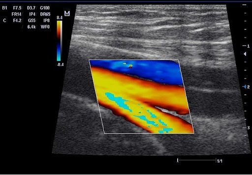
Doppler ultrasound allows you to measure the reflection of sound waves from moving blood particles. The measurement results are subjected to computer processing, after which the doctor receives a two-dimensional color image of the spectrum of the reflected signal, which clearly reflects problem areas with difficulties in blood flow.
The price of the ultrasound examination procedure in St. Petersburg
How much does a vascular scan cost and what does the cost of ultrasound of arteries and veins depend on? Our tariffs for Doppler ultrasound are based on:
- Medical staff experience
- Quality of the equipment used
| Services list | Price |
| Diagnostics | |
| Consultation (appointment) with a neurologist, professor, doctor of medical sciences, primary | 2000 rub. |
| Consultation (appointment) with a neurologist, professor, doctor of medical sciences, repeated (within 30 days) | 1000 rub. |
| Ultrasound Dopplerography of the vessels of the neck and head (USDG) | 2000 rub. |
| Treatment | |
| Massage and manual therapy (1 session) | 1500 rub. |
| Laser therapy (1 session) | 500 rub. |
| Magnetotherapy (1 session) | 500 rub. |
| Acupuncture (1 session) | 1000 rub. |
| Electrical stimulation (or electroanalgesia) (1 session) | 1000 rub. |
| Drug blockade | 1500 rub. |
Ultrasound and Doppler Ultrasound
Alexander Pavlovich Rechmedin
Ultrasound Doctor – Expert Highest category Experience over 20 years. Read reviews
Shmarin Alexey Nikolaevich
Doppler ultrasound and duplex scanning - differences between dopplerography and duplex scanning
The issue of prevention and treatment of vascular diseases remains one of the most pressing today. In the field of angiosurgery, many methods have been developed for the treatment of vascular pathologies, but, as is known, for an angiosurgeon the most important thing in making a diagnosis and prescribing treatment is preliminary ultrasound diagnosis of blood vessels.
Ultrasound is a very common term in the modern world. But, speaking about vascular ultrasound, there are many unclear terms that give rise to misconceptions among patients who are not directly related to them. Several methods are used to diagnose vascular diseases: Doppler ultrasound
(ultrasound Doppler method),
USDS
(ultrasound duplex scanning method) and CDK (color Doppler mapping method). These studies do not harm the body and are considered a safe type of diagnosis.
In this article we will try to figure out how ultrasound ultrasound
from ultrasound, and what is the difference between Doppler ultrasound (
USDG
) and duplex scanning method (
USDS
).
DIAGNOSTICS USING USG METHOD
One of the simple and accessible ways to study venous and arterial patency is Doppler ultrasound. At the heart of ultrasound
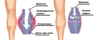
lies the Doppler effect, that is, to obtain the necessary data, changes in reflected sound waves from moving blood cells are recorded. The Doppler ultrasound method is prescribed when it is necessary to determine vascular patency, either to obtain an assessment of blood flow, or to identify pathologies of the venous valve.
When performing Doppler ultrasound, veins and arteries are not visible; the blood flow velocity and patency are assessed based on the obtained Doppler effect values. According to the results of the ultrasound
Headaches, hypertension, and varicose veins are successfully diagnosed. But if identifying impaired vascular patency is done with ease, then eliminating the causes of such pathology using Doppler ultrasound is very difficult. This is due to the fact that this diagnostic method does not allow visualization of the vascular walls and their possible curvatures, which affect the speed and quality of blood flow.
Initially, the images shown by the ultrasound device were made in a flat and thin projection of the organ being examined. Modern diagnostic medicine uses equipment that allows obtaining three-dimensional images of organs in real time and in motion.
The Doppler ultrasound method is one of the most common methods for diagnosing diseases of the arteries and veins. As a rule, ultrasound
prescribed to patients with suspected vascular obstruction or venous valve insufficiency. The Doppler effect, which gave rise to Doppler ultrasound, allows us to understand this pathology. With its help, a change in the reflection of a sound wave from the movement of blood cells is recorded, which makes it possible to subsequently diagnose the speed of blood flow, varicose veins, and identify the cause of headaches and hypertension.
Please note: Ultrasound
easily detects blood flow disorders, but determining its cause is not so easy!
As already mentioned, the USDG
does not visualize the walls of blood vessels and their pathological bends, but they affect the speed of blood flow and its quality.
Preparation and completion of the vascular Doppler ultrasound procedure
During the procedure, the patient must be at rest, lying motionless on the couch. A gel is applied to the desired area and then examined with a special ultrasound sensor. The doctor may ask you to breathe more quickly, hold your breath, or turn your head. It is strictly forbidden to move without a specialist’s request. You can't talk either. The sensor transmits a signal and the results of the procedure are displayed on the monitor.
The procedure itself is completely painless and will not bring you discomfort. But you need to be prepared for the fact that it is quite long - from 20 minutes. up to 1 hour.
Additional preparation for vascular ultrasound is not required
The moving object in Doppler ultrasound is the formed elements of blood. Ultrasound waves are reflected from them, and the sensor, which is stationary during one measurement, picks up the signal. The sensor contains two piezocrystals, thanks to which it is simultaneously a source of ultrasonic waves and their receiver.
The speed of blood flow is calculated using a special formula that takes into account the frequency shift. It is important to note that the calculations take into account both the frequencies of the ultrasonic beams themselves and the angle between the velocity vector of the formed elements and the signal trajectory.
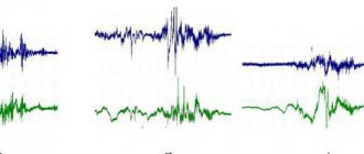
However, in modern medicine, the diagnostician does not have to make calculations independently. The device itself determines the difference in signal frequencies, and then conveniently converts ultrasound into an image. As a result, the doctor sees information presented as an enveloping arc or spectrogram.
Doppler ultrasound does not always work the same way. For example, for an adult patient, transcranial ultrasound (that is, examination of the level of blood supply to the brain) is possible only if a sensor is available. The sensor is used on so-called “acoustic windows”. These are the areas of the head where the cranial bones have the lowest density. Acoustic windows are also found in the area of natural openings. So, they are located in the orbits and in the suboccipital region, in the area of the temporal bones.
Methodology
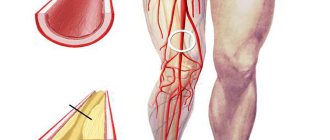
If the doctor sent the patient for ultrasound scanning of the arteries of the lower extremities, then the procedure proceeds as follows:
- the patient stands on a special elevation in front of the device and spreads his legs shoulder-width apart;
- the leg being examined is turned towards the device;
- Using a special sensor, the doctor examines the vessels in the groin area;
- an important point is correct breathing during the procedure: it is during inhalation and exhalation that different colors of blood are visible on the monitor, which characterize the blood flow;
- then the veins and arteries in the calf muscles and under the knees are checked.
When performing an ultrasound scan of the patient’s neck and head, the following manipulations are expected:
- the patient takes a position lying on his back, turning his head away from the device monitor;
- a special conductor substance is applied to the neck area;
- the doctor moves the sensor over the neck and head, carefully studying the condition of the blood vessels on the monitor screen.
There are a number of prerequisites before this procedure:
- one day before the procedure, avoid drinking tea, coffee and alcoholic beverages;
- do not smoke before the procedure;
- in some cases, when taking heart medications, doctors may temporarily stop using them;
- remove any jewelry from your neck and ears.
Ultrasound scanning of the veins of the lower extremities, what is it?
This is an ultra-modern type of diagnostics, which is used to study diseases such as vascular atherosclerosis, varicose veins, thrombosis, etc. The examination takes about 15 minutes and does not require special preparation . As a result, the doctor can assess the quality of the walls of the veins in the legs, identify possible blockages and prescribe appropriate treatment.
UZDS VDA, what is it? This is an examination of the veins of the aortic arch . The results of such a study help to identify disturbances in the functioning of the main cardiac vessel and prescribe comprehensive, timely treatment.
The procedure itself is painless and within 15-20 minutes the doctor can get results about the condition of the aorta.
The entire diagnostic procedure is painless for the patient; the doctor may ask you to change position to get a better picture on the monitor. The duration of the duplex ultrasound procedure takes from 10 minutes to half an hour.
Indications for ultrasound examination
The ultrasound examination procedure is prescribed to patients suffering from:
- headaches and migraines;
- various dizziness, including those associated with sudden turns of the head or changes in body position;
- if you suspect stenosis (narrowing) of the arteries.
It is worth conducting Doppler ultrasound of blood vessels in the presence of symptoms such as:
- noises in the ears and head;
- attacks of general weakness;
- bad feeling;
- “flies” before the eyes;
- “lack” of air;
- vegetative-vascular dystonia.
It will be useful to undergo an ultrasound of the veins and blood vessels in the following cases:
- with constantly elevated cholesterol levels in the body;
- different degrees of obesity;
- arterial hypertension;
- osteochondrosis.
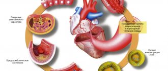
It will help more accurately diagnose the patient’s condition with diabetes, coronary heart disease, angina pectoris and myocardial infarction. Indications for Doppler sonography also include vertebrobasilar circulatory insufficiency, transient ischemic attack, stroke, cerebrovascular disease, numbness of the extremities, and speech impairment.
What is ultrasound examination (ultrasound)?
When it comes to vascular examination, ultrasound usually means duplex scanning - a method that combines Dopplerography and vascular scanning, resulting in a two-dimensional image of the vessel, on which you can see not only pathologies, but also the degree of their development. Another option for ultrasound examination is triplex scanning.
, which allows you to get an even more informative image in color. Unlike Doppler Doppler Ultrasound, these vascular ultrasound methods allow one to obtain not only information about the patency of veins and arteries, but also provide a detailed assessment of their condition.
When is an ultrasound performed?
Ultrasound is an accessible and relatively inexpensive research method, and it is prescribed for diagnosing diseases and pathologies of various kinds, however, ultrasound examination of blood vessels, which is more advanced and more expensive, is usually prescribed for already diagnosed vascular diseases to monitor their treatment. Ultrasound is often prescribed when ultrasound results are unsatisfactory or to clarify the details of the identified pathology. This is the most effective way to identify vascular pathologies of the lower extremities.
How is the ultrasound procedure performed?
The vascular ultrasound procedure does not require prior preparation. It is necessary to refrain from drugs and drinks that affect vascular tone, which can distort the test results. The area being examined is lubricated with gel so that oxygen does not interfere with the scanner’s reading. During an ultrasound, the sonologist moves a sensor over the surface being examined and records the readings in the conclusion. During a duplex study, a two-dimensional image of veins and arteries is obtained, which allows one to judge changes in their structure and find out the cause of problems with vascular patency.
Benefits of Doppler Ultrasound
This research method is painless, has no age restrictions, and there is no radiation exposure. In addition, it belongs to the category of non-invasive: before starting the procedure, there is no need to introduce any special drugs into the body; one external sensor is enough. Even patients in serious condition can undergo such a study. In modern medicine, ultrasound scanning can be part of duplex/triplex scanning, which significantly expands the capabilities of this diagnostic method.
Contraindications for vascular diagnostics
This examination technique is completely harmless to patients, and its high information content allows you to make an accurate diagnosis at an early stage of the development of pathologies and begin the treatment process in a timely manner. The study of blood vessels using Doppler ultrasound is the most effective diagnostic method, which has no side effects or contraindications, and does not expose the patient’s body to radiation.
The ultrasound examination procedure must be performed with the mandatory participation of an experienced neurologist.
Interpretation of results in comparison with the norm
An important stage of any diagnostic method is the correct interpretation of the data obtained, as well as their comparison with the established norm. Only a phlebologist, vascular surgeon or other specialist in this field can evaluate the results of duplex scanning of blood vessels.
Ultrasound scanning of the veins of the lower extremities has a number of criteria by which the data is assessed:
- The diameter of the superficial and deep veins, as well as their patency.
- The degree of pathological obstruction.
- The condition of the venous valves, their performance, the presence of pathologies, the degree of damage to functionality.
- The presence of venous blood clots, their size, qualitative composition, degree of mobility.
- The lumen of blood vessels, the condition of the walls, the degree of deformation, the presence of pathology and inflammatory foci, the thickness of the membranes of blood vessels.
- Pulsation index.
- Vascular resistance.
- General condition of perforating veins.
- The maximum and minimum speed of blood flow in the system.
Vascular duplex scanning (USDS) is a multifunctional and extensive diagnostic study that operates on many data. With its help, doctors have the opportunity to thoroughly study all the details of the functioning of the circulatory system in the lower extremities of each patient.
The main task of such diagnostics is to identify the development of a pathological process or confirm the diagnosis.
Experts have identified certain indicators (standards) that need to be guided by when interpreting the data obtained during the study:
- The ankle-brachial index is the norm of at least 0.9. Shows blood pressure levels in the shoulder and ankle. The index determines the difference between two indicators. If the index value drops to 0.3 or less, this indicates the presence of pathology.
- The speed of blood flow in the femoral joint is 100 cm/sec.
- The speed of blood flow in the lower leg is 50 cm/sec.
- The resistance of the femoral artery is 2 m/sec.
- The pulsation index of the greater tibial artery is normal 1.8 m/sec.
- The type of blood flow in large vessels is normal - mainline. Any changes in the system current indicate the presence of restrictions. Thus, turbulent blood flow is characteristic of stenosis. The collateral type indicates a complete absence of blood flow in a certain place.
- Pulsation index is normal 180 m/sec or higher. This parameter indicates the state of the vascular lumen.
Regardless of the results of the study, it is important to consult with your doctor. The specialist will make a diagnosis and prescribe therapy to the required extent.
If the disease is detected in the initial stage, then physiotherapeutic procedures and medications will be prescribed that can quickly eliminate the pathology and restore the functionality of the patient’s veins and vessels.
If more complex treatment or surgical intervention is required, then a clear strategy will be drawn up, strict adherence to which will help to avoid all sorts of complications.