Research Center
Contrasting coronary vessels is the most reliable method for choosing treatment tactics for patients with myocardial ischemia. Complications with this procedure are quite rare.
Diagnostics involves the insertion of a catheter into the vessels of the heart and the supply of a contrast agent through it, so it can pose a potential danger to the patient.
To prevent undesirable consequences, careful examination and preparation is necessary.
Risks of coronary angiography for the patient
Since diagnosing coronary blood flow involves puncturing a peripheral artery of the thigh or shoulder, inserting a catheter through it, moving it through the aorta and coronary vessels, and supplying a contrast iodine-containing substance, this may be accompanied by a negative reaction of the body.
The risk of complications increases if the patient suffers from:
- arterial hypertension;
- diabetes mellitus;
- widespread atherosclerosis;
- acute coronary insufficiency;
- tendency to allergic reactions;
- kidney pathology;
- weakness of the contractile function of the heart;
- rhythm disturbance;
- obesity or underweight;
- chronic alcoholism;
- systemic vasculitis;
- infectious disease.
Depending on the stage of coronary angiography, it can cause the following complications:
- puncture of a peripheral artery - bleeding, hematoma, false aneurysm, fistula between artery and vein, wall dissection, thrombosis, embolism, vascular spasm, infection, allergy to painkillers;
- contrast – allergy, anaphylaxis, intoxication, kidney damage;
- administration of heparin - a decrease in blood clotting ability and, as a result, bleeding;
- insertion of a catheter - arrhythmia, embolism with parts of a cholesterol plaque, dissection of the aorta or coronary vessels, heart attack, stroke.
We recommend reading about coronary angiography of the heart vessels. You will learn about indications and contraindications for the procedure, preparation and performance of coronary angiography, recommendations during the recovery period.
And more about bypass surgery of the heart vessels.
Possible complications after vascular reconstruction through the arm
The incidence of adverse effects of coronary angiography ranges from 0.05% (severe rhythm disturbances, vascular accidents) to 20 - 40% (allergy and violations of the integrity of the vascular walls). The occurrence of complications may depend on the presence of background predisposing factors or be a consequence of a violation of the procedure technique.
Air embolism
It happens in 0.2% of cases, it is associated with the passage of air bubbles into the bloodstream. Occurs when the integrity of the balloon at the end of the catheter or other technical errors in catheterization are violated.
With coronary angiography, air emboli are visible during the period of contrasting the coronary arteries. There are no symptoms, or pain in the heart appears, pressure drops, the rhythm of contractions is lost, until the heart stops.
Treatment involves aspiration (removal) of bubbles from vessels or injection of a solution under pressure to crush them. Patients are prescribed oxygen therapy, pain relief and antiarrhythmic drugs.
Hematoma and other vascular complications
The appearance of visible swelling of tissues and thickening of the skin at the site of arterial puncture occurs quite often. A large accumulation of blood in the hematoma cavity can lead to:
- pain,
- increased heart rate,
- bleeding,
- compression of nerve endings,
- deep vein thrombosis,
- blockage of the pulmonary artery.
Hematoma at the puncture site after coronary angiography
For prevention, you need to carefully apply a pressure bandage, increase the time spent on bed rest after the procedure, and use heparin in smaller doses. Treatment is carried out using sufficient compression of the punctured vessel or by surgery.
With a false aneurysm, blood passes through a defect in the arterial wall during heart contractions into the hematoma cavity, and partially returns back during diastole. Occurs when the puncture is incorrect or there is insufficient compression of the artery after removing the catheter. Manifests:
- swelling of tissue at the site of puncture of the vessel;
- the formation of a hematoma that pulsates in time with the rhythm of the heart;
- pain on palpation;
- noise on auscultation.
The pseudoaneurysm may rupture, causing severe pain and increasing swelling. If compression of the nerve fibers occurs, weakness in the limb persists for several months. Patients are advised to rest and discontinue medications that inhibit blood clotting. For large aneurysms, thrombin is injected into the cavity or surgical removal is performed.
Pseudoaneurysm of the heart on echocardiography
Less common complications of vascular puncture are the formation of an arteriovenous fistula with edema, deep vein thrombosis and limb ischemia. There is also dissection of the arterial wall against the background of atherosclerotic changes. The occurrence of arterial blockage is possible in patients with thin vessels, concomitant obliterating diseases, aneurysms or a blood clot in the heart cavity.
Heart attack
The causes of acute coronary blood flow disturbance may be:
- formation of a blood clot at the top of the catheter;
- destruction of atherosclerotic plaque and movement of its parts along the artery;
- prolonged blocking of blood flow with a catheter at the site of narrowing of the vessel;
- dissection of the coronary artery;
- blockage of the branch when advancing the catheter to the bifurcation site;
- spasm in response to mechanical irritation of the vascular wall.
It is important to note that coronary angiography does not always cause the classic ECG picture of myocardial infarction (less than 1% of cases), but when studying specific enzymes, an increase in their level is observed in approximately 5 - 40% of patients. At the same time, asymptomatic and atypical cases of the disease are often found.
Arrhythmia
Life-threatening rhythm disturbances and cardiac impulse conduction during coronary angiography include ventricular tachycardia and ventricular fibrillation. They are associated with changes in blood flow during the procedure and injury to the conduction system of the heart.
In most patients, restoration of normal rhythm occurs independently at the end of the study. In cases of severe circulatory impairment due to arrhythmia, electrical pulse therapy is indicated. To prevent this complication, patients with electrical instability of the myocardium are prescribed beta-blockers.
Stroke
Factors that can lead to a stroke include:
- blockage by a blood clot formed due to arterial injury;
- dissection of the aortic walls;
- cholesterol embolus;
- arterial hypotension;
- administration of heparin (provokes intracerebral hemorrhage).
More often, this complication occurs in cerebral atherosclerosis, in elderly and senile patients who have had transient attacks in the past, cerebral ischemia, or have suffered a stroke. Manifestations of acute disturbance of blood flow in the brain include:
- weakness in the limbs,
- nausea and vomiting,
- headache,
- speech disorder,
- dizziness,
- convulsions,
- mental disorder.
Nephropathy
For 1 to 3 days after using the contrast agent, the excretory capacity of the kidneys may be impaired. Nephropathy occurs more often in the following conditions:
- elderly and senile age;
- kidney disease in the past;
- diabetes;
- dehydration;
- shock or vascular collapse;
- circulatory failure with congestive processes in the internal organs;
- myocardial infarction;
- anemia;
- the use of drugs that destroy the renal parenchyma (non-steroidal anti-inflammatory drugs, antibiotics from the aminoglycoside group);
- injection of a large amount of contrast agent or rotary contrast.
Kidney damage can be reversible, but a third of patients develop renal failure. To prevent it, you need to take 0.5 liters of water before coronary angiography and at least 2.5 liters after.
In heart failure and edema syndrome, the amount of fluid is determined by daily diuresis.
Patients with reduced glomerular filtration may require hemofiltration to prevent nephropathy.
How to avoid complications
It is possible to prevent negative consequences with careful selection of patients for diagnosis. It is contraindicated in the presence of:
- severe renal and heart failure;
- arrhythmias with the threat of developing ventricular fibrillation or complete atrioventricular block;
- decompensated diabetes mellitus;
- bacterial endocarditis;
- malignant course of hypertension or symptomatic hypertension;
- allergic reactions and drug intolerance to iodine-containing drugs;
- discirculatory encephalopathy, stroke with a persistent neurological defect;
- obliterating lesions of the limbs;
- acute period of myocardial infarction;
- exacerbation of diseases of internal organs;
- infectious process.
Ultrasound of the heart and peripheral vessels
To identify these diseases, a prerequisite is preparation for coronary angiography, which includes assessment of the functional class of angina pectoris and heart failure, collection of information about concomitant diseases and previous vascular accidents. Patients are shown:
- ECG in 24-hour Holter monitoring mode;
- Ultrasound of the heart and peripheral vessels, kidneys;
- chest x-ray;
- general clinical blood and urine tests;
- blood test for viral hepatitis, HIV and syphilis, coagulogram, electrolyte composition, kidney tests, cardiac-specific enzymes, lipid profile.
We recommend reading about cardiac catheterization. You will learn about the indications and contraindications for cardiac catheterization, features of the procedure, as well as possible complications and the cost of the procedure.
And more about CT angiography.
Coronary angiography is an invasive test, so complications may include damage to the vessels through which the heart is accessed. Adverse consequences also include thromboembolic complications, myocardial infarction and stroke, kidney damage, and heart rhythm disturbances.
To prevent negative reactions, a thorough examination is required before prescribing a contrast procedure for the coronary arteries of the heart.
Watch the video about errors during CT coronary angiography:
Source: https://pskov-is.ru/bolit-ruka-posle-koronarografii/
Complications of the procedure
In practice, complications are extremely rare - no more than 1%. The literature reports 0.19 to 0.99% complications following this study.
- Bleeding and reapplying a pressure bandage. After the examination, the doctor who performed the procedure will definitely come to you. He will come in as often as the situation requires.
- Allergic reactions to contrast. There may be nausea, vomiting, and rash. Problems go away on their own, or allergy shots are prescribed.
- Myocardial infarction, arrhythmias, heart pain – no more than 0.05%. A loved one is allowed to be in the room next to the patient. Two doctors will be required to observe: the department doctor and the doctor who performed the coronary angiography. Such complications will be diagnosed in time.
- Contrast-induced nephropathy (acute kidney damage) is accompanied by a short-term increase in creatinine in the blood due to contrast. Creatinine is a product of protein metabolism and an important indicator of kidney function. The contrast is removed within 24 hours without harm to the kidneys.
- Perforation and rupture of the coronary artery. Occurs in 0.22% of patients. This complication develops in patients with advanced atherosclerosis of the coronary arteries. (Emergency Medicine Practice Journal, 2014). In more than 99% of patients, the complication can be eliminated on the operating table.
Why does my arm hurt after coronary angiography?
hello, I’m 33 years old, the first time this started was 3 years ago, I started having a strong heartbeat, a pre-fainting state, high blood pressure and shaking a lot, this happened again a year later for no reason, and after that it became regular once a month or once every 2 weeks, I didn’t complain about my heart before, but now pain appeared in the left shoulder, arm under the shoulder blade and the left leg is pulling, colitis in the area of the heart, I don’t know how to correctly say walking pains, only pain in the chest, after a few minutes the shoulder already hurts.
Definition
Coronary angiography is a procedure that uses radiography to see the blood vessels of your heart. Coronary angiography is part of a general group of procedures known as cardiac catheterization.
Cardiac catheterization procedures can both diagnose and treat diseases of the heart and blood vessels. Coronary angiography, which can help diagnose heart disease, is the most common type of heart catheter procedure.
During a coronary angiography, a type of dye that is visible on an X-ray machine is injected into the blood vessels of your heart. The X-ray machine quickly takes a series of images (angiography), offering a detailed look at the inside of the blood vessels. If necessary, the doctor may perform procedures such as angioplasty during a coronary angiography.
Why is this being done
Your doctor may recommend that you have a coronary angiography if you have:
- Symptoms of coronary artery disease, such as chest pain (angina)
- Chest, jaw, neck, or arm pain that cannot be explained by other tests
- New or increasing chest pain (unstable angina)
- Heart defect you were born with (congenital heart defect)
- Heart failure
- Other blood vessel problems or chest injury
- Heart valve problem that requires surgery
You may also need an angiogram if you are about to have non-heart surgery, but you are at high risk of having a heart problem that occurs during surgery.
Because there is a small risk of complications, angiography is usually done after non-invasive heart tests have been performed, such as an ECG, echocardiography or stress test.
Risks
Coronary angiography. During the procedure.
Like most procedures performed on the heart and blood vessels, coronary angiography has some risks. Serious complications are rare, though. Potential risks and complications include:
- Heart attack
- Move
- Trauma arterial catheterization
- Heart rhythm disturbances (arrhythmias)
- Allergic reactions to paint or drugs used during the procedure
- Tears in your heart or artery
- Kidney damage
- Excessive bleeding
- Infection
- Blood clots
- Exposure to radiation from X-rays
How do you prepare
In some cases, coronary angiography is performed as an emergency. More often, however, they are planned in advance, giving you time to prepare.
Angiograms are performed in the catheterization (cathode) laboratory of the hospital. You usually go to the hospital on the morning of the procedure. Your health care team will give you specific instructions and talk with you about any medications you are taking. General recommendations include:
- Do not eat or drink after midnight the day before your angiography. Angiograms will often continue into the morning hours.
- Take all your medications to the hospital with you - in their original bottles. Ask your doctor if you should take your regular medications in the morning.
- If you have diabetes, ask your doctor whether you should take insulin or other oral medications before the angiography.
Before the angiogram procedure begins, the team's doctor will review your medical history, including allergies and medications you are taking.
The team may perform a physical examination and check vital signs such as blood pressure and pulse. You will empty your bladder and change into a hospital gown.
You may need to remove contact lenses, glasses, jewelry, and hair clips.
What can you expect
During the procedure
For the procedure, you lie on your back on the x-ray table. Because the table may be tilted during the procedure, seat belts may be attached to the chest and legs. X-ray cameras can move through and around your head and chest to take pictures from different angles.
An intravenous (IV) line is inserted into a vein in the arm. You may be given a sedative through an IV to help you relax, as well as other medications and fluids. You will be awake during the procedure so you can follow the instructions. Throughout the procedure, you may be asked to take deep breaths, hold your breath, cough, or place your arms in various positions.
Electrodes on your chest monitor your heart throughout the procedure. A blood pressure cuff monitors your blood pressure and another device, a pulse oximeter, measures the amount of oxygen in your blood. You may be given drugs (anticoagulants) to help prevent your blood from clotting at the catheter and in your coronary arteries.
A small amount of hair may be shaved from the groin or arm where the catheter is to be inserted. The area is washed and disinfected, and then numbed with an injection of local anesthetic. A small cut is made at the entry site and a short plastic tube (sheath) is inserted into the artery. The catheter is inserted through the sheath into the blood vessel and carefully threaded to the heart or coronary arteries.
Inserting the catheter should not be painful, and you will not feel it moving through your body. Tell your doctor if you have any discomfort.
Dye (contrast agent) is injected through the catheter. When this happens, you may have a brief flushing or warm sensation. Don't be alarmed if you feel your heart skipping beats - this is a common occurrence during an angiography. But again, what is your doctor's command if you feel pain or discomfort.
Definition. After the procedure.
The dye is easy to see on x-rays, and as they move through the blood vessels, your doctor can observe its flow and identify any blockages or narrowing in the area.
Depending on what your doctor finds during the angiography, you may have additional catheter procedures at the same time, such as balloon angioplasty or stenting, to open up the narrowed artery.
Having an angiogram takes about one hour, although it can be longer, especially when combined with other cardiac catheter procedures. You can add a few more hours to the preparation and post-care procedure.
After the procedure
When the angiography is complete, the catheter is removed from your arm or groin and the incision is closed with manual pressure, a clamp, or a small plug.
You will be taken to a recovery area for surveillance and monitoring. When your condition is stable, you will return to your room, where you will be monitored regularly. You will need to lie down for several hours to avoid bleeding. During this time, pressure can be applied to the incision to prevent bleeding and promote healing.
Sometimes, the plastic sheath that was first inserted into the blood vessel remains in place for several hours or even overnight if you have had angioplasty or stenting. If you received anticoagulants during the procedure, removing the sheath too soon may cause severe bleeding.
You may be able to go home the same day, or you may have to stay in the hospital for a day or longer. Drink plenty of fluids to help flush the dye from your body.
If you feel for her, there is something.
Ask your healthcare provider when you should resume taking your medications, bathing or showering, returning to work, and resuming normal activities.
Call your doctor if:
- You notice bleeding, new bruising, or swelling at the catheter site
- You develop increasing pain or discomfort in the catheter
- You have signs of infection such as redness, drainage, or fever
- There is a change in temperature or color of the leg or arm that was used for this procedure
- You will feel weak or weak
- You develop chest pain or shortness of breath
If the catheter site actively begins bleeding or swelling, apply pressure to the site, and contact emergency medical services.
results
Angiography can show doctors what's wrong with your blood vessels. He can:
- Show how many of your coronary arteries are blocked or narrowed by fatty plaques (atherosclerosis)
- Determining where blockages are located in blood vessels
- Show how much blood flow is blocked through your blood vessels
- Check the results of previous coronary artery bypass surgery
- Check blood flow through the heart and blood vessels
Knowing this information can help your doctor determine which treatment is best for you and how much of a risk your heart poses to your health.
Depending on your results, your doctor may decide, for example, that you would benefit from having coronary angioplasty to help unblock blocked arteries.
It is also possible that angioplasty or stenting may be done during the angiography to avoid the need for another procedure.
Source: https://remson58.ru/posle-koronarografii-bolit-ruka-delat/
Coronary angiography of the heart vessels - dangerous and non-dangerous consequences
Coronary heart disease (CHD) is the absolute leader in the world in the number of deaths. IHD is diagnosed when the blood supply to the heart is partially or completely disrupted due to damage to the coronary arteries. Most often, IHD is caused by progressive atherosclerosis, which impairs the patency of blood vessels.
Research methods
In the arsenal of modern medicine there are various methods for intravital study of the blood vessels of the human heart.
The most informative include:
- ultrasound Dopplerography of blood vessels (USDG),
- Cardiography of cardiac vessels with contrast agent,
- magnetic resonance imaging (MRI),
- angiography of the blood vessels of the heart,
- MSCT of coronary vessels (with and without contrast).
Cardiac ultrasound (US) is the basis of both Dopplerography and cardiography. MRI is a scan of blood vessels using a magnetic field and radiofrequency pulses. The essence of angiography is a contrast X-ray examination of the vessels of the heart. MSCT examination is carried out using a multislice computed tomograph.
The method is part of angiography. It got its name because it can be used to study the coronary vessels of the heart. In the medical literature you can find another name - coronary angiography.
Coronary angiography is often used in ischemic heart disease because it has earned a reputation as a reliable test of blood vessels in this disease.
In this regard, many heart patients and their relatives have a well-founded interest in how coronary angiography of the affected vessels is performed in coronary heart disease. Of interest are the possible negative consequences that such diagnostics of the arterial vessels of a diseased heart may have on human health.
Coronary angiography consists of two stages:
- preparatory,
- diagnostic procedure.
Preparation
The doctor must tell the person undergoing coronary angiography about the purposes of diagnosis, the procedure, and possible complications. The patient must inform the doctor about all diseases.
- The patient is given an electrocardiogram (ECG).
- Blood tests required:
- general,
- biochemical,
- for clotting,
- for the presence of a number of infections (HIV, hepatitis B and C, syphilis).
Be sure to do tests to determine the tolerability of the X-ray contrast agent and medications used in the procedure.
Contraindications
For a number of diseases, coronary angiography cannot be done:
- Contraindicated in people with uncontrolled hypertension, in whom stress during a coronary angiography procedure can cause a hypertensive crisis.
- Not performed after a recent stroke to avoid recurrent brain damage.
- Another prohibition is associated with decompensated diabetes mellitus, when there is serious damage to internal organs and the possibility of a heart attack cannot be ruled out.
- Internal bleeding or very low blood clotting is another reason to avoid coronary angiography.
- Kidney damage due to various diseases does not allow for coronary angiography, since a sharp deterioration in the patient’s condition is possible after the administration of an X-ray contrast agent.
- High temperature also makes coronary angiography impossible.
- Intolerance to the substance used for contrast during the procedure.
Procedure
Coronary angiography is performed on an outpatient or inpatient basis in the cardiology ward of a hospital.
- It is done on an empty stomach, before it you need to go to the toilet to empty your intestines and bladder.
- The places where the puncture of the vessel is punctured (wrist, armpit, groin, etc.) are shaved.
- In addition to the surgeon performing the operation, a resuscitator and an anesthesiologist are present in the room.
- Before the procedure, the patient takes a sedative so as not to worry too much and keep the heartbeat normal.
- During the operation, the patient lies on the operating table (on his back), his body is fixed so that damage to the vessel does not occur as a result of involuntary movement.
- After applying local anesthesia, a puncture of the vessel is made, through which access to the coronary arteries will be provided.
- An introducer plastic tube is inserted into the puncture site. It has a built-in hemostatic valve to prevent backflow of blood, an additional channel for collecting blood for analysis and administering medications.
- Through the introducer, the surgeon inserts a catheter, which is advanced into the area of the artery to be examined.
- After reaching the required position, a radiopaque agent containing iodine isotopes is injected by the catheter.
- The computer on the monitor shows a shadow image of a vessel in which a radiopaque substance is present.
- The study is done from several angles in order to obtain maximum information about the condition of the vessel or vessels of the heart.
- The results of the examination are recorded on digital media.
- After completing the procedure, the surgeon removes the catheter and introducer from the patient's circulatory system and applies a special bandage to the puncture site to stop the bleeding.
Depending on the scope of the study, the duration of the procedure ranges from 20 minutes to an hour.
Possible complications
Modern high-tech methods for studying heart vessels are quite safe. However, coronary angiography of the heart vessels can also have undesirable consequences, since the human body is complex, and it is impossible to foresee and calculate absolutely everything, even with an experienced surgeon and advanced medical equipment.
The cardiovascular system
The most severe consequences for the heart, brain and blood vessels are:
- myocardial infarction,
- stroke,
- perforation of blood vessels or heart cavity.
The probability of a heart attack is estimated at 1:1000. The risk of heart attack during or after coronary angiography is higher in patients with severe lesions of the coronary arteries.
Lower chance of stroke (7 in 10,000). It can occur in a patient if the movement of blood to the brain is blocked by a blood clot, cholesterol plaque, or air.
In 3-6 cases out of 1000, perforation or dissection of the coronary vessels or aorta is possible. The probability of injury to the iliac or femoral artery is estimated at 4:1000.
Vascular injuries are dangerous because retroperitoneal bleeding can occur, in which blood gradually accumulates in the retroperitoneal space. Moreover, blood loss occurs without visible external manifestations.
Complications that do not pose a direct threat to human life are more common.
In people suffering from diabetes mellitus with narrow lumens of blood vessels in the leg, when introducing an introducer and catheter that does not match the size of the vessel, thrombosis of the vessel of the lower extremity may develop. In this case, additional treatment will be required, either surgery to remove the blood clot or drug therapy.
If an artery and a vein are simultaneously damaged by a needle, an arteriovenous fistula can form. Probability 1:100. To eliminate it, surgery is required.
Hematomas often occur at the puncture site. If they are small in size, they will dissolve on their own.
If the hematoma is large in size, it can connect to the lumen of the artery, which leads to the appearance of a false aneurysm of the vessel. In most cases, surgery is not required.
Possible heart rhythm disturbances during diagnosis. More often the rhythm decreases (bradycardia). Less common are cases of increased heart rate (tachycardia) and uneven rhythm (arrhythmia).
Another common complication is a drop in blood pressure, which can be caused by various reasons related to the functioning of the cardiovascular system.
Allergic reactions
It is possible that an allergy may develop in response to the introduction of a radiopaque substance, sedatives, anticoagulants or antiplatelet agents, or anesthetics into the body. Therefore, preparation for coronary angiography includes careful checking of the patient's response to all drugs intended for the procedure.
If the check is not done properly, anaphylactic shock is possible, which threatens the patient's life. This happens extremely rarely, but more often a skin reaction is observed (rash, itching, redness).
Kidneys
An organ that may be damaged during coronary angiography.
In people with chronic renal failure, diabetes, or advanced age, the kidneys may not respond well to X-ray contrast agents.
Acute renal failure may develop. Severe complications require medical attention; for minor dysfunction, drinking plenty of fluids after coronary angiography is recommended.
Respiratory system
The most serious consequence is pulmonary edema. May develop due to heart failure and a severe allergic reaction. The risk of pulmonary edema is low, especially with careful preparation.
Thrombocytopenia
During coronary angiography, heparin is used, which reduces blood clotting. After a few days, thrombocytopenia may develop, triggered by heparin. Thrombocytopenia is a pathology characterized by a low number of platelets in the blood and increased bleeding.
Infections
Pathogenic pathogens enter the patient’s body at the site of puncture of the vessel.
To reduce the likelihood of infection, it is better to use an electric razor for shaving before diagnosis rather than razor blades, which can leave small scratches.
Strict compliance with hygiene requirements in the operating room is mandatory for medical staff.
After diagnosis, the puncture site should not be wetted with water for at least two days.
The easiest form of coronary angiography is considered to be when the catheter is inserted through the radial artery. If there are no complications, the person returns home within a few hours.
When access is made through the femoral artery, the patient remains in the hospital for a day.
A person who has undergone coronary angiography should monitor their well-being. If alarming symptoms appear (pain, weakness, low blood pressure, swelling at the puncture site), you should consult a doctor, and not expect that it will go away on its own, and not self-medicate.
Additional information about coronary angiography can be obtained from the video:
You can learn more about pathologies of the coronary arteries from the video:
Loading…
Source: https://dlja-pohudenija.ru/serdcze/posledstviya-koronarografii-sosudov-serdcza-metody-issledovaniya-i-vozmozhnye-oslozhneniya
How is the procedure done?
We will consider the progress of the entire procedure of coronary angiography of the heart vessels “from the patient’s side.”
Hospitalization and preparation
The patient enters the department in the evening or comes in the morning at the appointed time for the study. He should have blood tests on hand (the doctor will specify which ones), electrocardiography and the results of an ultrasound of the heart.
In the reception department or in the ward, the patient will receive an information consent, which must be signed (unless you change your mind about undergoing the study). Coronary angiography is performed on an empty stomach, the duration of the entire procedure is from 30 minutes to 2 hours. The patient is discharged the next day. In the morning before discharge, all tests will be taken.
This procedure can be carried out in two ways (we are talking about a standard planned diagnostic method): through the vessels of the arm and through the femoral artery.
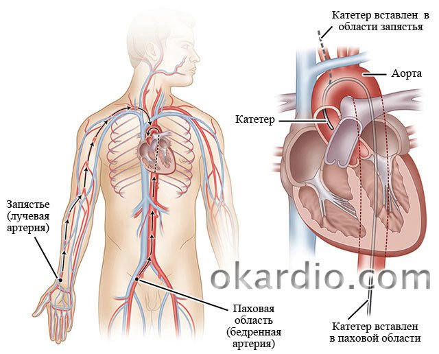
Methods for inserting a catheter for coronary angiography of the heart vessels
Before coronary angiography, an injection (premedication) will be given to relieve nervous tension.
Usually the patient is conscious during the study and communicates with the doctor. In rare cases, it is necessary to put the patient into a state of medicated sleep - then an anesthesiologist will be present for the study.
What happens in the operating room itself?
- In both cases, local anesthesia (lidocaine and other means) is initially given.
- A vessel is pierced on the thigh or arm, and a catheter or tube is inserted into the vessel. Initially, you need to reach the ostia of the coronary artery (this is where the coronary artery exits the aorta).

The surgeon inserts a tube into the patient's right arm vessel. Click on the photo to see it clearly - The doctor inserts a catheter directly into the mouth of the coronary arteries. At the other end (where they entered through the skin), a syringe with contrast is attached to the catheter. So they introduce him. The contrast fills the heart arteries and is washed away by the blood flow. The entire procedure is video recorded. The doctor monitors the progress of the process on the screen. The monitor can be rotated so that the patient can also see his own arteries. You will be able to talk with the doctor.

The surgeon injects contrast from a syringe through the catheter. Click on the photo to see it clearly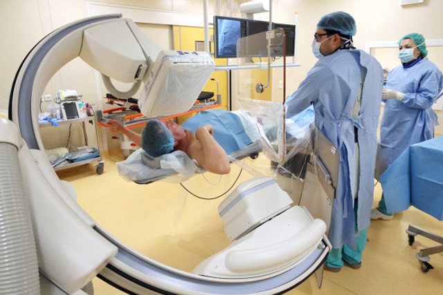
The doctor watches the progress of the process on the screen - After the procedure is completed, the doctor applies physical pressure with his hands to the puncture area. This is necessary to stop the bleeding.
- Then a sterile pressure (very tight) bandage is applied and the patient is transferred to the ward.
After the procedure, the surgeon applies a tight bandage to the patient. Click on the photo to see it clearly
After coronary angiography
The patient is not recommended to get out of bed for 5 to 10 hours. This difference is understandable - after all, some patients take medications that thin the blood. And not in all cases it is possible to cancel them before the procedure.
You can eat immediately after the procedure. A surgeon will come into the room to discuss all the nuances of the study.
The recording of the coronary angiography procedure is carefully and repeatedly studied and analyzed by doctors. A copy of the video will be given to you immediately in the operating room.
If there are no complications, the patient is discharged the next day. You can start working in a day.