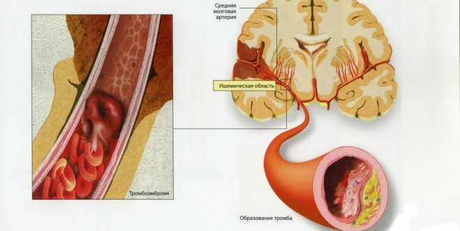
The disease is a type of necrosis of cerebral tissue. A distinctive feature of this type of pathological condition is its deep location in the vessels of the brain and its small diameter, which does not exceed 2 cm. The ischemic focus formed during a heart attack over time transforms into a lacuna - a cavity. This circumstance later determined the name of the disease.
In 1838, the term “lacunar infarction” was introduced by the French physician Decambre. By this he meant lesions in the gray matter of the brain that resemble areas of softening, similar to cavities. The emergence of accurate diagnostic techniques, such as MRI and CT, has made it possible to thoroughly study this type of necrosis and supplement information about it.
Lacunar cerebral infarction has some features:
- According to statistics, it accounts for 15% of all types of heart attacks;
- Elderly people suffering from arterial hypertension are more susceptible to it;
- 100% detection of pathological health is possible only with the help of an autopsy;
- The vessels affected by lacunar infarction have an average diameter of 40–900 microns and are located in the deep part of the brain.
Among the main causes of this type of heart attack are:
- Arterial hypertension both in case of resistance and in case of hypertensive crisis;
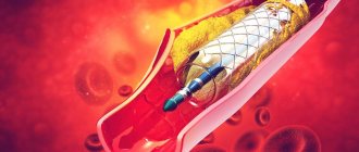
- Atherosclerotic damage to brain cells;
- Congenital pathology or defect in the development of cerebral vessels;
- Heart rhythm disorders such as atrial fibrillation;
- Diabetes mellitus, which can lead to diabetic angiopathy of cerebral vessels;
- Smoking, which promotes blood viscosity and predisposes the tubular formations of the circulatory system to spasm and narrowing.
- Lacunar cerebral infarction goes through several stages:
- Acute, which is 3 days;
- Acute, for up to 28 days;
- Early recovery, which is 6 months after a heart attack;
- Late rehabilitation, which lasts about 2 years.
This division into periods is not mandatory. Therefore, this classification is considered conditional.
Basic treatment tactics
Drug treatment depends on the severity of the disease.
To prevent the formation of blood clots, the patient is often prescribed aspirin (acetylsalicylic acid). The following medications may also be prescribed:
- To restore blood microcirculation, Nicergoline can be prescribed, for example, in the form of injections with further transfer to treatment with tablets.
- For cardioembolic cerebral infarction, Warfarin is often prescribed.
- Neurotrophics help eliminate oxygen starvation of the brain and restore its affected structures. These include: Cerebrolysin, Actovegin.
- In case of development of dementia, anticholinesterase drugs are prescribed: Proserin, Neuromidin.
- For parkinsonism, medications are prescribed to reduce hand tremors, for example, Cyclodol.
- Since cerebral infarction most often develops against the background of high blood pressure, the doctor prescribes appropriate medications to stabilize it, taking into account concomitant diseases, for example, diabetes or kidney disease. To treat arterial hypertension, you should not take strong medications, especially this note applies to elderly patients. The decrease in blood pressure should be gradual.
The rehabilitation period after a cerebral infarction depends on the severity of the disease and usually lasts from six months to two years. During this period, care and creation of a healthy atmosphere for the patient by relatives is important.
A patient after an ischemic stroke must adhere to the following actions:
- Compliance with a special anti-cholesterol diet.
- Vigorous physical activity.
- Rejection of bad habits.
- Rehabilitation includes following your doctor's recommendations for treating high blood pressure. In addition, if there is a pronounced risk of embolism, the patient is prescribed drugs that make the blood more fluid.
Thus, lacunar cerebral infarction is a disease of a somewhat milder form in its consequences than a major stroke. However, it requires urgent hospitalization and examination, especially in case of relapse. Almost complete restoration of all body functions is possible, subject to appropriate treatment and compliance with preventive measures.
Prognosis and prevention
The future condition of the patient after suffering a lacunar infarction depends on many factors. They consist of:
- Age of the patient;
- Presence of concomitant diseases;
- Timeliness of treatment provided;
- Extension of the affected area.
A single lacuna after a heart attack raises favorable expectations from specialists. This means that the patient will gradually, with the course of rehabilitation measures, return lost functions. They may not return fully, however, most of them will return to normal. Despite this, residual effects may remain.
There is no specific prevention against heart attack. There is no vaccine or special pill that will cure a person from a heart attack. However, there are a number of measures that will help prevent the pathological process. It is important to understand that there is a risk group of people predisposed to the disease. Knowing this, it is possible to try to avoid the occurrence of lacunar infarction.
The main trigger mechanism, that is, the provoking factor of necrosis, is an unfavorable lifestyle, stress, constant load on the target organs, which are the brain, vasculature and heart. The most accessible and optimal medication for preventing heart attacks is Aspirin. This drug, known to everyone, has a very important property - antiplatelet.
Its use reduces the likelihood of a recurrence of a heart attack many times over. Therefore, often, Aspirin is the first drug that an ambulance gives to a patient who has suffered a heart attack. The patient is asked to chew 250 mg of the drug at a time, and then prescribed for a long period. You need to take the medicine only in its pure form. But besides this, you need to change your lifestyle. This includes the following principles:
- Follow a diet that does not contain foods high in sugar and cholesterol;
- Monitoring blood pressure and its correction if its indicators change from the norm;
- Abstinence from bad habits such as alcohol and smoking;
- Reduce stress factors, fatigue and stress;
- Don't forget about physical activity;
- Contact a doctor if alarming symptoms appear.
Author of the article: Neurologist of the highest category Tatyana Mikhailovna Shenyuk.
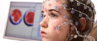
Diagnosis of cerebral infarction
The doctor may suspect a lacunar cerebral infarction based on the patient’s complaints. But a complete picture of the disease can only be obtained using the following research methods:
- First of all, this is computed tomography, which allows you to obtain a virtual multi-level slice of the brain;
- General laboratory analysis of blood and urine, as well as analysis of blood biochemistry, the presence of glucose in it, determination of cholesterol levels, coagulation indicators;
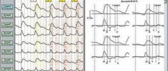
- ECG to detect heart rhythm disturbances;
- Ultrasound duplex scanning allows you to analyze the condition of the vessel walls, the presence of cholesterol plaques on them, and congenital defects.
Diseases that increase the risk of cerebral infarction
The following causes of lacunar cerebral infarction are distinguished:
- Constant high blood pressure, chronic hypertension. The disease can occur both against the background of arterial crises and with stable indicators.
- Atherosclerosis of the cerebral arteries. It can occur as a result of an improper diet containing animal fats. With this disease, plaques form on the vascular walls, obstructing the flow of blood.
- Diabetes.
- Tendency to form blood clots.
- Infection of brain tissue.
- Systemic vasculitis.
- Congenital features of brain tissue.
- Previous myocardial infarction.
Smoking, which causes spasm of blood vessels, as well as obesity are also risk factors.
Causes and sources of occurrence
Lacunar ischemic stroke may be a further development of arterial hypertension, since with such a disease the risk of its occurrence is quite high. Pathological changes in the blood vessels of the brain can also lead to disease and related conditions. Among scientists, it is believed that a fairly compelling reason is the presence of atherosclerotic vascular lesions.
The most common risk factors for developing this type of stroke are:
- hypertension;
- diabetes mellitus, especially type 2;
- cerebral atherosclerosis during its development;
- presence of manifestations of systemic vasculitis;
- the presence of inflammation in the brain tissues, their infectious nature in the anamnesis;
- myocardial infarction;
- disturbances in heart rhythm or vascular conduction;
- heart defects of a rheumatic nature;
- the presence of hyaline dystrophy.
The largest affected area in lacunar ischemic stroke, as a rule, does not exceed 1.5 - 2 cm in diameter. To summarize, the most important role in the occurrence of LII can be given to a violation of the rheological properties of the blood, as well as various pathologies that affect the heart and/or main arteries.
Up to 30% of all cases of ischemia that occur are localized in the vertebrobasilar region. Localization of lacunar ischemic stroke in its zone has, as a possible consequence, a fatal outcome with the highest probability. The vertebrobasilar system accounts for up to 30% of the total blood flow that occurs within the human brain. It consists of:
- The circumflex arteries, which supply blood to the lateral areas of the brain.
- The largest arteries of the brain, which are localized in the extra- and intracranial parts of the brain.
- Paramedian arteries, which directly branch from the main arterial trunks.
Localized in the vertebrobasilar region, lacunar ischemic stroke is a particularly complex and dangerous condition. The complexity of the areas of the brain that it can affect determines the dangerous clinical picture of possible disorders.
Ischemic stroke in the vertebrobasilar basin itself has several characteristic types and possible foci:
- in the area of the posterior cerebral artery;
- directly in the basilar artery;
- in the basin of the left part of the vertebrobasilar basin;
- on its right side.
What does a cerebral infarction lead to?
The consequences of lacunar cerebral infarction depend on the following factors:
- The part of the brain in which the hemorrhage occurred;
- The size of the focus;
- How quickly and professionally was medical care provided?
Most often the disease leads to the following deviations:
- Memory and speech impairment.
- Uncontrolled bowel movements and urination.
- Movements become constrained, and unsteadiness of gait is observed.
- Dementia develops. Years of observations indicate that after a year the patient develops parkinsonism, and subsequently vascular dementia.
- Coordination of movements is also impaired after stroke.
Probable changes due to ischemic stroke are expressed as follows:
- Mental disorders. The patient may fall into long-term depression, realizing that he has become a burden to his loved ones. There may be signs of aggressiveness or, conversely, tearfulness. Frequent mood swings.
- Lack of sensory sensation in the muscles of the face, arms or legs. Motor impairments can be expressed in weakness of the leg (you will have to move with the help of a cane) or arm (up to the inability to hold tableware independently). Nerve fibers recover even more slowly, so the patient requires long-term rehabilitation, one of the goals of which is to restore sensitivity in the limbs.
- Memory and speech impairment. The patient forgets faces, names, telephone numbers and addresses. Cannot speak normally, uttering incoherent phrases.
- Lack of coordination of movements. After an attack of illness, the gait often changes, the person often falls, and becomes dizzy.
Treatment
In the hospital!
This condition, despite its milder course compared to other types of IS, requires hospitalization in the neurological department for inpatient treatment. Monitoring of physiological parameters is mandatory: blood pressure, heart rate, ECG, respiratory rate, body temperature, blood glucose, hemoglobin oxygen saturation in arterial blood.
It is also necessary to protect GM from oxidative stress, or neuroprotection using neurometabolic drugs. To enhance regenerative and reparative processes, nootropics (GABA or choline derivatives), vascular and metabolic nootropics are used.
In order to improve the rheological properties of blood (reduce its viscosity), an appropriate drug group is used. Treatment is complemented by the appointment of vitamin therapy, especially vitamins E (antioxidant effect), group B (regulation of nervous system functions), RR (redox processes).
Micro-stroke “on your feet” and treatment at home
It must be said that sometimes minor symptoms (for example, numbness of the face and arm, slight weakness in an arm or leg, difficulty speaking, which went away within 1-2 days) could be signs of a minor stroke. You can assume a micro-stroke in the legs based on the results of an MRI (tomograms show small single cysts in the substance of the brain) and an episode of the appearance of the above complaints in the past, according to the patient.
When finding out the reasons for the appearance of such complaints in the past, the patient may associate them with severe psycho-emotional stress and experiences, or they may be causeless.
Treatment at home is unacceptable for any type of stroke. It may be fraught with the fact that, under the “mask” of a micro-stroke, the onset of a more serious brain disease may be missed, which requires timely identification and prescription of targeted therapy aimed at eliminating the immediate cause.
When assuming a previous vascular episode, the main task is not to miss a more serious recurrent vascular accident, especially since the risk of its occurrence is higher in those who have already had this happen. Get examined, take preventive treatment (if required) and see a doctor.
What vascular agents and neuroprotectors are most active in cerebral ischemia?
To support and develop collateral blood flow, drugs such as Cavinton, Nicergoline, Cinnarizine, Eufillin are used. In modern neurology, these drugs are not recommended due to the identified “steal syndrome” (increased blood flow through dilated collaterals further increases the ischemic area, since blood flows into a new direction).
The need to activate metabolism and energy production in damaged brain cells requires drugs with neuroprotective and antioxidant effects. For this purpose: Glycine, Semax, Cerebrolysin, Nootropil, Mexidol, Cortexin are used.
The modern level of medicine takes into account the evidence base of clinical effectiveness. And, unfortunately, it does not exist for these drugs. However, many neurologists consider the use to be practically effective and justified.
To shorten the recovery period after a lacunar stroke and ensure complete restoration of the nervous system, medications such as:
- nootropics - improve blood circulation in the brain, increase oxygen delivery to nerve cells and speed up metabolism (“Cerebrolysin”);
- multivitamin complexes - especially B vitamins, which have a beneficial effect on the nerves, in particular on their membrane;
- means for strengthening the vascular wall - "Trental".
Lacunar ischemic stroke of the brain: life prognosis
Survival rate:
70-80% - in the first months; about 50% - within 1 year after the impact.
This type of stroke is considered the least dangerous, provided that the clinical picture is visualized in a timely manner, which is why the prognosis in most cases sounds optimistic.
Rapid recovery is possible due to a small area of damage, which allows healthy neurons to successfully perform a compensatory function.
Driving a car with lacunar ischemic stroke is possible after complete recovery of the body, which once again confirms that patients maintain the same quality of life.
But such patients require the presence of other people in the car to control their actions, since studies by Canadian scientists have confirmed that “stroke patients” make twice as many errors compared to a healthy group of drivers. In turn, this can lead to the creation of an emergency situation on the road, incl. irreparable accident.
Clinical picture of the disease
In most cases, the attack can be sudden, very quickly leading to blockage of the arteries, but it can last up to five days. Precursors may include ischemic attacks that cause disruption of cerebral circulation in small cerebral arteries.
During an attack, you may experience high blood pressure and an irregular heart rhythm. Single cysts - lacunae do not lead to paralysis of the body. Detection of the lacunar form of cerebral infarction is complicated by the implicit manifestation of symptoms:
- Impaired motor skills are expressed in the inability to move the limbs; they become less sensitive.
- Loss of coordination. The patient may complain that everything is floating before his eyes, there is an unsteady gait and disorientation.
- The lacunar form of the disease is characterized by neurological disorders, expressed in the deterioration of memory, the ability to analyze, there is no logic of reasoning, and other processes of the brain are disrupted.
To immediately contact a doctor, one of the following symptoms of a stroke is sufficient:
- A sharp manifestation of weakness in the legs, fainting, loss of consciousness;
- Distortion of the face, numbness of half the body, limbs, impaired skin sensitivity;
- Difficulty in speech and impairment of its perception;
- The patient begins to feel dizzy and finds it difficult to maintain coordination of movements;
- Severe pain in the head appears;
- Cramps, increased muscle tone.
The severity of the damage depends on the size of the part of the brain affected by the stroke. Symptoms of the disease may appear during sleep and not be accompanied by loss of consciousness. Unlike extensive cerebral hemorrhages, the lacunar form is not accompanied by loss of vision. The prognosis of this disease is the most favorable compared to other forms of cancer.
Symptoms and first signs
LI begins, as a rule, simultaneously, which distinguishes it from other types of ischemic stroke, in which the condition worsens gradually, moreover, when the person is still sleeping or in the morning. There are no disturbances in consciousness, higher mental functions, visual fields, or epileptic seizures with this type of stroke. There is evidence of more than twenty syndromes occurring in LI, but only five of its variants are most often found:
- The so-called “pure motor stroke” occurs more often than others (about 60%) and is manifested by disturbances in the motor sphere (paresis of the limbs and tongue of the central type on one side). Both mild hemiparesis and hemiplegia (unilateral paralysis of the limbs), expressed equally in the arm and leg, are possible. In this case, patients complain of numbness in the affected limbs, but sensitivity disorders are not detected by available methods. As a rule, the posterior limb of the internal capsule (IC) or the base of the bridge is ischemic; rarely, the corona radiata (CR), the pedicle of the GM or the base of the medulla oblongata;
- “Sensorimotor stroke” is the next most common after motor LI. In this type, motor and sensory disorders are combined on one side of the patient’s body. The posterior leg of the AC or PV is affected, rarely the knee or anterior leg of the AC, or the thalamus, and the lesions are the largest.
- “Pure sensory stroke” is manifested by numbness with sensitivity disorders (pain and temperature suffer) on the one hand - hemihypesthesia. In this case, the area of ischemia affects the thalamus.
- The symptom complex of “dysarthria and clumsy hand” is characterized by impaired pronunciation of words, weakness in the hand and paresis of the central facial muscles on one side. The base of the bridge or the anterior leg of the VC is involved.
- “Ataxic hemiparesis” is central hemiparesis (on one side), as well as ataxia (loss of coordination) in the affected limbs. The posterior leg of the VC, the base of the pons, or the PV are affected.
In general, if a heart attack forms in clinically “silent” areas, then the course is possibly asymptomatic and such a heart attack is found on computed tomography or magnetic resonance imaging.
Treatment of lacunar cerebral infarction
The etiopathogenetic component of lacunar stroke therapy is aimed at normalizing and maintaining adequate blood pressure, preventing cardioembolism, and correcting lipid metabolism. Patients with hypertension and cardiac pathology are simultaneously supervised by a cardiologist.
They are prescribed antihypertensive therapy, antithrombotic drugs (warfarin, heparin, acetylsalicylic acid, clopidogrel). Treatment with heparin and warfarin is indicated for patients with cardioembolic etiology of lacunar stroke and a high probability of its recurrence (after myocardial infarction, atrial fibrillation, the presence of an artificial heart valve, etc.).
Acetylsalicylic acid is used in the presence of microangiopathy of cerebral vessels, taken orally in an individual dose, and can be prescribed in combination with dipyridamole. Correction of blood lipid composition is carried out with the help of statins (lovastatin, simvastatin, etc.).
In order to restore cerebral hemodynamics and microcirculation, nicergoline can be used as a cerebral antispasmodic; vinpocetine and pentoxifylline are recommended. In case of cognitive deficit, to prevent dementia, neurotropic therapy is carried out, including neuroprotectors (ipidacrine, amantadine, choline), ginkgo biloba pharmaceuticals, nootropics (memantine, piracetam).
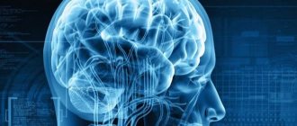
Magnetic resonance imaging
In the acute period:
- T1: slightly hypointense signal
- T2/FLAIR: hyperintense signal
- DWI: diffusion restriction in the acute period can be visualized as a lesion not visible on other sequences
In the chronic stage, a previous lacunar infarction has signal characteristics similar to the MR signal from the cerebrospinal fluid in all sequences. On T2/FLAIR, a ring-shaped zone of marginal gliosis may be visualized in the periphery.
Consequences
ABOUT
Complications after a lacunar cerebral infarction can be very diverse. They will depend on the following range of factors:
- Areas of brain damage;
- Size of the damaged area;
- Timeliness of diagnostic measures and adequacy of treatment.
- The consequences of lacunar infarction can lead to such negative complications as:
- Coordination disorder;
- Intellectual impairment such as dementia;
- Decreased memory;
- Decreased ability to analyze.
- Problems with urination and defecation;
- Speech defects;
- Changes in a person's mental state. The patient is characterized by tearfulness, hysteria, and depression.
Consequences and prognosis
It was previously thought that lacunar infarction did not leave permanent damage to brain tissue, but recent studies show that 50% of people suffer from complications.
The consequences depend on the degree of damage to the brain tissue, i.e. on how quickly blood flow in the affected vessel can be restored. This ensures that the area of the brain that is not damaged is preserved.
Repeated lacunar infarction leads to the following consequences:
- plegia (complete paralysis) of one limb;
- hemiplegia (complete paralysis of the right or left half of the body);
- speech disorders (both the ability to speak and the ability to understand);
- inability to read, write;
- intellectual-mnestic disorders;
- various types of consciousness disorders.
If a patient develops consequences after a lacunar cerebral infarction, an important part of the therapeutic course is rehabilitation treatment under the guidance of a specialist. The goal of rehabilitation is to ensure the restoration of the greatest possible number of functions of the affected part of the brain.
There are various programs to improve speech, speech therapy, impulse rehabilitation; patients explore alternative movements that provide maximum self-sufficiency.
Diagnosis and treatment
When a patient comes to see a neurologist with complaints of headache, muscle weakness, increased blood pressure, cramps, increased bowel movements and urination, the doctor first prescribes an MRI. This method makes it possible to identify lesions in the brain. Often, in addition to lacunae, cysts and other neoplasms are also found. The patient should consult a doctor in time so as not to miss the onset of the development of more serious diseases, such as cancer.
In addition to CT and MRI, the doctor prescribes tests for cytological examination, as well as an autopsy. This makes it possible to determine the presence of arterial hypertension. If the tests for hypertension are positive, then the main therapy is directed specifically at it. The main task of the doctor is to eliminate the cause, and only then deal with the manifestations and consequences. If hypertension is left untreated, the risk of developing recurrent lacunar infarction increases.
The patient is treated in a hospital and is later discharged to home rehabilitation. The patient is taking medications that lower blood pressure. In addition, complete peace and absence of stressful situations are indicated. Any sudden changes in pressure can worsen the condition. The doctor also prescribes medications that are designed to improve blood circulation. This promotes the rapid restoration of damaged brain structures.
A therapeutic diet plays an important role. The body of a patient with lacunar stroke should not be overloaded with carbohydrates, animal proteins, fried and fatty foods. The diet should include porridge, boiled chicken and beef. In addition, you need to limit your consumption of legumes. During rehabilitation, the patient should not eat flour products or sweets.
Rehabilitation lasts approximately 6 months. If the patient promptly seeks professional medical help, then all brain functions are completely restored and hypertension goes away. The cause is eliminated and speech becomes intelligible, coordination of movements when walking is improved, frequent bowel movements and frequent urge to urinate cease to bother the patient. To prevent the re-development of lacunar infarction, drugs are prescribed that control blood pressure and eliminate arterial hypertension.
Appendix c. patient information
Patients with a malignant infarction in the middle cerebral artery in the acute period of the disease are deeply disabled. After discharge from the hospital where the patient was treated for stroke, comprehensive rehabilitation in a specialized center is indicated, aimed at partial regression of the neurological deficit.
The nature of rehabilitation and the number of courses is determined by a rehabilitation specialist. In a non-rehabilitation center on an outpatient basis, the patient is under the supervision of a neurologist at the place of residence, who determines treatment. Activities aimed at regressing neurological deficits (physical therapy, massage, speech therapy sessions), prevention and treatment of extracranial complications continue.
Causes of lacunar cerebral infarction
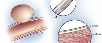
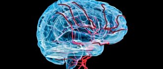
Lacunar stroke occurs due to disruption of blood flow through one of the perforating arterial vessels of the brain. In 80% of cases, the infarction zone is located in the white cerebral matter of the subcortical structures and internal capsule, in other cases - in the pons and brainstem.
In most cases, cerebral infarction of the lacunar type occurs against the background of chronic arterial hypertension and is associated with changes in the wall of perforating vessels caused by it - cerebral microangiopathy. Morphologically, this may be hyalinosis, intravascular deposition of lipid deposits, fibrous replacement of the muscular and elastic structures of the vascular wall, fibrinoid necrosis.
Such changes entail a significant narrowing and occlusion of the lumen of the artery, as a result of which the blood supply to the area of cerebral tissue fed by it is disrupted. Ischemia and necrosis develop in this zone. Over time, a lacuna forms in place of the dead cells.
Cerebral microangiopathy accounts for about 75% of lacunar infarctions. Among its etiofactors, along with hypertension, are atherosclerosis, diabetes mellitus, alcoholism, chronic obstructive pulmonary disease, chronic renal failure with an increase in the concentration of creatinine in the blood, and in rare cases, infectious and autoimmune vascular lesions.
What is lacunar stroke
There are several types of stroke. Each type is characterized by a specific localization. Lacunar stroke is one of the causes of infarction (cessation of blood flow) of the brain, provoking the formation of small cavities in the gray matter. They are called lacunae. These cavities are round or irregular in shape and measure from 1 mm to 2 cm. The lacunae of the brain vessels are filled with fibrin or blood.
Lacunar ischemic stroke is part of the group of cerebrovascular lesions of the brain. The condition is defined as a pathology of unknown origin. The lacunar form is also called “mute”. This is due to the fact that the pathology may not manifest itself for years. A person can live normally with small gaps, unaware of the changes that have occurred in the brain. The asymptomatic course of the disease often leads to numerous heart attacks, which result in cognitive impairment and severe dementia.
Articles on the topic
- Classification of cerebral strokes by degree - ischemic, hemorrhagic and apoplectic
- The difference between a heart attack and a stroke - the first signs and types of pathologies, drug therapy, consequences
- Eye stroke - first manifestations and types of pathology, drug therapy and preventive measures
Distinctive features
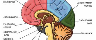
This type of stroke was first described as a manifestation of hypertensive encephalopathy (impaired brain function due to high blood pressure) in 1965. A distinctive feature of the lacunar form is disturbances in the capillaries, and not in the basilar artery, as in other types of stroke. Small vessels are located inside the brain and are responsible for the blood supply to the organ. The size of these capillaries is 30-40 microns. Lacunar pathology does not cause damage to the cerebral cortex. Cavities can be located in the area:
- main nerve nodes;
- pons;
- white matter;
- cerebellum;
- visual thalamus;
- internal capsule.
Causes
Research to identify the etiology of the lacunar form of the disease has been conducted since the end of the 19th century. Several hundred autopsies of the deceased were performed. The most common reasons include:
- Uncompensated arterial hypertension. The condition is characterized by a crisis or sudden changes in pressure in the absence of therapy.
- Atherosclerosis due to circulatory disorders, which leads to the development of hypertension.
- Diabetes mellitus type 2.
- Infectious or allergic inflammation of the arteries, leading to a lack of nutrition to the brain.
- Diseases characterized by increased blood clotting and the risk of thrombosis (burns, dehydration, erythremia (increased concentration of red blood cells), extensive trauma, shock).
- Minor bleeding in the area close to the lacuna. As a result, the lumen of the vessels becomes sticky and blood circulation becomes difficult.
- Genetic changes in the structure of the arterial wall.
Disease prognosis
The prognosis for lacunar cerebral infarction, in contrast to other forms of ischemic stroke, is most favorable. In a single case of the disease, almost complete restoration of all functional capabilities of the body occurs. Residual sensory or motor impairments bring some discomfort, but you can learn to live with it.
In case of relapses, a lacunar state of the brain can be diagnosed. This complication is observed in the majority of patients with vascular dementia. The patient often withdraws into himself and falls into a depressive state.
After an attack of illness, a person experiences problems with memory, often does not remember the address of residence, does not recognize relatives, is incapable of analyzing situations, and becomes sloppy.
Features of treatment
Complex treatment of lacunar stroke has the following goals:
- decreased blood pressure;
- restoration of normal nutrition of brain tissue;
- prevention of complicated situations (embolism, thrombosis).
This can be dealt with by competent and timely prescribed therapy, including:
- taking medications;
- rehabilitation;
- therapeutic nutrition.
Antihypertensive drugs are used to lower blood pressure:
- ACE inhibitors (lisinopril, enalapril);
- vasodilators (Diltiazem, Nifedipine);
- diuretics (Furosemide, Indapamide), etc.
An important direction in the treatment of lacunar cerebral infarction is antithrombotic therapy using Heparin, Aspirin, Warfarin, etc.
To restore brain structures and improve microcirculation, Nootropil, Vinpocetine, Akatinol, B vitamins, etc. are prescribed. Antidepressants (Amitriptyline, Fluoxetine) are used to relieve depressive conditions. Statins are used to combat atherosclerosis and lower cholesterol. To prevent complications, the patient is prescribed Aspirin, which can thin the blood.
A rehabilitation program for patients who have suffered a lacunar stroke is necessary to restore motor and speech functions and lost sensitivity. It includes:
- physiotherapy;
- exercise therapy;
- massage;
- speech training, etc.
Important information: What are the prognosis for recovery after ischemic stroke of the brain stem?
If all medical recommendations are followed, the prognosis is considered favorable.
Prevention
Primary prevention of lacunar infarction involves timely correction of arterial hypertension, regular intake of aspirin in patients with cardiovascular pathology in need of antiplatelet therapy, and adequate treatment of chronic cerebrovascular insufficiency.
To prevent relapse of lacunar stroke, long-term use of aspirin is recommended; in the presence of severe occlusion of the brachiocephalic arteries, surgical treatment of cerebrovascular insufficiency is possible - carotid endatherectomy, creation of extra-intracranial anastomosis.