There's so much you can't find on the net. Even the question about the color of blood and veins is often accompanied by assumptions and fiction, although most people actually know the answer. Yes, everything is simple here - the blood is red, only in different shades, depending on the amount of hemoglobin in it and oxygen enrichment. Everything is taught in biology and BJD at school: arterial blood
(oxygen-rich, coming from the heart)
is bright scarlet
, and
venous
(which gives oxygen to the organs, returning to the heart) is
dark red
(burgundy). The veins that are visible under the skin are also red when blood flows through them inside. After all, the blood vessels themselves are quite transparent. But still, many people have questions such as “Why does blood come in different colors and what does this depend on?” and “Why are veins blue or cyan?”
The red color of blood can have different shades. Oxygen carriers, i.e. red blood cells (erythrocytes), are a shade of red depending on hemoglobin, an iron-containing protein found in them that can bind with oxygen and carbon dioxide to carry them to the desired location. The more oxygen molecules connected to hemoglobin, the brighter the red color the blood is. That’s why arterial blood, which has just been enriched with oxygen, is so bright red. After the release of oxygen to the cells of the body, the color of the blood changes to dark red (burgundy) - such blood is called venous.
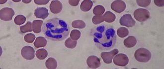
Of course, the blood contains other cells besides red blood cells. These are also leukocytes (white blood cells) and platelets. But they are not in such significant quantities compared to red blood cells as to affect the color of the blood.
Blood color in anemia and cyanosis
In fact, of course, although the veins carry dark burgundy blood, unlike the bright scarlet arterial blood, they are not at all blue in color. They are red, like the color of the blood that flows through them. And you shouldn’t believe in the theory that you can find on the Internet that the blood actually runs through the vessels is blue, but when cut and in contact with air it instantly turns red - this is not so. Blood is always red, and why is described above in the article.
The veins only appear blue to us. This is explained by the laws of physics about the reflection of light and our perception. When a beam of light hits the body, the skin reflects some of all the waves and therefore looks light, well, or different, depending on melanin. But it transmits the blue spectrum worse than red. But the vein itself, or rather the blood, absorbs light of all wavelengths (but less, in the red part of the spectrum). That is, it turns out that the skin gives us a blue color for visibility, and the vein itself gives us red. But, interestingly, the vein actually reflects even a little more red than the skin in the blue spectrum of light. But why then do we see veins blue or cyan? And the reason, in fact, lies in our perception - the brain compares the color of the blood vessel against the bright and warm tone of the skin, and in the end shows us blue.
Why don’t we see other vessels through which blood flows?
If a blood vessel is located closer than 0.5 mm to the surface of the skin, then it generally absorbs almost all blue light, and reflects much more red light - the skin looks healthy pink (ruddy). If the vessel is much deeper than 0.5 mm, then it is simply not visible, because the light does not reach it. Therefore, it turns out that we see veins that are approximately located at a distance of 0.5 mm from the surface of the skin, and why they are blue has already been described above.
Why can't we see arteries from under the skin?
In fact, about two-thirds of the blood volume is permanently in the veins, which means they are larger than other vessels. In addition, arteries have much thicker walls than veins, because they have to withstand greater pressure, which also prevents them from being sufficiently transparent. But even if the arteries were visible from under the skin as well as some veins, it is assumed that they would have approximately the same color, despite the fact that the blood running through them is brighter.
What color are veins actually?
If you've ever cooked meat, you probably already know the answer to this question. Empty blood vessels are reddish-brown in color. There is not much difference in color between arteries and veins. They differ mainly when viewed in cross section. Arteries are thick-walled and muscular, while veins have thin walls.
As for aristocrats, the expression “blue bloods” arose due to the paleness of their skin. Until the twentieth century, tanning was not in fashion, and the aristocrats themselves, especially women, hid from the sun, which protected their skin from premature aging and looked appropriate for their status, that is, they differed from the serfs who “plowed” all day in the sun. We now understand that pale skin color with a blue tint is actually a sign of less health.
But scientists also claim that there are about 7,000 people in the world whose blood has a blue tint. They are called kyanetics (from the Latin cyanea - blue). The reason for this is not the same hemoglobin. Their protein contains more copper than iron, which during oxidation acquires a blue tint instead of the red we are accustomed to. These people are considered more resistant to many diseases and even injuries, as their blood is said to clot several times faster and are not susceptible to many infections. In addition, there are different theories about the origin of kianeticians, including that they are descendants of aliens. There is not much information about them on the Internet, but there are articles in foreign publications where the birth of such children is explained by the abuse of rudimentary drugs long before conception. As they say, “Don’t smoke, girl, the children will be green!”, but the results from birth control may turn out blue (meaning the color of blood).
Noun, g., used. very often Morphology: (no) what? blood, what? blood, (see) what? blood, what? blood, about what? about blood and on blood 1. Blood is a red liquid that moves through the blood vessels in your body and nourishes your body... ... Dmitriev's Explanatory Dictionary
BLOOD, blood, about blood, in blood, many. blood, blood, wives 1. units only A red liquid that circulates in the animal body, delivering nutrients to tissues and removing their breakdown products. Deoxygenated blood. Blood flows from the wound.... ... Ushakov's Explanatory Dictionary
A fluid that circulates in the circulatory system and carries gases and other dissolved substances necessary for metabolism or formed as a result of metabolic processes. Blood consists of plasma (a clear, pale yellow liquid) and... ... Collier's Encyclopedia
And, prev. about blood, in blood, kind. pl. blood, w. 1. Liquid tissue that moves through the blood vessels of the body and provides nutrition to its cells and metabolism in it. Deoxygenated blood. Arterial blood. □ [Semyon] stabbed himself in the left... ... Small Academic Dictionary
blood
- and, sentence; about blood/vi, in blood/; pl. genus. blood; and. see also blood, bloody, bloody 1) Liquid that moves through the blood vessels of the body and provides nutrition to its cells and metabolism in it. Venous blood ... Dictionary of many expressions
I (sanguis) liquid tissue that transports chemicals in the body (including oxygen), due to which the integration of biochemical processes occurring in various cells and intercellular spaces occurs into a single system ... Medical encyclopedia
BLOOD
- BLOOD, a liquid that fills the arteries, veins and capillaries of the body and consists of a transparent pale yellowish color. the colors of plasma and the formed elements suspended in it: red blood cells, or erythrocytes, white, or leukocytes, and blood plaques, or ... Big Medical Encyclopedia
The human system consists of two main sections: the superior vena cava system (v. cava superior) brings venous blood from the upper half of the body and upper extremities to the right atrium; it is composed of two nameless veins (venae innominatae s.... ...
The system of lower vertebrates is significantly different from the human venous system and approaches its structure in the human embryo. In fish, the main veins stretch along the sides of the body: the anterior and posterior cardinal veins (venae cardinales) of the right and ... ... Encyclopedia of Brockhaus and Efron
Women red, vital fluid that circulates in the animal body, in the veins, by the power of the heart. The blood consists of light, yellowish fluid and thick liver; scarlet, veiny, arterial blood circulates in the fighting veins; black, subcutaneous, venous ... Dahl's Explanatory Dictionary
The blood of absolutely all representatives of humanity is red. Even people of “blue blood” are no exception. This color is provided by red blood cells. About a third of their component is hemoglobin. It is formed in the process of contact of iron atoms with a protein, scientifically called globin. Iron oxide (Fe2+) gives hemoglobin its rich red color.
There are 2 types of blood:
- arterial;
- venous.
Arterial blood is characterized by a scarlet color. As it moves through the lungs, it is saturated with oxygen, due to which the formation of “oxyhemoglobin” occurs, which affects the color and makes it so bright.
Venous blood, on the contrary, is dark in color. Sometimes it is purple, almost black. Unlike arterial blood, such blood, moving through the vessels and capillaries, on the contrary, loses a significant part of the oxygen, which is replaced by carbon dioxide. It is carbon dioxide that makes its shade darker.
A little experience will help prove this. A small amount of venous blood will be required, which we will monitor. Only extracted from the vein, it will have a characteristic dark color, and after standing for a while and coming into contact with oxygen, it will turn scarlet.
If you have to take a blood test for the first time, do not be alarmed by its excessively dark color.
Many adults know practically nothing about how their body works, believing that such information given to them at school is completely useless to them. In fact, the average person really doesn’t need the exact names of many processes and complex functions. However, at the same time, each of us needs to have at least some understanding of the basic mechanisms of our body and the features of their activity. Such knowledge will help you pay attention in time to any problems in the functioning of organs and systems, and also, if necessary, provide help to yourself and others. Today we will talk about how arterial and venous blood differ, what the circulatory system is, and the circulatory circles.
Our blood moves through a closed system, which is called the circulatory system, and consists of two circles - small and large.
Pulmonary circulation
In this system, blood moves from the heart to the lungs and back. In this case, venous blood moves from the right cardiac ventricle into the pulmonary artery, as well as into the pulmonary capillaries. There it leaves carbon dioxide and absorbs oxygen, after which it moves through the pulmonary veins, flowing into the left atrium. This blood then enters the systemic circulation and saturates all organs of the body with oxygen.
Dividing our circulatory system into two circles at once helps to separate arterial blood from venous blood, in other words, blood enriched with oxygen from that which has already been used and is saturated with carbon dioxide. Accordingly, thanks to this structure, our heart faces much less stress, as if it were pumping both types of blood through common blood vessels.
Blood enters the right atrium by passing through a pair of venous trunks, namely the superior vena cava, which carries venous blood from the upper part of the body, and the inferior vena cava, which supplies used blood from below. After this, the blood passes into the right heart ventricle, from where it enters through the pulmonary artery into the lungs.
Systemic circulation
Once in the lungs, the blood is saturated with oxygen and goes into the left atrium, and then into the left ventricle. When the left ventricle contracts, blood flows into the aorta. This section consists of a pair of large iliac arteries; they move downwards, supplying blood to the limbs. Also, a number of blood vessels depart from the aorta and its arch, carrying blood to the head, torso, as well as the chest and arms.
Arterial and venous blood
Many are sure that arterial blood always carries exclusively oxygen, and venous blood always carries carbon dioxide. However, in the pulmonary circulation the system works the other way around, used blood is carried through the arteries, and fresh blood through the veins.
Circulatory system
If we take all the arteries, as well as the veins of the circulatory system of an ordinary person, then their total length will be approximately one hundred thousand kilometers, and the total area will be approximately six to seven thousand square meters. Thanks to such a significant number of blood vessels, our body has the opportunity to fully undergo all metabolic processes.
Blood vessels are located throughout the body, they can be easily seen in the folds, for example, veins are quite easy to see in the area of the elbows. The arteries run a little deeper, so you can’t just see them. Due to the high elasticity of the vessels, they do not compress during natural flexion of the limbs.
The diameter of the largest artery, the aorta, is approximately two and a half centimeters, and the smallest capillaries do not exceed a diameter of eight thousandths of a millimeter.
All organs that actively participate in metabolic processes are directly connected to the circulatory system. So the aorta branches into a significant number of arteries, which ensures the distribution of blood flow across several vascular networks, which are located, as it were, in parallel. Each such mesh effectively communicates with each individual organ, saturating it with blood. Thus, the aorta provides nutrition to the kidneys and adrenal glands, spleen and digestive tract. In the lumbar region, the aorta divides into two branches, one going to the genitals, and the second to the lower extremities.
Blood, rich in oxygen, releases its nutrients through the thin walls of the capillaries, saturating the tissue fluid with them. In return, the waste products of the cells enter the blood.
If we talk about venous blood, which carries depleted blood back to the heart, then in the area of the lower extremities it collects to the femoral veins, which then form the iliac vein, and it already gives rise to the inferior vena cava. From the side of the head, venous blood passes through the jugular veins, they are located on both sides, and from the arms it moves through the subclavian veins. They then fuse with the jugular veins to form the innominate veins, one on each side. Such vessels merge into the large superior vena cava.
Also one of the parts of the systemic circulation is the portal vein; it is part of the system into which venous blood enters from the digestive tract. Before entering the inferior vena cava, such blood passes through a network of capillaries in the liver.
Despite the apparent complexity of the circulatory system, it all ideally works like a clock, providing every cell of our body with nutrients.
The vascular system maintains constancy in our body, or homeostasis. It helps him in the adaptation process, with its help we can withstand significant physical stress. Prominent scientists, since ancient times, have been interested in the structure and operation of this system.
If we imagine the circulatory apparatus as a closed system, then its main components will be two types of vessels: arteries and veins. Each performs a specific set of tasks and carries different types of blood. How venous blood differs from arterial blood will be discussed in the article.
The task of this type is the delivery of oxygen and nutrients to organs and tissues. It flows from the heart, rich in hemoglobin
.
The color of arterial and venous blood is different. The color of arterial blood is bright red.
The largest vessel through which it moves is the aorta. It is characterized by high speed of movement.
If bleeding occurs, stopping it requires effort due to its pulsating nature under high pressure. The pH is higher than that of the venous one. On the vessels through which this type moves, doctors measure the pulse
(on the carotid or radial).
Deoxygenated blood
Venous blood is that which flows back from the organs to return carbon dioxide
. It contains no useful microelements and carries a very low concentration of O2. But it is rich in metabolic end products and contains a lot of sugar. It is characterized by a higher temperature, hence the expression “warm blood.” It is used for laboratory diagnostic procedures. Nurses administer all medications through veins.
Human venous blood, unlike arterial blood, has a dark, burgundy color. The pressure in the venous bed is low, the bleeding that develops when the veins are damaged is not intense, the blood oozes out slowly, and is usually stopped with a pressure bandage.
To prevent its reverse movement, the veins have special valves that prevent backward flow; the pH is low. There are more veins in the human body than arteries
. They are located closer to the surface of the skin and are clearly visible visually in people with a light color type.
Why are tests taken from a vein?
This is due to the type of blood in the veins - saturated with metabolic products and vital functions of organs. If a person is sick, it contains certain groups of substances, remains of bacteria and other pathogenic cells. In a healthy person, these impurities are not detected.
By the nature of the impurities, as well as by the level of concentration of carbon dioxide and other gases, the nature of the pathogenic process can be determined.
The second reason is that venous bleeding when a vessel is punctured is much easier to stop. But there are times when bleeding from a vein does not stop for a long time. This is a sign of hemophilia, a low platelet count. In this case, even a minor injury can be very dangerous for a person.
How to distinguish venous bleeding from arterial bleeding:
- Assess the volume and nature of leaking blood. The venous flows out in a uniform stream, the arterial flows out in portions and even in “fountains”.
- Determine what color the blood is. Bright scarlet indicates arterial bleeding, dark burgundy indicates venous bleeding.
- Arterial is more liquid, venous is thicker.
Once again about the differences
The table provides a comparative description of what arterial and venous blood is.
Attention!
The most common question is which blood is darker: venous or arterial? Remember - venous. It is important not to confuse this when you find yourself in an emergency situation. In case of arterial bleeding, the risk of losing a large volume in a short period of time is very high, there is a threat of death, and urgent measures must be taken.
First aid
When providing first aid for bleeding, it is important to determine its type and, depending on this, act.
- If an artery in the arm or leg is affected, a tourniquet must be applied above the affected area. While the tourniquet is being prepared, press the artery above the wound to the bone. This is done with a fist or by pressing hard with your fingers. Elevate the injured limb.
Place a soft cloth under the tourniquet. You can use a scarf, rope, or bandage as a tourniquet. The tourniquet is tightened until the bleeding stops. You need to place a piece of paper under the tourniquet to indicate the time of application of the tourniquet.
ATTENTION. For arterial bleeding, the tourniquet can be held for two hours in the summer, and half an hour in the winter. If medical help is still not available, loosen the tourniquet for a few minutes while holding the wound with a clean cloth pad.
If a tourniquet cannot be applied, for example, if the iliac artery is injured, make a tight tampon with a sterile or at least clean cloth. The tampon is wrapped with bandages.
- In case of venous bleeding, a tourniquet or tight bandage is applied below the wound. The wound itself is covered with a clean cloth. The affected limb needs to be raised higher.
For these types of bleeding, it is good to give the victim painkillers and cover him with warm clothes.
- In case of capillary bleeding, the wound is treated with hydrogen peroxide, bandaged or covered with a bactericidal adhesive plaster. If it seems to you that the blood is darker than a normal wound, then the venule may be damaged. Venous blood is darker than capillary blood. Proceed as if you had damaged a vein.
IMPORTANT. Capillary bleeding is dangerous if blood clotting is poor.
The health and sometimes life of a person depends on proper assistance during bleeding.
Blood constantly circulates throughout the body, providing transport of various substances. It consists of plasma and a suspension of various cells (the main ones are erythrocytes, leukocytes and platelets) and moves along a strict route - the system of blood vessels.
Circulation circles
At the beginning of the article, it was noted that blood moves in the vascular system. From the school curriculum, most people know that movement is circular, and there are two main circles:
- Big (BKK).
- Small (MCC).
Mammals, including humans, have four chambers in the heart.
. And if you add up the length of all the vessels, you get a huge figure - 7 thousand square meters.
But it is precisely this area that allows you to supply the body with O2 in the required concentration and not cause hypoxia, that is, oxygen starvation.
BCC begins in the left ventricle, from which the aorta emerges. It is very powerful, with thick walls, with a strong muscle layer, and its diameter in an adult reaches three centimeters.
It ends in the right atrium, into which 2 vena cava flow. The ICC originates in the right ventricle from the pulmonary trunk, and closes in the left atrium with the pulmonary arteries.
Oxygen-rich arterial blood flows in a large circle, it is directed to each organ
. As they progress, the diameter of the vessels gradually decreases to very small capillaries, which give away everything useful. And back, through venules that gradually increase their diameter to large vessels, such as the superior and inferior vena cava, the depleted venous flows.
Once in the right atrium, through a special opening, it is pushed into the right ventricle, from which the small circle, pulmonary, begins. The blood reaches the alveoli, which enrich it with oxygen. Thus, venous blood becomes arterial!
Something very surprising happens: arterial blood moves not through the arteries, but through the pulmonary veins, which flow into the left atrium. Blood saturated with a new portion of oxygen enters the left ventricle and the circles are repeated again. Therefore, the statement that venous blood moves through the veins is incorrect; here everything works the other way around
.
Fact!
In 2006, a study was conducted on the functioning of the BCC and MCC in people with poor posture, namely scoliosis. Attracted 210 people under 38 years of age. It turned out that in the presence of scoliotic disease, there is a disruption in their work, especially among adolescents. In some cases, requiring surgical treatment.
In some pathological conditions, blood flow may be disrupted, namely:
- organic heart defects;
- functional;
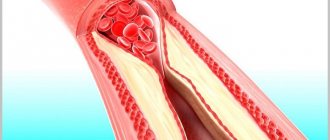
- pathologies of the venous system: , ;
- , autoimmune processes.
Normally there should be no mixing
. During the newborn period, there are functional defects: open oval window, open Batalov duct.
After a certain period of time, they close on their own, do not require treatment and are not life-threatening.
But severe valve defects, reversal of the main vessels, or transposition, absence of a valve, weakness of the papillary muscles, absence of a heart chamber, combined defects are life-threatening conditions.
That is why it is important for the expectant mother to undergo screening ultrasound examinations of the fetus during pregnancy.
.
Thick blood: causes and treatment in women during pregnancy
The pregnancy period requires the female body to exert all its strength. An increase in blood viscosity in expectant mothers is a physiological phenomenon. This is how nature protects a woman from possible large blood loss during childbirth. This becomes a problem when laboratory parameters significantly deviate from the norm.
Pathological thickening of blood during pregnancy may be due to
- insufficient intake of water from food;
- deficiency of vitamins, microelements, minerals, since the formation of the fetus requires them in large quantities;
- enzyme deficiency;
- increased work of the spleen;
- taking iron supplements;
- excess protein and carbohydrates in the diet.
In addition, pathology of the liver, kidneys, intestines, blood loss, increased coagulability, severe pain also contribute to blood thickening and can lead to miscarriages.
Lack of attention to excessively viscous blood during pregnancy is dangerous
- the formation of thrombosis, heart attacks and strokes, varicose veins in the mother;
- fading of pregnancy, its premature termination, delayed fetal development, hypoxia.
Healthy blood is the key to the healthy functioning of the entire body, so it is extremely important to maintain its condition within normal limits. In case of any deviation, you should definitely consult a doctor and carry out the necessary treatment.
Thick blood is not a disease, it is a symptom of one of many diseases, the prognosis for treatment of which is not always favorable. A change in the natural state of the blood, its structure and consistency may be a signal of pathological changes in the myocardium or blood flow. By understanding the causes and treatment of thick blood in women, you can prevent the occurrence of more serious diseases.
Increased thickness is a deviation from the norm caused by an imbalance of plasma and blood cells. In the female half of the population, who do not suffer from diseases or system failures, the hematocrit varies from 0.36 to 0.46. Blood is thicker than water and, in the absence of pathological changes, it moves 5 times slower. Plasma has a viscosity from 1.4 to 2.2 units. And the density of a woman’s total composition is 3.9 - 4.9 units. The concentration of blood cells always exceeds the plasma volume. Density - 1,050 - 1,064 g/ml. Viscosity among representatives of different sexes does not differ significantly; this is due to physiological differences. A man's blood is thicker.