Normal pulse
The pulse is the most important indicator of the vital functions of the body; it characterizes the state of human health, the functioning of all its systems and organs, primarily the cardiovascular system. This indicator must be monitored in the intensive care unit, and the pulse is checked when visiting a doctor.
Also, every person should know what a normal arterial pulse should be and be familiar with the algorithm for measuring it.
How is a pulse wave formed?
Determining a person's pulse
During cardiac contraction, blood spreads like a wave through the vessels, from central to peripheral. The walls of blood vessels experience blood pressure as it passes through arteries, veins, and capillaries. Fluctuations in the walls of the arteries are the arterial pulse; it is the most diagnostically significant. It is the arterial pulse that is taken into account during medical visits or when assessing hemodynamics. In addition, it can be venous and capillary.
Arterial can be determined both on the central arteries - the aorta, carotid arteries, and on the peripheral ones - radial, brachial, tibial. On the radial artery of the wrist, the difference between the pulse and heart rate is literally a second and is practically not felt, but if the study is carried out on the artery of the foot, then with simultaneous auscultation of the heart, one can clearly hear first the heart contraction, and then, a little later, the pulse impulse.
This algorithm is easily explained by the distal location of the vessel, because blood needs time to “get” from the heart to the artery of the foot. 60-90 beats per minute is the normal heart rate of an adult. A rate below 60 beats can be regarded as bradycardia, above 90 beats as tachycardia. A person's resting pulse is on average 70-75 beats per minute. The slowdown is observed in athletes and older people. Particular attention should be paid to changes in heart rate in athletes, as well as during pregnancy.
Pulse and its parameters
Pulse is a jerky vibration of the walls of blood vessels that occurs as a result of the ejection of blood from the heart into the vascular system. There are arterial, venous and capillary pulses. Of greatest practical importance is the arterial pulse, usually palpable in the wrist or neck.
Pulse measurement. The radial artery in the lower third of the forearm immediately before its articulation with the wrist joint lies superficially and can be easily pressed against the radius. The muscles of the hand that determines the pulse should not be tense. Place two fingers on the artery and squeeze it with force until the blood flow completely stops; then the pressure on the artery is gradually reduced, assessing the frequency, rhythm and other properties of the pulse.
In healthy people, the pulse rate corresponds to the heart rate and is 60-90 beats per minute at rest. An increase in heart rate (more than 80 per minute in a lying position and 100 per minute in a standing position) is called tachycardia, a decrease (less than 60 per minute) is called bradycardia. The pulse rate at the correct heart rhythm is determined by counting the number of pulse beats in half a minute and multiplying the result by two; in case of cardiac arrhythmias, the number of pulse beats is counted for a whole minute. With some heart diseases, the pulse rate may be lower than the heart rate - pulse deficiency. In children, the pulse is more frequent than in adults, in girls it is slightly more frequent than in boys. At night the pulse is lower than during the day. A rare pulse occurs with a number of heart diseases, poisoning, and also under the influence of medications.
Normally, the pulse quickens during physical stress and neuro-emotional reactions. Tachycardia is an adaptive response of the circulatory system to the body’s increased need for oxygen, promoting increased blood supply to organs and tissues. However, the compensatory reaction of a trained heart (for example, in athletes) is expressed in an increase not so much in the pulse rate as in the strength of heart contractions, which is preferable for the body.
Pulse characteristics. Many diseases of the heart, endocrine glands, nervous and mental illnesses, increased body temperature, and poisoning are accompanied by increased heart rate. During palpation examination of the arterial pulse, its characteristics are based on determining the frequency of pulse beats and assessing such qualities of the pulse as rhythm, filling, tension, height, speed
.
Pulse rate
determined by counting pulse beats for at least half a minute, and if the rhythm is incorrect, within a minute.
Pulse rhythm
assessed by the regularity of pulse waves following one after another. In healthy adults, pulse waves, like heart contractions, are observed at regular intervals, i.e.
The pulse is rhythmic, but with deep breathing, as a rule, the pulse increases during inhalation and decreases during exhalation (respiratory arrhythmia). An irregular pulse is also observed with various cardiac arrhythmias
: pulse waves follow at irregular intervals.
Pulse filling
determined by the sensation of pulse changes in the volume of the palpated artery. The degree of filling of the artery depends primarily on the stroke volume of the heart, although the distensibility of the arterial wall is also important (it is greater, the lower the tone of the artery
Pulse voltage
determined by the amount of force that must be applied to completely compress the pulsating artery. To do this, the radial artery is compressed with one of the fingers of the palpating hand and, at the same time, the pulse is determined with another finger distally, recording its decrease or disappearance. There are tense or hard pulses and soft pulses. The degree of pulse tension depends on the level of blood pressure.
Pulse height
characterizes the amplitude of the pulse oscillation of the arterial wall: it is directly proportional to the magnitude of the pulse pressure and inversely proportional to the degree of tonic tension of the artery walls. With shock of various etiologies, the pulse value decreases sharply, the pulse wave is barely palpable. This pulse is called threadlike.
studopedia.ru>
How is the pulse “trained”?
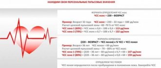
Heart rate in athletes
The heart rate of athletes varies depending on how long they have been in professional sports and what their training is aimed at. For example, for athletes who play sports for a long time, 50 beats per minute may be normal. For skiers, cyclists, and biathletes, the norm is 40-45 beats per minute, since the athletes’ hearts have “learned” to work in an energy-saving, rational mode.
At this frequency, the heart muscle receives more oxygen, nutrients, and rests more. During training, the pulse of well-trained athletes can increase to 200-210 beats, without negative consequences for health. While in a person who is far from sports, such frequency leads to adverse consequences and complications.
Such a large increase in athletes is due to the fact that a trained heart under heavy loads is able to sharply increase the frequency of contractions to ensure the best blood supply to the body. Heavy weight athletes also experience a sharp increase in heart rate when lifting heavy weights.
Rate variability is sharply reduced: how to treat
If there is a sharp decrease in heart rate variability, you must:
- reduction of load (reduction of physical activity, elimination of anxiety, conflicts);
- refusal of stressful changes (for example, change in climate, work, diet, business trip, participation in competitions);
- sleep at least 8 hours at night, if necessary, rest during the day;
- transition to a healthy diet - vegetables, fruits growing in the area of residence, lean meat, fish, dairy products, a minimum of processed and canned food, no spicy, fatty foods, alcohol, strong tea, coffee;
- provide an opportunity for relaxation for the nervous system - breathing exercises, walks in nature, light massage sessions, aromatherapy, listening to calm music.
If there are heart diseases, the doctor reviews treatment regimens; they can be supplemented with drugs that improve metabolism in the myocardium (for example, Preductal, Mildronate).
Pregnancy
Pulse during pregnancy
A normal pregnancy is a physiological state, therefore all changes in the body of a pregnant woman are not a pathology, but a type of normal. During pregnancy, a woman’s cardiovascular system experiences greater stress due to the increased volume of circulating blood, increased cardiac output, and hormonal changes. This cannot but affect such an indicator as the pulse wave frequency. It is logical that with increased load on the heart, the frequency will increase somewhat.
If before pregnancy a woman’s pulse was 82-89 beats, then the normal frequency during pregnancy can be 100-110 beats per minute. As a rule, during pregnancy this indicator increases by 10% from the initial value before pregnancy. The highest frequency is observed in the period from 28 to 32 weeks of pregnancy. After 32 weeks, this figure may decrease slightly. The frequency increases when walking, doing gymnastics, and in women during late pregnancy in a supine position.
History of pulse diagnostics
- Pulse studies for diagnostic purposes in Alexandria during the Ptolemaic dynasty (from which Cleopatra came) were used by Herophilus of Chalcedon and Erasistratus. Herophilus was the author of the work "Peri sphigmon pragmateias", which was considered the best treatise of antiquity on the pulse. Herophilus believed that the pulse is “the movement of the arteries” and with the help of the pulse one can recognize “the existence of illness in the body and foresee future ones.” The terms “systole” and “diastole” belong to him.
- In the 1st century AD, the physician Archogenes from Syria was popular in the Roman Empire. By the term “sphygmos” Archigen understood the normal movement of the arteries and heart, distinguished between systole and diastole and identified four beats: systole-diastole and two pauses. Archigen proposed a classification of the pulse according to the duration of diastole (long, small, medium), according to the nature of vessel movements (fast, rare, strong), according to pressure tone (strong, weak, medium), according to the strength of the pulse beat, according to resting time, according to the condition of the wall vessel (hard, soft, medium), by evenness or unevenness, by regularity or irregularity, by fullness or thickness, by rhythm. He distinguished dicrotic, ant-like, gazelle-like, and wavy pulses.
- The famous surgeon Rufus of Ephesus, who long before Harvey described the mechanics of blood circulation, called the pulse of healthy people “eurythmic” (ancient Greek εὐρυθμία - proportionality), the painful one - “pararhythmic” (παρά - near). He described extrasystole, dicrotic, alternating pulse and threadlike (lat. pulsus vermicularis) in agonizing patients.
- Galen wrote 7 books (334 pages) about the pulse, identified 27 types of pulse, and divided each type into three more varieties. He described sinus and respiratory arrhythmias. It was by the pulse that he diagnosed the stomach disease of Emperor Marcus Aurelius.
- The physician Aetius from Amida, who worked in Alexandria and Constantinople, in his book “Tetrabiblion” described the characteristics of the pulse in anemia, dehydration, and malaria.
- The doctor Archimatheus from Salerno described the method of palpation of the pulse, which is widely used today.
- Paracelsus suggested palpating the pulse in the arms, legs, as well as the cervical and temporal arteries, chest and armpits.
Ancient China
The pulse diagnostic method originated many centuries BC. Among the literary sources that have reached us, the most ancient are works of ancient Chinese and Tibetan origin. The ancient Chinese include, for example, “Bin-hu Mo-xue”, “Xiang-lei-shi”, “Zhu-bin-shi”, “Nan-ching”, as well as sections in the treatises “Jia-i-ching”, “Huang-di Nei-ching Su-wen Lin-shu” and others.
The history of pulse diagnostics is inextricably linked with the name of the ancient Chinese healer - Bian Qiao (Qin Yue-Ren). The beginning of the pulse diagnostic technique is associated with one of the legends, according to which Bian Qiao was invited to treat the daughter of a noble mandarin (official). The situation was complicated by the fact that even doctors were strictly prohibited from seeing and touching persons of noble rank. Bian Qiao asked for thin string. Then he suggested tying the other end of the cord to the wrist of the princess, who was behind the screen, but the court doctors disdained the invited doctor and decided to play a joke on him by tying the end of the cord not to the princess’s wrist, but to the paw of a dog running nearby. A few seconds later, to the surprise of those present, Bian Qiao calmly stated that these were not the impulses of a person, but of an animal, and this animal was suffering from worms. The doctor’s skill aroused admiration, and the cord was confidently transferred to the princess’s wrist, after which the disease was determined and treatment was prescribed. As a result, the princess quickly recovered, and his technique became widely known.
Hua Tuo successfully used pulse diagnostics in surgical practice, combining it with clinical examination. In those days, it was prohibited by law to perform operations; the operation was performed as a last resort, if there was no confidence in a cure using conservative methods; surgeons simply did not know diagnostic laparotomies. The diagnosis was made by external examination. Hua Tuo passed on his art of mastering pulse diagnosis to diligent students. There was a rule that only a man could learn perfect mastery of pulse diagnostics by learning only from a man for thirty years. Hua Tuo was the first to use a special technique for examining students on the ability to use pulses for diagnosis: the patient was seated behind a screen, and his hands were inserted into the slits in it so that the student could see and study only the hands. Daily, persistent practice quickly produced successful results.
middle Asia
The Central Asian doctor Abu Ali Hussein ibn Abdallah ibn Sina created the “Treatise on the Pulse” (“Risolai Nabziya”)
- A copy of the manuscript “The Canon of Medicine” (made in 1143 in Baghdad). Institute of Manuscripts of the National Academy of Sciences of Azerbaijan in Baku
Modern era
Until the 18th century, doctors did not count the pulse, limiting themselves only to assessing its qualities. At the beginning of the 18th century, the British physician Sir John Floer ordered a watchmaker to watch with a hand that ran for one minute. He was convinced of their practical convenience and published the book “The Phisican's Pulse Watch” in 1707, but the first Russian doctor P. Posnikov counted the pulse using an hourglass. They began to use a stopwatch to count the pulse only in the 19th century. It is believed that counting the pulse in seconds and minutes was proposed by astronomer Johannes Kepler.
Pulse diagnostics in modern medicine
Various versions of techniques, one way or another related to the analysis of heartbeats and pulse waves, are widely used in modern medicine. At the same time, both “traditional” methods are being developed, similar to those used in historical medicine [4], and hardware ones (when devices are used to analyze the rhythm of the heart: a heart rate monitor, a pulse oximeter, an electrocardiograph, etc.). Thus, today pulse research can be divided into 2 branches:
- manual examination
of manifestations of heart function; - hardware studies
of heartbeat rhythm.
Hardware methods include, for example, analysis of heart rate variability[5], the foundations of which were laid in the USSR and Russia in the 60s. XX century. The pioneer of the method in the USSR is most often called R. M. Baevsky. Similar methods of analysis have gained recognition throughout the world[6].
In practical medicine, there are a number of areas related to the analysis of rhythm
cardiac function[7]:
- screening for gross cardiac pathology, monitoring cardiac function in seriously ill patients and in the operating room;
- routine diagnosis of conduction disorders;
- assessment (prognosis) of the risk of acute cardiovascular pathology, including death;
- screening for various cardiomyopathies;
- control of the so-called “cardiotoxicity” of pharmacological drugs and substances;
- functional control in general medical and sports practice.
Heart rate variability analysis has also become widespread for assessing stress levels.
[8].
Today, the cognitive aspects
of heart rhythm are being studied, where the state of the mental sphere and the structural features of the heart rhythm are connected[7].
Characteristic
Heart rate characteristics
Frequency is not the only characteristic of a pulse wave. There are other equally important properties of the pulse. Let's look at them in more detail:
- Rhythm. The intervals between oscillations of the vessel walls should be the same, this characterizes the rhythm of the pulse wave. Arrhythmia indicates any disturbances in cardiac conduction or other diseases. Sometimes there are single ari during extrasystoles - extraordinary heart contractions, but normally they are single, and after them the rhythm is restored.
- Uniformity. A uniform pulse wave is characterized by the correct rhythm and the same height. The height of the pulse waves should be the same; it characterizes the magnitude of the oscillations of the vessel walls. If there is a different height and arrhythmia of the pulse waves, then such a pulse is called uneven.
Pulse symmetry
- Symmetry. When measuring the pulse on the radial artery, it should be simultaneously palpated in both arms. If this does not happen, or it appears later on one hand, this indicates its asymmetry and is regarded as a pathology. It can be a consequence of heart defects, pathology of the upper limb and various other diseases.
- Filling. This characteristic is assessed by the height of pulse oscillations. Normally, the pulsation is complete.
- Voltage. It is determined by pressing the radial artery with your fingers until the pulse wave is no longer detectable. The more effort you have to put into this, the greater the filling. Soft, insufficiently tense, occurs with low blood pressure, in asthenics, and adolescents. High tension is typical for high blood pressure numbers and for older people. A thread-like pulse, with low tension and filling, occurs with a critical decrease in blood pressure, states of shock, blood loss, and coma.
Arterial pulse studies
BLOCK OF INFORMATION
Arterial pulse is a rhythmic oscillation of the wall of arterial vessels under the influence of heart contractions.
Places for detecting pulse.
The pulse is determined by palpation of the radial, carotid, popliteal arteries, and the arteries of the feet, which are located superficially. Most often, the pulse is determined at the radial artery. Very carefully you should determine the pulse in the carotid artery, where the carotid sinus is located (its irritation causes changes in heart rate up to asystole and changes in blood pressure).
Basic properties of the pulse.
1. Normal heart rate is 60-80 beats per minute (last 60-90 beats/min). A heart rate decrease of less than 60 beats/min is called bradycardia.
An increased heart rate of more than 80 (90 beats/min) is called tachycardia. The pulse rate is counted per minute.
2. pulse rhythm is:
- rhythmic, if pulse fluctuations occur at regular intervals
- arrhythmic - incorrect alternation of pulse waves.
3. Pulse voltage is determined by the force with which the radial artery must be pressed so that its pulse fluctuations completely stop. Pulse voltage depends on the value of systolic blood pressure.
- If the blood pressure is normal, the artery is compressed with moderate force, therefore the normal pulse is of moderate (average, satisfactory) tension.
- With high blood pressure, the pulse is very tense (it is more difficult to compress the artery).
- With low blood pressure, the artery contracts easily - the pulse will be weak.
4. Pulse filling depends on the amount of blood in the artery and blood pressure.
If cardiac output is normal, the pulse will be of medium filling; if there is bleeding or dehydration, the pulse will be weak filling.
5. More often, the tension and filling of the pulse are noted together and called the pulse value, for example. pulse of strong tension and filling, pulse of satisfactory filling and tension. pulse is weak and tense.
When examining the pulse:
1. First, determine the first two properties (frequency and rhythm) for 1 minute, then the remaining two properties of the pulse (tension and filling).
2. Then assess the pulse . that is, they compare it with normal parameters and draw a conclusion. For example, if during the study: pulse is 95 beats/min, rhythmic, strong tension and filling. They assess it as tachycardia and probably high blood pressure. It is necessary to measure your blood pressure and report the results to your doctor.
3. The data obtained from the pulse examination are graphically recorded daily in the medical history on a temperature sheet using red paste. Column “P” (pulse) shows pulse values from 50 to 160 beats/min. for pulse rate values from 50 to 100, the “price” division in the temperature sheet is equal to 2, and for pulse rate values over 100, the “price” division is equal to 4. The pulse is noted once a day. On the line that is located between o and “Evening” the value of the pulse rate is marked with red paste in the form of a dot. Then a point is placed corresponding to the next day's heart rate value. these points are connected to each other.
Technique for determining the pulse on the radial artery (PS)!!!
Purpose: diagnostic, assess the state of the cardiovascular system
Indication: monitoring the patient's condition
Places for examining the pulse: radial artery, ulnar, carotid, temporal, popliteal, femoral, dorsum of the foot.
Conditions: After 10-15 minutes of physical rest (physiological tachycardia is excluded).
Pulse parameters: rhythm, frequency, tension, filling, magnitude
Prepare: Watch (stopwatch), paper, pen with red ink, temperature sheet.
- Explain the procedure to the patient and obtain his consent/
- Wash and dry your hands
- find the place where the pulse is detected.
- Give the patient a comfortable position - sitting or lying in a relaxed, comfortable position, in a calm state
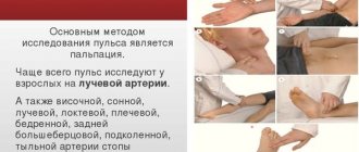
- Place your 1st, 3rd, 4th fingers on the area of the radial artery (the outer side of the forearm, the area of the wrist joint, the edge corresponding to the patient’s first finger) of the patient’s relaxed, calmly lying hand, your 1st finger should be on the side of the back of the forearm, lightly press the tissues of the forearm to brushes Feel the elastic pulsating waves associated with the movement of blood through the vessel.
- Determine the number of pulses contracted in 1 minute, while simultaneously determining the rhythm of the pulse.
- Determine the pulse tension (compare the force pressure of the fingers when examining the pulse on both hands, especially in elderly people and
- pregnant women).
- Write down the result and, if necessary, tell it to the patient, his relatives, and the doctor.
Pulse assessment.
- PS normally 60-80 beats per minute
- PS more than 80 beats per minute - rapid - tachycardia.
- PS less than 60 beats per minute – reduced – bradycardia.
Attention!!! Data from the pulse rate study are marked daily in the temperature sheet with red paste in the form of dots on the line between columns “U” and “B”. Column “P” (pulse) shows the pulse rate values. The “price” of one division on the “P” (pulse) scale up to 100 is 2 beats per minute, after 100 – 4 beats per minute
studopedia.ru>
Special Features
Paradoxical pulse
When measuring the algorithm and determining properties, pathological characteristics such as pulse deficiency and paradoxical pulsus should be taken into account. Pulse deficiency is a lower pulse rate compared to the heart rate. Pulse deficiency is a pathology. Pulse deficiency occurs in diseases of the heart and blood vessels, conduction disorders, atrial fibrillation, and extrasystole.
Paradoxical pulse - decreased pulsation during inspiration. Paradoxical pulse occurs in severe forms of pulmonary diseases, SVC obstruction, cardiac disease and tamponade. With these diseases, there is a pronounced drop in SBP by more than 10 mmHg, which leads to the formation of paradoxical pulsus.
Why measure your pulse?
What is the significance of vascular wall vibrations for diagnosis? Why is this so important?
The pulse makes it possible to judge hemodynamics, how effectively the heart muscle contracts, the fullness of the vascular bed, and the rhythm of heartbeats.
In many pathological processes, the pulse changes, and the pulse characteristic no longer corresponds to the norm. This allows us to suspect that not everything is in order in the cardiovascular system.
View gallery
Measurement algorithm
Pulse measurement
- Measurements should be carried out in a calm, relaxed state;
- Prepare a stopwatch or mechanical watch in advance;
- Place fingers 2,3,4 on the wrist of the other hand on the inner surface of the wrist joint on the thumb side;
- Lightly press your fingers against the skin of the wrist, determine the most pronounced place of pulsation;
- Determine the frequency, rhythm, uniformity, height, filling and tension of pulse waves in 1 minute. In conditions of time shortage, when calculating the frequency, you can count the number of pulse waves for 30 seconds or 15 seconds, and then multiply the obtained data by 2 or 4, respectively.
What are the types of rhythm disturbances?
Arrhythmias due to changes in the functioning of the sinus node (the area of the myocardium that generates impulses leading to contraction of the heart muscle):
- Sinus tachycardia - increased contraction frequency.
- Sinus bradycardia - decreased contraction frequency.
- Sinus arrhythmia is the contraction of the heart at irregular intervals.
Ectopic arrhythmias. Their occurrence becomes possible when a focus appears in the myocardium with activity higher than that of the sinus node. In such a situation, the new pacemaker will suppress the activity of the latter and impose its own rhythm of contractions on the heart.
- Extrasystole is the appearance of an extraordinary cardiac contraction. Depending on the location of the ectopic focus of excitation, extrasystoles are atrial, atrioventricular and ventricular.
- Paroxysmal tachycardia is a sudden increase in heart rate (up to 180-240 heart beats per minute). Like extrasystoles, it can be atrial, atrioventricular and ventricular.
Impaired conduction of impulses through the myocardium (blockade). Depending on the location of the problem that prevents the normal movement of the nerve impulse from the sinus node, blockades are divided into groups:
- Sinoauricular block (the impulse does not go beyond the sinus node).
- Intraatrial block.
- Atrioventricular block (the impulse does not pass from the atria to the ventricles). With complete atrioventricular block (III degree), a situation becomes possible when there are two pacemakers (the sinus node and the focus of excitation in the ventricles of the heart).
- Intraventricular block.
Separately, we should dwell on the flickering and fluttering of the atria and ventricles. These conditions are also called absolute arrhythmia. In this case, the sinus node ceases to be a pacemaker, and multiple ectopic foci of excitation are formed in the myocardium of the atria or ventricles, setting the heart rhythm with a huge contraction frequency. Naturally, under such conditions the heart muscle is not able to contract adequately. Therefore, this pathology (especially from the ventricles) poses a threat to life.
View gallery
