Treatment of vein phlebitis should be timely.
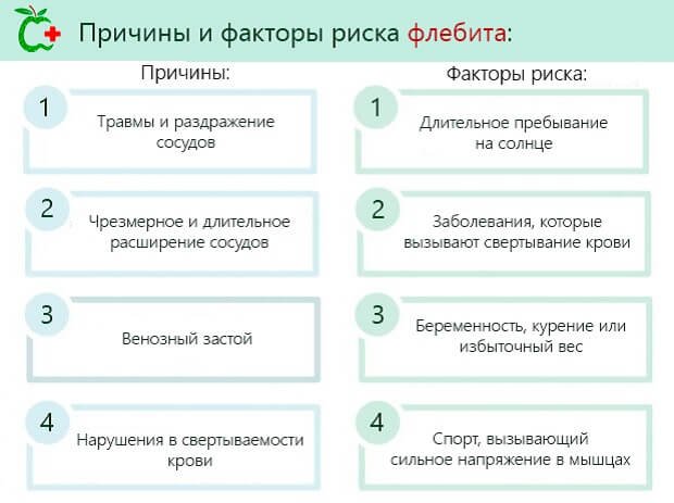
Phlebitis is an inflammatory process of the vein walls. During the development of the disease, the walls of blood vessels in the arm or leg, after a certain period of inflammation, are destroyed.
Find the answer Are you having a problem? Enter “Symptom” or “Name of the disease” into the form, press Enter and you will find out all the treatment for this problem or disease.
The site provides reference information. Adequate diagnosis and treatment of the disease is possible under the supervision of a conscientious doctor. Any medications have contraindications. Consultation with a specialist is required, as well as detailed study of the instructions! Here you can make an appointment with a doctor.
The disease comes in acute and chronic forms.
Phlebitis often accompanies varicose veins. It appears after unsuccessful injections or the influence of pathogenic microorganisms.
Treatment methods for phlebitis of the veins on the arm
During the treatment of phlebitis of the vein in the arm, conservative methods are used:
- Use of non-steroidal anti-inflammatory drugs;
- Antibacterial medications;
- Use of anticoagulants;
- Local measures - elastic bandage to restore blood flow.
If an infection is added to simple inflammation, then treatment consists of complex effects:
- Relief of the inflammatory focus;
- Prevention of spasms and hypertonicity of the walls;
- Increased venous blood flow;
- Qualitative improvement of blood viscosity;
- Fighting the formation of blood clots;
- Stabilization of the tone of the smooth muscles of the veins;
- Getting rid of puffiness and normalizing lymph circulation.
If an infection appears, then after determining the type of pathogen, specialized treatment measures are prescribed. Heparin and troxevasin ointments are used in the form of local preparations.
During the treatment of post-injection phlebitis of the veins in the arm, non-steroidal anti-inflammatory drugs are used, both orally and with the help of ointments.
Self-medication of phlebitis is fraught with complications for the patient, but poses a direct threat to his life.
If a focus of inflammation occurs on the arm after an injection or due to other reasons, it is necessary to seek specialized help for comprehensive treatment.
Therapeutic procedures
Treatment of post-injection disease of the affected vein is carried out conservatively and/or radically after assessing the totality of symptoms. Most often, if the patient seeks help within the first three days, the inflammatory process can be treated with medication. Treatment must be carried out in a hospital setting under the supervision of doctors, since there is a high probability of developing thromboembolism or phlegmon.
Conservative therapy is aimed at antibacterial treatment and detoxification, relieving inflammation in the area of post-injection lesions, increasing blood flow by stabilizing fibrous changes in the vein walls. Drug treatment of phlebitis includes the use of the following drugs:
- Non-steroidal drugs that relieve inflammation: Ibuprofen, Nimesulide, Butadione, etc. They are used in the form of tablets and topical ointments, no more than 2-3 times a day.
- Drugs that enhance blood flow dynamics: Aescusan, Troxevasin, Heparin, Glivenol. Medicines in this group are administered every 5-6 hours.
- Indirect anticoagulant drugs aimed at preventing blood clots: Warfarin, Aspecard. Medicines help reduce the viscosity of the blood stream.
- Fibrinolytic drugs aimed at dissolving blood clots: Streptokinase, Urokinase. They are used only when the condition worsens, when blood clots (thrombi) appear. Medicines act on the formed blood clot and help reduce the concentration of prothrombin.
- Antibacterial drugs: Aspirin, Butadione. Medicines are aimed at reducing the risk of blood poisoning. As a rule, they are inserted directly into the vessel using a catheter needle.
Treatment is carried out using anti-inflammatory drugs and anticoagulants in the form of tablets, ointments and injections, both intramuscularly and intravenously (a catheter needle is inserted into the veins of the other arm). When the inflammatory process is complicated, endolymphatic insertion of a catheter needle is used so that medications act more quickly on the affected area. Gauze bandages soaked in a silver solution, bandages with Heparin ointment, and Vishnevsky ointment are also applied locally. Local treatment alternates with the application of semi-alcohol compresses. However, if the wound does not dry out, but, on the contrary, its edges soften, then this indicates the occurrence of a purulent process.
If the patient seeks help on the first or second day, then the use of hyperthermic measures is allowed. On the third day, the inflammation process intensifies, physiotherapeutic procedures are strictly contraindicated. They are replaced by applying cold to the damaged area. Cold will prevent the inflammatory process from developing.
Causes of phlebitis
Phlebitis can be superficial or internal.
The first form is not dangerous, but the second leads to the formation of blood clots in the vessels, which is fraught with consequences. Phlebitis most often affects the vessels of the legs, there are cases of its appearance on the arms, the inflammatory process affects different parts of the walls , they are distinguished:
- Periphlebitis is mostly inflammation of the tissue around the lesion in combination with phlebitis and thrombosis.
- Endophlebitis is inflammation of the inner surface of a vessel, a consequence of infection or injury to the wall.
- Panphlebitis - damage to all parts of the vein.
More often on the hands there is endophlebitis - a lesion after a catheter, the needle even slightly irritates the walls of the vessel, the nerve endings contained in it. As a result, a spasm occurs, reducing the outflow of blood, promoting its thickening.
In the same way, substances introduced into a vessel can affect its walls and worsen the quality of blood. Post-injection phlebitis is complicated by thrombophlebitis, which is manifested by the formation of clots.
Phlebitis of the hand may result from infection. During or after a puncture, an infection enters the injection site, which leads to inflammation. If this process is not stopped, an abscess or phlegmon develops, which requires surgical intervention.
The cause of phlebitis on the arm will not be injections and IVs, but a long-lasting bruise, but this is rare.
How dangerous
Whatever the phlebitis, it always provokes the formation of a blood clot. And this leads to the emergence.
Phlebitis of the deep veins in the acute phase is considered very dangerous.
. This disease creates the risk of a blood clot breaking off, which moves along the vessels and can enter the heart cavity or pulmonary part. Blockage of the most important artery leads to the appearance of, which can be fatal. Decreased vascular tone and high blood viscosity can contribute to the development of this diagnosis.
The post-injection complication associated with thrombophlebitis is also dangerous because it can interfere with the outflow of blood in the inflamed area, which will provoke the development of gangrene and necrosis of cells and tissues.
Other complications are also possible:
- phlegmon;
- sepsis;
- abscess.
Based on the seriousness of the possible factors, it should be understood that in order to treat phlebitis, every effort must be made to prevent sad consequences.
Inflammation of superficial veins
There is another method for classifying phlebitis, which affects superficial vessels:
- Allergic phlebitis - exposure to allergens, sluggish without bright bursts.
- Infectious – a consequence of the influence of infections.
- Painful – often occurs in women after childbirth.
- Migratory is a chronic form, the foci of which can appear in different places of the body.
Each type of phlebitis of the superficial veins occurs as a result of a specific preceding cause, for example:
- Phlebeurysm;
- Diseases in which the walls of blood vessels are stretched, which is a favorable environment for the manifestation of phlebitis;
- Many injections, frequent use of a catheter;
- Violation of norms for the use of medical procedures;
- The presence of a focus of infection - purulent formations, boils, inflammation of internal organs.
- Injuries, heavy physical labor;
- Sedentary, sedentary lifestyle;
- Pregnancy and the ensuing consequences;
- Artificially provoked - during sclerotherapy, inflammation of the superficial wall of the vein is specially provoked.
Preventive measures
The vascular system is highly susceptible to various pathologies, mostly of an inflammatory nature. To prevent the development or re-occurrence of post-injection phlebitis, it is necessary to eliminate as much as possible those factors that could potentially lead to this disease. For the most part, it is enough to follow the basics of a healthy lifestyle, but prevention of this disease also includes the following points:
- Systematic but moderate physical activity and staying in the fresh air for at least 1-2 hours a day.
- People with varicose veins need to systematically use ointments and gels that alleviate the course of the disease.
- The correct daily routine is full sleep at night, no overexertion - physical and psycho-emotional.
Attention! Diseases of the infectious group and purulent processes contribute to the development of such pathology and therefore their treatment must be started immediately.
Post-injection phlebitis develops rapidly - its first symptoms are noticeable throughout the day. If signs of the disease are detected, you should immediately see a phlebologist or, if there is no such specialist in the hospital, a vascular surgeon. Treatment of phlebitis is simple; if the condition is not advanced, then conservative methods are used.
Post-injection form of pathology
Post-injection phlebitis of the hands occurs as a result of the use of catheters that injure the walls of the veins.
Many factors influence the degree and nature of injury:
- The material from which the instrument is made;
- Length, needle diameter;
- Time of continuous use;
- Volume, speed and concentration of the infused substance;
- Compliance with hygiene standards.
The reason will be the increased concentration of components injected using a dropper; it plays the role of an irritant.
For example:
- Doxycycline hydrochloride solution;
- Calcium chloride;
- Potassium;
- Glucose.medicines.
The use of drugs causes a spasm that affects nerve tissue, the lumen of the vein narrows, and inflammation develops. If an infection is added to everything, phlebitis worsens, then urgent therapy will be required.
Often phlebitis after injections occurs due to the use of IVs outside the hospital walls, when:
- They get out of binge drinking on their own at home.
- When performing active detoxification processes.
- IV injections for suicide attempts.
- Use of aggressive components by drug addicts.
During these moments, endophlebitis occurs, which leads to inflammation of the lining inside the vein, then a progressive process appears and severe consequences occur.
In determining the diagnosis, they rely on clinical signs and histological studies, using them to determine the degree of replacement of smooth muscle cells with fibrous formations, which characterizes chronic phlebitis based on post-injection.
Test with answers on “Nursing” Part 6
Test tasks “Nursing” were developed by teachers based on the National Standards of the Russian Federation “Technologies for performing simple medical services”, and are intended for independent preparation of students for certification testing. These test tasks are designed in such a way that you need to choose one correct answer out of five proposed ones.
401. With a subcutaneous injection, you can enter the volume of the drug
1) 0.1 - 0.2 ml; 2) 0.1 - 5 ml; 3) 0.1 - 7 ml; 4) 0.1 - 2 ml; 5) up to 10 ml.
402. After a subcutaneous injection
1) remove the needle by pressing a napkin/cotton ball to the injection site; 2) remove the needle without pressing the injection site with a napkin/cotton ball; 3) remove the needle, ask the patient to hold the napkin/cotton ball at the injection site for 1-2 minutes, pressing the ball with the finger of the other hand; 4) remove the needle, ask the patient to hold the napkin/cotton ball at the injection site for 5-7 minutes, pressing the ball with the finger of the other hand; 5) remove the needle and massage the injection site.
403. When performing a subcutaneous injection, the needle is positioned
1) cut up; 2) it doesn't matter; 3) cut down; 4) cut to the right; 5) cut to the left.
404. To treat the site of subcutaneous injection, you need to take napkins/cotton balls with a skin antiseptic
1) at least one; 2) at least three; 3) depending on the contamination of the patient’s skin, but not less than two; 4) at least four; 5) there is no correct answer.
405. Heparin is most often used to administer
1) subcutaneous fat layer of the lateral sides of the abdomen no more than 2.5 cm from the navel; 2) the outer lateral surface of the middle third of the shoulder; 3) the anterior and anterolateral surface of the thigh; 4) suprascapular region; 5) all of the above are true.
406. The needle is positioned relative to the skin when administering heparin subcutaneously
1) at an angle of 15°; 2) at an angle of 30°; 3) at an angle of 45°; 4) at an angle of 75°; 5) at an angle of 90°.
407. Collection of information from the patient about drug intolerance is carried out for the purpose of prevention
1) hypovolemic shock; 2) a sharp increase in blood pressure; 3) an attack of epilepsy; 4) allergic reaction; 5) hypotension.
408. Which of the following complications occurs most often with insulin injections?
1) necrosis; 2) thrombophlebitis; 3) lipodystrophy; 4) abscess; 5) hematoma.
409. Before administering a drug to prevent the drug from entering the vessels during intramuscular injection, it is necessary
1) apply a cotton ball; 2) check the angle of insertion; 3) pull the piston up; 4) check the insertion depth; 5) fix soft tissues.
410. Complication that does not occur during intravenous injection
1) lipodystrophy; 2) sepsis; 3) necrosis; 4) hematoma; 5) air embolism.
411. Failure to comply with the rules of asepsis when performing an intramuscular injection can lead to the following complication
1) Quincke's edema; 2) urticaria; 3) abscess; 4) anaphylactic shock; 5) drug embolism.
412. If an infiltrate occurs at the site of intramuscular injection, it is necessary to apply
1) ice pack; 2) warming compress; 3) aseptic dressing; 4) venous tourniquet; 5) heparin ointment.
413. When performing injections, complications arise associated with violation of asepsis and antisepsis
1) air embolism; 2) abscess; 3) allergic reaction; 4) fat embolism; 5) lipodystrophy.
414. The needle was inserted up to the cannula and broke so that its end is not visible. Choose how to remove it
1) surgically; 2) tweezers; 3) fingers; 4) magnet; 5) with another needle.
415. When air gets into the vessel, a complication develops
1) air embolism; 2) thrombophlebitis; 3) necrosis; 4) infiltration; 5) fat embolism.
416. Severe immediate allergic reaction to the administration of a medicinal substance
1) Quincke's edema; 2) anaphylactic shock; 3) urticaria; 4) allergic dermatitis; 5) polynosis.
417. Which drug is used first in emergency care for anaphylactic shock?
1) adrenaline; 2) strophanthin; 3) diphenhydramine; 4) cordiamine; 5) ketorol.
418. Complication of intravenous injection leading to instant death
1) air embolism; 2) hematoma; 3) necrosis; 4) sepsis; 5) thrombophlebitis.
419. What complication may arise when introducing unheated oil solutions subcutaneously or intramuscularly?
1) fat embolism; 2) hematoma; 3) necrosis; 4) infiltrate; 5) thrombophlebitis.
420. What complications during injection occur due to the nurse’s fault?
1) infiltrates; 2) abscesses; 3) embolism; 4) allergic reactions; 5) all of the above are true.
421. What complication may arise when a needle is inserted into the patient’s tissue up to the cannula?
1) infiltrate; 2) abscess; 3) needle breakage; 4) allergic reaction; 5) damage to nerve endings.
422. Fat embolism occurs when an oil solution enters
1) under the skin; 2) into a vein; 3) into the muscle; 4) on the mucous membrane; 5) into the artery.
423. During blood sampling in the treatment room, the patient briefly lost consciousness. On examination, blood pressure is 80/60, pulse is fast, breathing is shallow. What happened to the patient
1) shock; 2) cardiac asthma; 3) fainting; 4) collapse; 5) allergic reaction.
424. If a drug is administered by mistake, everything must be done except
1) apply a warm compress; 2) apply an ice pack; 3) reassure the patient; 4) inject 0.9% sodium chloride solution 50-80 ml into the injection site; 5) immediately inform the doctor.
425. Name the signs of a post-injection complication - thrombophlebitis
1) pain along the vein; 2) formation of infiltrate along the vein; 3) hyperemia of the skin along the vein; 4) low-grade fever; 5) all of the above are true.
426. Choose a definition for the term “abscess”
1) hemorrhage under the skin; 2) compaction at the injection site; 3) purulent inflammation of soft tissues with the formation of a cavity filled with pus; 4) inflammation of the vein with the formation of a blood clot; 5) tissue necrosis.
427. Choose a definition for the term “hematoma”
1) hemorrhage under the skin; 2) compaction at the injection site; 3) purulent inflammation of soft tissues with the formation of a cavity filled with pus; 4) inflammation of the vein with the formation of a blood clot; 5) tissue necrosis.
428. Select a definition for the term “infiltrate”
1) hemorrhage under the skin; 2) compaction at the injection site; 3) purulent inflammation of soft tissues with the formation of a cavity filled with pus; 4) inflammation of the vein with the formation of a blood clot; 5) tissue necrosis.
429. Select a definition for the term “thrombophlebitis”
1) hemorrhage under the skin; 2) compaction at the injection site; 3) purulent inflammation of soft tissues with the formation of a cavity filled with pus; 4) inflammation of the vein with the formation of a blood clot; 5) tissue necrosis.
430. Choose a definition for the term “necrosis”
1) hemorrhage under the skin; 2) compaction at the injection site; 3) purulent inflammation of soft tissues with the formation of a cavity filled with pus; 4) inflammation of the vein with the formation of a blood clot; 5) tissue necrosis.
431. What complication cannot happen after a subcutaneous injection
1) infiltrate; 2) abscess; 3) fat embolism; 4) thrombophlebitis; 5) allergic reaction.
432. What complication cannot occur after an intramuscular injection
1) allergic reaction; 2) abscess; 3) fat embolism; 4) thrombophlebitis; 5) damage to nerve trunks.
433. What complication cannot happen after an intravenous injection
1) allergic reaction; 2) hematoma; 3) air embolism; 4) thrombophlebitis; 5) there is no correct answer.
434. After the injection, an infiltrate appeared. Please indicate possible reason
1) use of a blunt needle; 2) introduction of unheated oil solutions; 3) erroneous introduction of an irritating substance; 4) incorrect choice of injection site; 5) all of the above are true.
435. After the injection, a hematoma occurred. Please indicate possible reason
1) use of a blunt needle; 2) introduction of unheated oil solutions; 3) erroneous introduction of an irritating substance; 4) incorrect choice of injection site; 5) incorrect injection technique.
436. After performing the injection, an abscess occurred. Specify what cannot be the cause of this complication
1) use of a blunt needle; 2) introduction of unheated oil solutions; 3) sharp muscle contraction during intramuscular injection; 4) incorrect choice of injection site; 5) violation of asepsis rules.
437. The needle broke during the injection. Please indicate possible reason
1) use of a blunt needle; 2) incorrect choice of human body position; 3) sharp muscle contraction; 4) incorrect choice of injection site; 5) all of the above are true.
438. After the injection, tissue necrosis occurred. Please indicate possible reason
1) unsuccessful venipuncture; 2) the oil solution entered the artery; 3) erroneous administration of an irritating drug; 4) air entered the artery; 5) all of the above is true.
439. After the injection, thrombophlebitis occurred. Please indicate possible reason
1) frequent venipuncture; 2) the oily solution entered the vein; 3) erroneous administration of an irritating drug; 4) wrong choice of needle; 5) all of the above is true.
440. The needle is positioned relative to the skin when performing an intramuscular injection
1) at an angle of 15°; 2) at an angle of 30°; 3) at an angle of 45°; 4) at an angle of 75°; 5) at an angle of 90°.
441. What length of needle is used for intramuscular injection?
1) 55 - 60 mm; 2) 38 - 40 mm; 3) 15 - 20 mm; 4) 10 - 15 mm; 5) 5 - 10 mm.
442. Depth of needle insertion when performing intramuscular injection
1) two thirds of the needle; 2) depending on the location of the vessel; 3) only the needle cut; 4) the entire length of the needle; 5) one third parallel to the skin.
443. After performing an intramuscular injection
1) remove the needle, pressing a napkin/cotton ball to the injection site, without lifting your hand with the ball, lightly massage the injection site; 2) remove the needle without pressing the injection site with a napkin/cotton ball; 3) remove the needle, ask the patient to hold the napkin/cotton ball at the injection site for 1-2 minutes, pressing the ball with the finger of the other hand; 4) remove the needle, ask the patient to hold the napkin/cotton ball at the injection site for 5-7 minutes, pressing the ball with the finger of the other hand; 5) remove the needle and massage the injection site.
444. Select from the following the place of intramuscular administration of drugs
1) the outer and anterior surface of the thigh in the upper and middle third; 2) the inner surface of the forearm; 3) anterior abdominal wall; 4) middle outer third of the shoulder; 5) lower third of the thigh.
445. Choose the correct procedure when opening an ampoule
1) file the ampoule, wipe the cut area with a dry sterile cotton ball and break off the end of the ampoule; 2) treat the ampoule with a cotton ball moistened with alcohol, file and break off the end of the ampoule; 3) file the ampoule and break off the end of the ampoule without processing; 4) file the ampoule, treat it with a cotton ball moistened with alcohol, and break off the end of the ampoule; 5) the order of actions does not matter much.
446. Before administration, a sterile oil solution must be heated to a temperature
1) 45°C; 2) 38°C; 3) 32°C; 4) 28°C; 5) 24°C.
447. To treat the injection field you can use
1) ethyl alcohol 96°C; 2) ethyl alcohol 30°C; 3) a skin antiseptic approved for treating the injection field; 4) iodine; 5) brilliant green.
448. When performing venipuncture, the needle is positioned with a bevel
1) down; 2) left; 3) to the right; 4) up; 5) doesn't matter.
449. To treat the skin when performing an intravenous injection, swabs with alcohol are required
1) at least one; 2) at least three; 3) depending on the contamination of the patient’s skin, but not less than two; 4) at least four; 5) there is no correct answer.
450. The needle is positioned relative to the skin when performing an intravenous injection
1) at an angle of 30°; 2) parallel to the skin; 3) at an angle of 15°; 4) at an angle of 10°; 5) at an angle of 5°.
451. What length of needles are used for intravenous injections?
1) 60 mm; 2) 40 mm; 3) 20 mm; 4) 15 mm; 5) 10 mm.
Article Rating
Symptoms and manifestation of the problem
The first signs of phlebitis are redness at the site of the catheter, redness, and swelling of the skin.
Often all these symptoms go away quickly after the catheter is removed.
But when the process worsens:
- The skin is hyperemic, which actively spreads along the injured artery.
- Severe swelling appears.
- The temperature rises extremely.
- During the examination, inflammation and infiltration of the subcutaneous tissue and soft tissues are noticeable.
- There is a noticeable increase in regional lymph nodes - axillary, elbow.
- The vein will become like a thick cord, akin to connective tissue.
The patient's condition worsens extremely due to elevated temperature, inflammation at the injection site, and after 2-3 days the lower third of the forearm and hand are affected. If treatment is not started immediately, the adjacent vessel will succumb to damage.
At this stage, deviations in the correctness of the established diagnosis are allowed, phlebitis is similar to phlegmon, the reason for this is blockage of the central venous trunk, resulting in a reflex spasm of the adjacent artery, which is perceived as arterial obstruction.
What indicates the development of pathology
With phlebitis, which occurs after injections of the patient, the following symptoms are observed: general weakness of the body, decreased physical activity. Also in the first days, characteristic symptoms of phlebitis are observed:
- Two to three hours after the injection, the area of the limb becomes too thick due to the accumulation of blood and protrudes outward. Any movement of the arm or leg causes pain in the vein.
- Upon palpation, tension is felt in the surrounding soft tissues; upon palpation, tension is felt; the arm or leg becomes “wooden.”
- Sharp intense pain in the limb has a pulsating character. The pain radiates to the fingers, shoulder or hip.
- A few hours after the post-injection lesion, the area near the vein becomes very swollen and swollen.
- On the first day, the affected area turns significantly red, and after another 12 hours, the arm or leg becomes a deep burgundy shade and turns blue over time.
- After a day or two days, the swelling increases significantly. The affected area swells completely: the swelling of the affected area of the vein rises to the forearm on the arm or to the thigh of the leg and covers the surrounding tissues.
If you do not take measures to eliminate the symptoms, then the next day the patient will not be able to bend the limb: it will be impossible to step on the leg or bend the arm at the wrist or elbow. If assistance is not provided to a patient with post-injection lesions in a timely manner, namely on the fourth day, pronounced hyperemia and infiltration of the vascular walls are observed. Body temperature gradually increases. After 5-6 hours the temperature rises to 39-40°C.
If measures are not taken to eliminate the symptoms, then the next day the patient will not be able to bend the limb.
On the fifth day after the injection, the symptoms intensify, post-injection inflammation affects nearby (elbows and armpits) lymph nodes. After six to seven days, suppuration of the vascular walls begins, and inflammation spreads to other arteries. With this symptom, drug therapy is no longer effective; surgery is necessary to clean the walls of blood vessels from pus.
Symptoms of chronic post-injection disease are expressed in sharp pain in the affected area during active physical mobility; some patients develop liver failure. The injured leg or arm differs in swelling from the other limb.
Diet for phlebitis of the upper extremities
Phlebitis is a disease that affects the veins and is accompanied by their inflammation. The root cause of the formation of this condition in the body is in most cases considered to be varicose veins; the causes may include infectious agents, excess body weight, and an unbalanced diet.
Since with phlebitis the venous wall suffers, to which unnecessary substances stick over and over again, the diet should be based on healthy foods, you should avoid eating foods:
- Fatty, canned, smoked foods;
- Fast food;
- Carbonated drinks, spirits;
- Animal fat;
- Lots of flour, confectionery, chocolate, chips, snacks;
- Margarine and butter.
For phlebitis of the upper extremities, you need to drink a daily dose of clean water. The range of foods consumed should be expanded; the main complication of phlebitis is clogging of the lumen of the veins, the formation of thrombotic masses, this occurs due to an increase in the density of the blood fluid.
There is a list of products that can cope with the problem of the formation of thrombotic masses:
- Lemon, which contains vitamin C and potassium, thanks to these elements, reduces blood density. You can take the zest or the pulp, and all together. It is not forbidden to use lemon with tea, water, pureed with sugar or honey.
- Ginger root, which is best used in the form of ginger tea, but you should not drink more than a liter of such tea per day; there are contraindications if a person suffers from kidney, liver, or heart diseases.
- Cranberries are used in their original form and in dried form. You can eat ripe fruits and make teas, decoctions, and juices based on them. Do not eat berries if you have gastritis or ulcers.
- Garlic prevents blood clotting; you can eat it in its pure form or as a food additive. Contraindications exist in the case of gastritis, stomach ulcers, hemorrhoids, and heart disease.
Due to the fact that an excess of the presented products can provoke the appearance of unwanted side effects, the intake and course of treatment, the need to use the product will be determined by the attending doctor.
Symptoms
For phlebitis, every doctor should know the symptoms and treatment. The clinical picture is determined by the localization of the pathological process and the form of the disease.
Surface form
Superficial phlebitis is most often diagnosed. When it occurs, the vessels that are located shallowly in the leg area become inflamed. Signs of phlebitis of the lower extremities are:
- Soreness. It is felt upon palpation.
- Pain. This symptom appears when inflammation is combined with thrombosis. Pain may occur with active movements. The cause is irritation of the nerves of nearby tissues. There are no pain receptors in the veins themselves.
- Vessel tension.
- The presence of dense vascular cords and nodes (with concomitant varicose veins).
- Swelling. It's local. There is no swelling of the entire limb. This symptom occurs due to stagnation of blood, sweating of blood vessels, release of plasma into the intercellular space and impaired lymph outflow.
- Redness and thickening of the skin.
- Presence of red stripes.
- Increase in local temperature. The affected area becomes hot to the touch. The reasons are increased blood flow in the capillaries and acceleration of metabolic processes.
- Increase in general body temperature. This symptom is most pronounced when the disease is infectious. Most often the temperature does not exceed 38ºC. With accompanying purulent processes, severe fever is possible. This symptom is characteristic of acute superficial phlebitis.
- Weakness.
- Malaise.
Important information: How to treat obliterating endarteritis of the vessels of the lower extremities (legs)
In the chronic course of phlebitis, symptoms occur periodically, and between them there are intervals in the form of remission.
Acute lesion
With acute inflammation of the vein, symptoms appear suddenly. This pathology is characterized by local and general symptoms in the form of a deterioration in the general condition of the sick person. Features of acute phlebitis of deep veins are:
- severe swelling;
- pain;
- increased body temperature;
- absence of tissue compaction and redness;
- milky white skin tone.
This pathology often develops into acute deep vein thrombophlebitis.
Pylephlebitis
With pylephlebitis, the portal vein is affected. This vessel collects blood from the abdominal organs. Symptoms of this pathology are:
- pain in the abdominal area (its nature depends on the underlying disease);
- symptoms of intoxication in the form of weakness, chills, malaise and fever;
- pain in the right hypochondrium;
- dyspeptic disorders (frequent, loose stools, vomiting, nausea, lack of appetite);
- yellowness of the skin (occurs due to the formation of abscesses);
- signs of portal hypertension in the form of flatulence, an enlarged spleen, swelling of the ankles, an increase in the size of the abdomen, gastrointestinal bleeding, bloody vomiting, melena (black stool mixed with blood) and translucent veins in the abdomen.
Brain shape
The most dangerous inflammation of the veins in the brain area. Symptoms of this pathology are:
- headache (worsens in a horizontal position of the body);
- increased blood pressure;
- swelling under the eyes (appears with concomitant thrombosis);
- dizziness;
- nausea;
- stunned;
- noise in the head.
In severe cases (with acute thrombophlebitis), focal symptoms appear in the form of movement disorders (paresis) and convulsive seizures. Visual disturbances are also possible.
Important information: How to treat internal carotid artery occlusion
Traditional methods for getting rid of phlebitis on the arm
There are several productive methods of traditional medicine that help solve the problem of inflammatory processes in the walls of the veins; recipes are usually used for this:
- Several horse chestnut fruits should be cut into smaller pieces, dried, and crushed in a mortar or coffee grinder to a powdery consistency. Additionally, the chestnut tree bark should be dried and crushed in the same way. Then you need to take a tablespoon of the resulting horse chestnut powder, a spoonful of the bark, pour two hundred milliliters of red wine (dry), then the infusion sits for 3 days. After 3 days, five hundred milliliters of olive oil is added to it, the mixture is heated over a fire until the wine evaporates, the remaining mass is applied to the affected areas as a compress.
- Dried tops (fifty grams) or fresh tops (100 grams) are doused with a liter of boiling water and left to settle for an hour. The resulting decoction should be consumed half a cup after meals three times a day.
- A tablespoon of dried and crushed hazel leaves is poured with five hundred milliliters of water and sent to a boil over medium heat. After boiling, you need to slightly reduce the gas and leave to simmer for another five minutes. After removing from the stove, you should leave the broth for about five minutes. Drink half a cup of infusion four times a day before meals.
- Crush the dried wormwood leaves and mix a tablespoon with a small amount of kefir until a mixture has the consistency of sour cream. Spread it on thick gauze, apply to the affected area, and leave overnight. Should be done for 4 days with a week break. Fern leaves can be used this way.
- Leaves of currant, bearberry, lingonberry, dried rowan fruits should be brewed like ordinary tea, drink half a mug in the mornings and evenings.
It is important to note that traditional methods act as additional preventive measures, but will never replace complete drug treatment, especially when it comes to the acute course of the disease. Such treatment can be carried out with the permission of the doctor; after making an accurate diagnosis, the blood can be greatly diluted, which is not a good indicator.
When is surgery needed?
If drug treatment of a post-injection disease is not successful, suppuration and blood clot formation begin, surgical intervention is necessary. The surgical operation takes place under local anesthesia within an hour. The operation includes removal of purulent formations.
To do this, the surgeon makes an incision along the inflamed vein and excises the pus and the edges of the wound. After which the affected area is bandaged. Sutures are not necessary for such an operation, because it will slow down the recovery of surrounding tissue.
Recovery after surgery takes 2-3 weeks. The patient feels tense. To relieve pain, you need to ensure complete rest and place the limb on an elevation to ensure blood flow. On the second day after surgery, it is allowed to bandage the injured limb. Twice a day, wrap your hands with an elastic bandage: in the morning after sleep and in the evening just before bed. The bandages are removed during the day in order to treat the wound with ointment.
Post-injection phlebitis is a fairly common disease during long-term intravenous therapy. It must be remembered that self-treatment in this case will only be harmful. Any physiotherapeutic procedures are prohibited; heating the inflamed area is not allowed. At the slightest sign of phlebitis, you need to contact a specialist who will prescribe proper treatment.
Treatment, diagnosis, causes and symptoms of this disease will be discussed below.
Outcome and complications of phlebitis
The central terrible complication of phlebitis is thrombophlebitis. It occurs as a result of an increase in the thickness of the blood, which makes it difficult for it to move through the veins that are subject to destructive effects.
The accumulation of blood clots on the venous wall may be triggered, or a thrombus or embolus may form. The most terrible consequence of these modifications will be the detachment of a blood clot or embolus from the wall and circulation through the blood. As a result, it is introduced into some organs, which leads to a disastrous outcome.
Thrombophlebitis, which has passed into the stage of acute development, can be dangerous due to the manifestation of thromboembolism of the pulmonary artery, that is, clogging of the lung vessels with a detached blood clot, which disrupts the breathing process.
The companions of phlebitis will be abscesses and phlegmon. But various unfavorable outcomes and complications of phlebitis are observed when treatment is not started on time. If you consult a specialist in a timely manner, inflammatory processes in the veins are easily eliminated and do not cause undesirable consequences. The main thing is to identify the cause of the lesion, and then try to lead a healthy lifestyle and be guided by the principles of proper nutrition.
Diagnostics
Only a doctor can determine whether a patient has developed a disease in the veins after an injection. To do this, a comprehensive study of the patient is carried out. First, the emphasis is on examining the clinical signs. Secondly, conducting a histological examination. It helps to detect whether cells in smooth muscle have been replaced by so-called fibrous tissue. Thirdly, taking tests to understand what type of post-injection thrombophlebitis doctors need to deal with.
Doctors pay special attention to those patients whose disease began to develop after surgery. The most dangerous is pulmonary thrombophlebitis, which results in pulmonary embolism. It is very difficult to diagnose and treat.
The easiest way to treat a disease that occurs as a result of mechanical damage to the veins, their walls, and inflammation that can be observed there. But this must be done before the disease spreads to the veins of the arms and legs, which is a fairly common occurrence with venous thrombophlebitis.
Basic therapy methods
Post-injection phlebitis should be treated comprehensively: using both conservative and radical methods. If a patient has post-injection phlebitis, it is recommended to refrain from surgical intervention during the first 72 hours. Treatment at this stage comes down to antibacterial and detoxification therapy under the systematic supervision of a doctor.
Self-medication of the disease may be unsafe and may pose a threat to the patient’s life.
Drug therapy
If a patient is diagnosed with post-injection thrombophlebitis, treatment begins with the use of the following groups of medications:
- Drugs from the group of non-steroidal anti-inflammatory drugs: ibuprofen, acetylsalicylic acid, nimesulide.
- Venodynamic drugs: use of Aescusan or Troxevasin.
- In severe cases of the disease, the doctor may decide to treat post-injection phlebitis through lymphotropic administration of antibacterial drugs.
- Local therapy involves the application of ointment dressings, as well as the use of Heparin ointment and silver solution.
Post-injection phlebitis should be treated by applying semi-alcohol compresses to the affected area. If the tissue under the bandage begins to soften, this is an alarming sign indicating the development of an inflammatory process.
If post-injection phlebitis is detected at an early stage of development, then it is advisable to use hypothermic procedures. In case of severe inflammatory process, it is recommended to abstain from elements of physiotherapy.
In what cases is radical therapy required?
If thrombophlebitis festeres, treatment can be radical. Surgical intervention is required in cases where ointment compresses and the use of antibacterial agents do not provide quality treatment.
During the radical treatment procedure, the opening of the abscess is required, followed by excision of the purulent edges of the wound. The doctor makes an incision along the affected vein and ligates it. Sutures are not required in the future, as they may slow down the patient's healing process.
The recovery period after radical treatment can take several weeks.