| Neutrophil granulocyte | |
| Textile | connecting |
| History of cell differentiation | Zygote → Blastomer → Embryoblast → Epiblast → Primary mesoderm cell → Prehemangioblast → Hemangioblast → Hemocytoblast → Common myeloid progenitor → Neutrophilic promyelocyte → Neutrophilic myelocyte → Neutrophilic metamyelocyte → Band neutrophil → Segmented neutrophil (neutrophilic granulocyte) |
| Neutrophil granulocyte at Wikimedia Commons | |
Neutrophil granulocytes
or
neutrophils
,
segmented neutrophils
,
neutrophilic leukocytes
- a subtype of granulocytic leukocytes.
They are called neutrophils because, when stained according to Romanovsky, they are intensely stained with both the acidic dye eosin and basic dyes, in contrast to eosinophils, stained only by eosin, and from basophils, stained only by basic dyes.
Characteristic
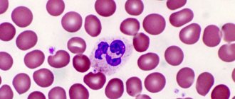
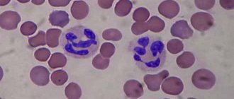
Mature neutrophils have a segmented nucleus, that is, they belong to polymorphonuclear leukocytes, or polymorphonuclears. They are classical phagocytes: they have adhesiveness, motility, chemotaxis, and the ability to capture particles (for example, bacteria).
Neutrophil chemotaxis is caused by protein fragments formed as a result of complement activation, factors of the fibrinolytic and kinin systems, as well as products of leukocyte, platelet and bacterial origin. Under the influence of chemotactic stimuli, the “edge state” (adhesion to endothelial cells) and diapedesis of neutrophils occur[1].
Mature segmented neutrophils are normally the main type of leukocytes circulating in human blood, making up from 47% to 72% of the total number of leukocytes in the blood. Another 1-5% are normally young, functionally immature neutrophils that have a rod-shaped solid nucleus and do not have the nuclear segmentation characteristic of mature neutrophils - the so-called band neutrophils.
Neutrophils are capable of active amoeboid movement, extravasation (emigration outside the blood vessels), and chemotaxis (predominant movement towards sites of inflammation or tissue damage).
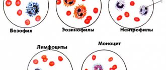
Kinds
The number of granulocytes in the blood is about 60% of the total number of white cells. These components, in turn, are divided into subspecies.
Thus, the following cells belong to granulocytes:
- Neutrophil granulocytes are the largest number of them in the blood of an adult or child. In essence, neutrophil granulocytes neutralize pathogenic organisms by “sacrificing” themselves.
- Basophils - cells instantly react to allergens, accelerate blood circulation and direct a large amount of fluid to the affected area.
- Eosinophilic granulocytes – act on parasitic organisms and prevent the development of an allergic reaction. Among the white bodies, these are the smallest and most mobile bodies.
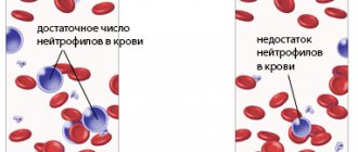
If certain granulocytes are low or high, this can negatively affect the functioning of the body, for example, when pathogenic organisms enter, the immune response may be too weak, which will lead to serious complications.
Types of granulocytes
Process of phagocytosis
Neutrophils are capable of phagocytosis, and they are microphages, that is, they are able to absorb only relatively small foreign particles or cells.
After phagocytosis of foreign particles, neutrophils usually die, releasing a large amount of biologically active substances that damage bacteria and fungi, increasing inflammation and chemotaxis of immune cells into the lesion.
Neutrophils contain large amounts of myeloperoxidase, an enzyme that can oxidize chlorine anion to hypochlorite, a strong antibacterial agent. Myeloperoxidase, as a heme-containing protein, has a greenish color, which determines the greenish tint of the neutrophils themselves, the color of pus and some other secretions rich in neutrophils.
Dead neutrophils, together with cellular detritus from tissues destroyed by inflammation and pyogenic microorganisms that caused inflammation, form a mass known as pus.
An increase in the proportion of neutrophils in the blood is called relative neutrophilosis
, or
relative neutrophilic leukocytosis
.
An increase in the absolute number of neutrophils in the blood is called absolute neutrophilosis
.
A decrease in the proportion of neutrophils in the blood is called relative neutropenia
.
A decrease in the absolute number of neutrophils in the blood is designated as absolute neutropenia
.
The first to meet microbe scouts are granulocytes patrolling our immune system.
Granulocytes , or, as they are also called, granular leukocytes, are the most numerous representatives of the leukocyte family, the number of which makes up 50-80% of the total number of white blood cells.
Granulocytes are formed in the bone marrow, then, having become a little accustomed and swimming in the bloodstream, they move into tissues and organs to their places of deployment, where they patrol the surroundings. As a matter of fact, they are very similar to our patrol service (PPS), i.e. on our policemen. They also drive UAZ cars around our agencies and “resolve” minor conflicts and scandals that do not require calling for reinforcements. They “go” to places where there are cuts, abscesses or penetration of microbes, thus maintaining the order and healthy condition of the area of the body or organ entrusted to them.
But bad luck, granulocytes, like PPS, do not really like to figure out who is right and who is wrong. They first hit the kidneys with a baton, and only then ask questions.
Having arrived at the scene of the crime, granulocytes wrap crime scenes with ribbons, put up barriers, and inside this “cordon sanitaire” they organize a real massacre. When the skin is damaged and there are open wounds, granulocytes rush to the scene and begin to devour everything indiscriminately, everything that seems suspicious to them and looks like an enemy.
Microbes devoured by granulocytes are killed by hydrogen peroxide, nitric oxide and hypochlorite, which are produced in the “stomach” of the granulocyte, called the lysosome.
Of course, often innocent “onlookers” - the cells of our body that happen to be nearby - also fall under the influence of such a “chemical attack”. Those same redness and swelling on the wounds are a consequence of granulocytes “cleaning” everyone indiscriminately, including our innocent tissues. It’s good at least that these rednesses are only a temporary and relatively harmless phenomenon for the body.
But, despite all their rudeness and clumsiness, granulocytes do an excellent job with their responsibilities of protecting our lungs and skin from all parasites, with minimal losses to the body. Without them, all our minor sores and scratches could easily develop into serious and dangerous diseases.
More serious and professional defenders of our body are macrophages . These are the special services of the Ministry of Internal Affairs and the FSB. And although they also belong to the family of leukocytes, as they say, the department is the same, but the capabilities are different. There are a hundred times fewer of them than the PPS of granulocytes, and they have the same weapons as granulocytes, but their “intelligence” and special training allow them to more competently cope with the impending threat.
In truth, macrophages traveling through blood vessels are called monocytes , and they are taken directly into macrophages when monocytes leave the blood into organs and tissues. They open their “representations” in the kidneys, liver, skin and lungs and “specialize” in microbes that especially like to enter our body in these places.
Clever macrophages are trained to “recognize” bacteria using receptor antennas. When faced with such an enemy microbe, the macrophage feels it with its receptors and “reads” information about the composition of its cell wall. The macrophage sends the received data for analysis to its “card index” - the cell nucleus, where a database of enemies and criminals is stored, and from there it receives clear instructions about the properties of the enemy and how to destroy it.
As soon as a macrophage recognizes an enemy, it immediately releases cytokines into the blood - intermediary molecules of a protein nature, which spread the news about the aggressor. In general, various types of cytokines are messengers, or, if you like, signalmen and military couriers. We'll talk about them in more detail a little later. For now, let's continue.
So, remember that our macrophages are intelligence agents? That is why, even after killing and dismembering the enemy, they retain a part of him, which in the future will be useful for establishing a more complete identity of the enemy, compiling his photo identikit and studying his qualities. Macrophages transfer this part to the intelligence structures of the body - a special category of lymphocytes - T-lymphocytes .
Lymphocytes: helpers and killers. Let's study! >>
^ Top
Antimicrobial functions
Neutrophils have a large set of antibiotic proteins, which are stored in two types of granules. Primary (azurophilic) granules are lysosomes containing acid hydrolases, myeloperoxidase and muramidase (lysozyme). In addition to lysozyme, lactoferrin was found in secondary (specific) granules. In addition to enzymes and lactoferrin, these granules contain high concentrations of antibiotic proteins - defensins, seprocidins, cathelicidins and protein that induces bacterial cell permeability [2].
Neutrophils play a very important role in protecting the body from bacterial and fungal infections, and a relatively lesser role in protecting against viral infections. Neutrophils play virtually no role in antitumor or anthelmintic defense.
Question 15. Structure and functional significance of granulocytes
Leukocytes of vertebrate animals are nucleated cells capable of active movement in the tissues of the body. The classification is based on taking into account the structural features of their cytoplasm.
Leukocytes, the cytoplasm of which contains specific granularity, are called granular, or granulocytes. Mature granular leukocytes have a nucleus divided into segments - segmented cells; in young ones it is unsegmented.
Therefore, it is customary to divide them into young forms (bean-shaped nucleus), rod (nucleus in the form of a curved rod) and segmented - fully differentiated leukocytes, the nucleus of which contains from 2 to 5-7 segments.
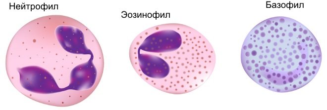
In accordance with the difference in the staining of cytoplasmic granules in the group of granulocytes, 3 types of cells are distinguished:
– basophils – granularity is colored violet with basic dyes;
– eosinophils – granularity is stained with acidic dyes in various shades of red;
– neutrophils – granularity is colored pink-violet with both acidic and basic dyes.
Neutrophils
– small cells (9-12 µm), the cytoplasm of which contains 2 types of granules: primary (basophilic), which are lysosomes, and secondary oxyphilic (containing cationic proteins and alkaline phosphatase).
Neutrophils are characterized by the finest (pulverized) granularity and the most segmented nucleus. They are microphages and carry out the phagocytic function of small foreign particles of any nature and the utilization of antigen-antibody complexes.
In addition, substances are released that stimulate the regeneration of damaged tissues.
Eosinophils
more often they contain a two-segment nucleus and large oxyphilic granules in the cytoplasm. Their diameter is 12-18 microns. The granules contain hydrolytic enzymes (microphages by function).
They exhibit antihistamine reactivity, stimulate the phagocytic activity of connective tissue macrophages and the formation of their lysosomes, and utilize antigen-antibody complexes.
But their main task is to neutralize toxic substances, so the number of eosinophils increases sharply during helminthic infestations.
Basophils
, 12-16 microns in size, contain medium-sized basophil granules, which contain heparin (prevents blood clotting) and histamine (regulates vascular and tissue permeability). They also participate in the development of allergic reactions.
The percentage ratio between individual types of leukocytes is called the leukocyte formula, or leukogram. For granulocytes, it looks like this:
– neutrophils – 25-40% – in pigs and ruminants; 50-70% – in horses and carnivores;
– eosinophils – 2-4%, in ruminants – 6-8%;
– basophils – 0.1-2%.
Netosis
In 2004, an important mechanism by which neutrophils exert protective functions was discovered, called NETosis.
(from the English NETosis (from NET - Neutrophil Extracellular Trap)) [3].
Netosis is the third major type of neutrophil cell death, along with apoptosis and necrosis.
With NETosis, the neutrophil goes through the stages of chromatin decondensation, production of reactive oxygen species (ROS - Reactive Oxygen Species), degranulation; this is followed by the release of a DNA network associated with ROS, histones, myeloperoxidase, and other pathogen-damaging molecules.
Pathogens, namely bacteria, fungi, parasites and viruses, become entangled in networks and die[4].
Neutrophil DNA traps are associated with the pathogenesis of various diseases such as sepsis, rheumatoid arthritis, thrombosis, lupus and other autoimmune diseases[5].
It has also been shown that other blood cells, such as monocytes, eosinophils, basophils, also have a similar mechanism called ETosis (from ET - Extracellular Trap) [6].
The neutrophil response (infiltration of the inflammatory focus by neutrophils, an increase in the number of neutrophils in the blood, a shift in the leukocyte formula to the left with an increase in the proportion of “young” forms, indicating increased production of neutrophils by the bone marrow) is the very first response to bacterial and many other infections.
The neutrophilic response in acute inflammation and infections always precedes the more specific lymphocytic response. In chronic inflammation and infections, the role of neutrophils is insignificant and the lymphocytic response predominates (infiltration of the inflammation site with lymphocytes, absolute or relative lymphocytosis in the blood).
Notes
- Royt A.
Cells that carry out the immune response // Immunology = Immunology / Royt A.; lane from English V. I. Kandrora, L. A. Pevnitsky. — 5th ed. - M.: Mir, 2000. - P. 38. - 592 p. — ISBN 5-03-003305-X. - Royt A.
Cells that carry out the immune response // Immunology = Immunology / Royt A.; lane from English V. I. Kandrora, L. A. Pevnitsky. — 5th ed. - M.: Mir, 2000. - P. 38. - 592 p. — ISBN 5-03-003305-X. - Brinkmann V., Reichard U., Goosmann C., Fauler B., Uhlemann Y., Weiss DS, Weinrauch Y., Zychlinsky A.
Neutrophil extracellular traps kill bacteria. (English) // Science (New York, NY). - 2004. - Vol. 303, no. 5663. - P. 1532-1535. — DOI:10.1126/science.1092385. - PMID 15001782. [fix] - Jenne CN, Wong CH, Zemp FJ, McDonald B., Rahman MM, Forsyth PA, McFadden G., Kubes P.
Neutrophils recruited to sites of infection protect from virus challenge by releasing neutrophil extracellular traps. (English) // Cell host & microbe. — 2013. — Vol. 13, no. 2. - P. 169-180. — DOI:10.1016/j.chom.2013.01.005. - PMID 23414757. [correct] - Xu J., Zhang X., Pelayo R., Monestier M., Ammollo CT, Semeraro F., Taylor FB, Esmon NL, Lupu F., Esmon CT
Extracellular histones are major mediators of death in sepsis. (English) // Nature medicine. - 2009. - Vol. 15, no. 11. - P. 1318-1321. — DOI:10.1038/nm.2053. - PMID 19855397. [correct] - Wartha F., Henriques-Normark B.
ETosis: a novel cell death pathway. (English) // Science signaling. - 2008. - Vol. 1, no. 21. - P. 25. - DOI:10.1126/stke.121pe25. - PMID 18506034. [correct]
Literature
- Mayansky A. N., Mayansky D. N.
Essays on the neutrophil and macrophage / Author. preface acad. Academy of Medical Sciences of the USSR A. D. Ado. Rec.: S. Belotsky, G. Nepomnyashchikh. — Novosibirsk: Science. Siberian Branch, 1983. - 256 p. - Mayansky A. N., Mayansky D. N.
Essays on the neutrophil and macrophage / Responsible. ed. V. P. Kaznacheev; USSR Academy of Sciences, Siberian Branch, USSR Academy of Medical Sciences, Siberian Branch, Institute of Clinical and Experimental Medicine. — 2nd ed., revised. and additional — Novosibirsk: Science. Sib. department, 1989. - 344 p. — ISBN 5-02-028718-0.
Norms of granulocytes in men, women, children
The normal number of granulocytes in the bloodstream is assessed by the most numerous group of cells - neutrophils (NEUT). If we talk about granulocytes themselves (GRA), the norms by age are as follows:
| Age | Normal: number of cells x10⁹ per liter of blood |
| Newborns | 6,0-26,1 |
| First day | 5,1-21,0 |
| A week | 1,5-10,0 |
| Up to a month | 1,0-9,0 |
| Up to six months | 1,0-8,5 |
| 1 year | 1,5-8,7 |
| Up to 4 years | 1,5-8,5 |
| Up to 8 years | 1,5-8,1 |
| Before 18 | 1,6-8,2 |
| Up to 25 years | 1,8-7,5 |
| Up to 50 and older | 1,5-7,7 |
These are average indicators that are assessed in a basic blood test taking into account reference values.